Özet
Kavernöz dönüşüm, çoğunlukla kronik ekstrahepatik portal ven tıkanıklığı sonrasında portal akımı taşıyan kıvrımlı kollaterallerin gelişmesidir. Kontrastlı BT, ana portal venin olağan yatağını, tıkanıklığın yayılımını ve portal çevresi damar ağını aynı incelemede göstererek kronik trombozu ve eşlik eden portal hipertansiyonu tanımaya yardım eder. Safra yolları boyunca uzanan kollateraller koledokta bası, kontur düzensizliği veya darlık oluşturabilir; sarılık ya da kolanjit nedeni gözden kaçabilir. Hilustaki kavernom damarları malign hiler lezyonlarla karışabileceğinden solid kitle görünümünü taklit edebilir.
Faz ve pencere
- Portal tanısal
- Ana portal ven yatağında daralma, kesintiye uğrama ya da izlenememe ile birlikte porta hepatis ve hepatoduodenal ligaman boyunca portal venle eş zamanlı kontrastlanan serpijinöz portoportal kollateraller aranır. Aynı fazda splenik ve üst mezenterik venlerin açıklığı, kollaterallerin safra yollarıyla ilişkisi ve portal hipertansiyon şantları değerlendirilir.
Önerilen pencereler: Anjiyo (G 600 / M 150), Batın (G 400 / M 50), Yumuşak doku (G 400 / M 40).
BT bulguları
- Ana portal venin beklenen seyrinde incelmesi, kesintiye uğraması veya lümeninin seçilememesi
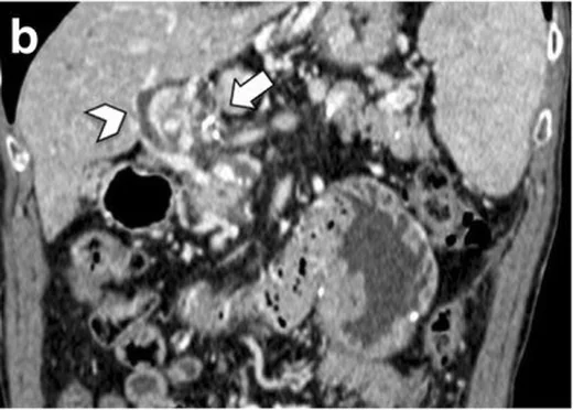
- Porta hepatiste ana ven yatağının çevresine yerleşen çok sayıda kıvrımlı, kontrastlanan portoportal kanal
- Kollaterallerin hepatoduodenal ligaman boyunca portal ven dallarına ve peripankreatik venöz ağa uzanması
- Kronik tıkanıklık zemininde ana portal ven yatağında ince fibrotik doku veya obstrüksiyon bulguları izlenebilir.
- Safra kesesi çevresi, koledok komşuluğu veya mide çevresinde genişlemiş venöz kollateraller
- Koledokta düzgün dış bası izlenimi, dalgalı kontur veya darlıkla birlikte proksimal safra yollarında genişleme
- Splenomegali, gastroözofageal varis, spontan portosistemik şant veya asit gibi portal hipertansiyon bulguları
- Dalak veni ya da üst mezenterik ven trombozunun ana portal ven tıkanıklığına eşlik etmesi
Normalde
Normal portal fazlı BT'de splenik ven ile üst mezenterik venin birleşmesinden oluşan ana portal ven, porta hepatiste tek ve devamlı lümen olarak izlenir; sağ ve sol portal dallara ayrılır. Aynı seviyede bu venöz eksenin yerini alan kıvrımlı damar kümesi bulunmaz ve koledok düzgün konturlu, çevre venöz pleksuslardan belirgin bası almamış görünür. Kıyaslamada ana portal venin devamlılığını, çevresindeki damar sayısını ve safra yolu konturunu birlikte takip et.
Normal BT ile kıyasla
Aynı bölgenin, kaynak veri setinde patoloji içermediği belirtilen bir BT incelemesini kesit kesit kaydırın; organ sınırlarını açarak anatomiyi hasta görüntüsüyle karşılaştırın. Küçük rastlantısal bulgular tümüyle dışlanmamıştır.
Kaynak: TotalSegmentator v2.01 veri seti (Wasserthal ve ark., Radiology: Artificial Intelligence 2023), CC BY 4.0, vaka s0541; organ sınırları veri setinin otomatik segmentasyonundan, birleştirilerek sadeleştirildi. Görüntüler 2,5 mm kesit aralığına yeniden örneklendi.
Ayırıcı tanı
- Akut portal ven trombozu
- genişlemiş ana portal ven lümeninde kontrastlanmayan dolum defekti görülür; kronik dönemdeki olgun periportal kollateral ağ baskın olmayabilir.
- Portal ven tümör trombüsü
- portal ven trombozunun ayırıcı tanısında malign portal ven invazyonu da düşünülmelidir.
- Hiler lenf nodu ya da tümör
- solid yumuşak doku portal venle aynı anda opasifiye olmaz; damar devamlılığı çok düzlemli kesitlerde izlenemez.
- Portal hipertansiyon varisleri
- özofagogastrik veya splenorenal kollateraller eşlik edebilir; kavernöz dönüşüm için ana portal ven yatağındaki kronik obstrüksiyon ve onu çevreleyen portoportal ağ aranır.
- Hepatik arteriyovenöz şant
- arteriyel fazda erken dolan damarlar ve erken portal ven opasifikasyonu görülebilir; yalnız portal fazdaki serpijinöz ağla karıştırılmamalıdır.
Tuzaklar
- Porta hepatisteki en geniş kollateral ana portal ven sanılabilir; damarı splenik-mezenterik ven birleşiminden intrahepatik portal dallara kadar ardışık kesitlerde izle.
- Hiler kollateral paketi tek parça yumuşak doku gibi görünebilir; kontrastlı ince kesitlerde ayrı lümenlerin portal venle eş zamanlı dolduğunu doğrula.
- Yetersiz portal ven opasifikasyonu, küçük kollateralleri görünmez kılabilir; damar açıklığı hakkında kesin yorum için faz zamanlamasını ve dalak veninin opasifikasyonunu kontrol et.
- Safra yolu genişlemesini otomatik olarak kollateral basısına bağlama; taş, kitle ve başka darlık nedenlerini ara; BT ince mukozal veya fibrotik değişiklikleri MRCP kadar iyi göstermez.
Kendini dene
Portal fazlı BT'de ana portal ven yatağı kayıp ve hilusta portal venle eş zamanlı dolan kıvrımlı damarlar görülüyor. En olası tanı hangisidir?
Cevabı göster
Kavernöz dönüşüm. Kronik ana portal ven obstrüksiyonunun çevresinde gelişen portoportal kollateral ağ kavernöz dönüşümün tipik görünümüdür. Akut trombozda genişlemiş ven içinde dolum defekti beklenir; lenfoma solid yumuşak dokudur, arteriyovenöz şantta ise erken arteriyel dolum ayırt ettirir.
Portal kavernom çevresinde koledokta düzgün dış bası ve proksimal kanal genişlemesi var. Safra yolu bulgularının en olası nedeni nedir?
Cevabı göster
Perikoledokal venöz bası. Portal kavernomla birlikte koledok çevresindeki venöz kollaterallerin dıştan basısı, portal hipertansiyona bağlı biliyer değişiklikleri açıklar. Portal kavernom tanısı için, bu safra yolu bulgularının primer sklerozan kolanjit, kolanjiyokarsinom veya taş hastalığı gibi başka bir patolojiye ait olmadığı ayırıcı tanı ile doğrulanmalıdır.
İlgili konular
Kaynaklar
Bu sayfadaki 39 cümle ve 2 quiz sorusu aşağıdaki kaynaklardan alıntılarla tek tek karşılaştırıldı; 4 cümle bu karşılaştırmada düzeltildi (2026-10-09).
- Portal cavernoma cholangiopathy: Update and recommendations on diagnosis and managementpmc.ncbi.nlm.nih.gov
- Biliary Obstruction following Transjugular Intrahepatic Portosystemic Shunt Creation in Patients with Variceal Bleeding.pubmed.ncbi.nlm.nih.gov
- Recanalization of the Portal Vein and Transjugular Intrahepatic Portosystemic Shunt (PVR-TIPS) as an Aid for Live Donor Liver Transplantation in Cirrhotic Patients with Portal Vein Thrombosis.pubmed.ncbi.nlm.nih.gov
- Hydatid disease of the liver with portal vein invasion and cavernous transformation: A case report and literature review.pubmed.ncbi.nlm.nih.gov
- Imaging and radiological interventions in extra-hepatic portal vein obstruction.pubmed.ncbi.nlm.nih.gov
- AGA Clinical Practice Update on Management of Portal Vein Thrombosis in Patients With Cirrhosis: Expert Review.pubmed.ncbi.nlm.nih.gov
- Hepatic morphology abnormalities: beyond cirrhosis.pubmed.ncbi.nlm.nih.gov
- Interventional Treatment of Acute Portal Vein Thrombosis.pubmed.ncbi.nlm.nih.gov
- Portomesenteric and portosystemic venous reconstructions in children using balloon-expandable endoprostheses.pubmed.ncbi.nlm.nih.gov
- [ELABORATION AND TREATMENT DECISION IN INCIDENTAL SPLANCHNIC VEIN THROMBOSIS].pubmed.ncbi.nlm.nih.gov
- Portal Vein Thrombosis: A Rare Finding in a Noncirrhotic Patient.pubmed.ncbi.nlm.nih.gov
- Liver transplantation in the setting of a spontaneous shunt between superior mesenteric vein and right renal vein.pubmed.ncbi.nlm.nih.gov
- Unusual aetiology of abdominal wall abscess: cholecystocutaneous fistula presenting 20 years after open subtotal cholecystectomy.pubmed.ncbi.nlm.nih.gov
- Portal vein thrombosis associated with psoriasis: a case report.pubmed.ncbi.nlm.nih.gov
- Pancytopenia With Hypocellular Bone Marrow Revealing Extrahepatic Portal Venous Obstruction and Cavernous Transformation in a Child: A Case Report of a Diagnostic Challenge.pubmed.ncbi.nlm.nih.gov
- Masked polycythemia vera presenting as non-cirrhotic portal hypertension with isolated gastric varices: a case report.pubmed.ncbi.nlm.nih.gov
- Clinical applications of 4D flow magnetic resonance imaging in hepatic vascular disorders: practical biomarkers, protocol design, and translational implementation.pubmed.ncbi.nlm.nih.gov
- Massive Esophagogastric Variceal Bleeding from Chronic Portal Vein Thrombosis in a Non-Cirrhotic Young Man: A Resource-Limited Endoscopic Management Dilemma.pubmed.ncbi.nlm.nih.gov
- Septic Pylephlebitis and Cavernous Transformation Presenting as Pyrexia of Unknown Origin: A Diagnostic Challenge.pubmed.ncbi.nlm.nih.gov
- Portal Vein Invasion Secondary to Hepatic Hydatid Disease: A Rare Case Report.pubmed.ncbi.nlm.nih.gov
- Spontaneous bacteremia and spontaneous bacterial peritonitis share similar prognosis in patients with cirrhosis: a cohort study.pubmed.ncbi.nlm.nih.gov
- Long-term safety and effectiveness of the "OptEase" vena cava filter.pubmed.ncbi.nlm.nih.gov
- Patterns of injury and violence in Yaoundé Cameroon: an analysis of hospital data.pubmed.ncbi.nlm.nih.gov
- The Role of Imaging in Portal Vein Thrombosis: From the Diagnosis to the Interventional Radiological Management.pubmed.ncbi.nlm.nih.gov
- Portomesenteric Reconstruction during Whipple Procedure Using Autologous Left Renal Vein Patch Graft in a Patient with a Gastric Cancer Recurrence.pubmed.ncbi.nlm.nih.gov
- Hemodynamic, Surgical and Oncological Outcomes of 40 Distal Pancreatectomies with Celiac and Left Gastric Arteries Resection (DP CAR) without Arterial Reconstructions and Preoperative Embolization.pubmed.ncbi.nlm.nih.gov
- Imaging diagnosis and staging of pancreatic ductal adenocarcinoma: a comprehensive review.pubmed.ncbi.nlm.nih.gov
- Benign focal liver lesions: The role of magnetic resonance imaging.pubmed.ncbi.nlm.nih.gov
- Portal cavernoma cholangiopathy: A systematic review of current understanding, clinical significance, and management.pubmed.ncbi.nlm.nih.gov
- Portal biliopathy, magnetic resonance imaging and magnetic resonance cholangiopancreatography findings: a case series.pubmed.ncbi.nlm.nih.gov
- Imaging of portal cavernoma cholangiopathy.pubmed.ncbi.nlm.nih.gov
- Right portal branch to ovarian vein anastomosis: An unusual portosystemic shunt secondary to pelvic venous congestion.pubmed.ncbi.nlm.nih.gov
- Life-Threatening Variceal Bleeding in Antisynthetase Syndrome: Nodular Regenerative Hyperplasia and Porto-Sinusoidal Vascular Disorder.pubmed.ncbi.nlm.nih.gov
- Long-term outcome of sinusoidal obstruction syndrome secondary to hematopoietic stem cell transplantation.pubmed.ncbi.nlm.nih.gov
- Portal vein recanalization and transjugular intrahepatic portosystemic shunt in chronic portal vein thrombosis: a narrative review.pubmed.ncbi.nlm.nih.gov
- Gun-sight technique for recanalization of chronic noncirrhotic, nonmalignant extrahepatic portal vein obstruction.pubmed.ncbi.nlm.nih.gov
- Peribiliary Inflammatory Phlegmon Masquerading as Cholangiocarcinoma in Janus Kinase 2 V617F-positive Primary Myelofibrosis: A Multimodality Imaging Diagnosis.pubmed.ncbi.nlm.nih.gov
- Contrast-Enhanced CT of the Coelom in a Female Bald Eagle (<i>Haliaeetus leucocephalus</i>): Development of a Preliminary CT-Based Anatomical Segmentation Workflow.pubmed.ncbi.nlm.nih.gov
- Schwannoma Likely Arising from the Pancreatic Head Nerve Plexus That Mimicked a Mucinous Cystic Neoplasm and Was Treated by Pancreaticoduodenectomy: A Case Report.pubmed.ncbi.nlm.nih.gov
- Duodenal imaging on the spotlight: from A to Z.pubmed.ncbi.nlm.nih.gov
- International Harmonization of Nomenclature and Diagnostic Criteria (INHAND): Non-proliferative and Proliferative Lesions of the Non-human Primate ( M. fascicularis ).pubmed.ncbi.nlm.nih.gov
- Medical hyperspectral imaging: an updated review of technology advancements and biomedical applications.pubmed.ncbi.nlm.nih.gov
- International Society of Paediatric Surgical Oncology (IPSO) Surgical Practice Guidelines.pubmed.ncbi.nlm.nih.gov
- EACR 2025 Congress: Innovative Cancer Science, 16-19 June 2025.pubmed.ncbi.nlm.nih.gov
- Diagnostic and Therapeutic Challenges of Chronic Portal Vein Thrombosis Secondary to Myeloproliferative Neoplasm in Sub-Saharan Region: A Series of Cases and Literature Review.pubmed.ncbi.nlm.nih.gov
- Budd-Chiari syndrome: imaging review.pubmed.ncbi.nlm.nih.gov
- Imaging Diagnosis of Splanchnic Venous Thrombosis.pubmed.ncbi.nlm.nih.gov
- Liver Imaging Reporting and Data System (LI-RADS): A Comprehensive Review.pubmed.ncbi.nlm.nih.gov
- When Cancer Clots: An Extensive Radiologic Analysis of Cancer-Associated Thromboembolism.pubmed.ncbi.nlm.nih.gov
- Prognostic Significance of Portal Vein Tumor Thrombus in Pancreatic Ductal Adenocarcinoma Treated With Chemoradiotherapy.pubmed.ncbi.nlm.nih.gov
- Splenic pathologies: A pictorial review of common, wandering, twisting and rare presentations.pubmed.ncbi.nlm.nih.gov
- Pathophysiological management of gastric varices: From hemodynamics to targeted therapies.pubmed.ncbi.nlm.nih.gov
- Coagulation profile and platelet count in pediatric patients with extrahepatic portal vein obstruction.pubmed.ncbi.nlm.nih.gov
- A rare case of esophageal variceal bleeding as a result of portal hypertension due to extra-hepatic portal vein obstruction and its management in a 7-year-old.pubmed.ncbi.nlm.nih.gov
- Cardiovascular Computed Tomography Angiographic Assessment of Simple Cardiac Shunts in Adults.pubmed.ncbi.nlm.nih.gov
- Chinese Guidelines for Clinical Diagnosis, Treatment, and Management of Cirrhosis (2025).pubmed.ncbi.nlm.nih.gov
- Imaging of inferior vena cava normal variants, anomalies and pathologies, Part 2: Acquired.pubmed.ncbi.nlm.nih.gov
- High-Frequency Miniprobe Endoscopic Ultrasonography Across the Gastrointestinal Tract.pubmed.ncbi.nlm.nih.gov
- Clinical Practice Guideline—Acute and Chronic Pancreatitis.pubmed.ncbi.nlm.nih.gov
- DUCT reveals architectural mechanisms contributing to bile duct recovery in a mouse model for Alagille syndrome.pubmed.ncbi.nlm.nih.gov
- Lymphatic Vessel Network Structure and Physiology.pubmed.ncbi.nlm.nih.gov
- Commentary: an eye on PET quantification.pubmed.ncbi.nlm.nih.gov
- High-affinity alphavbeta3 integrin targeted optical probe as a new imaging biomarker for early atherosclerosis: initial studies in Watanabe rabbits.pubmed.ncbi.nlm.nih.gov
- Arterial and biliary complications after transarterial chemoembolization for hepatocellular carcinoma.pubmed.ncbi.nlm.nih.gov
- Role of imaging in visceral vascular emergencies.pubmed.ncbi.nlm.nih.gov
- Liver Transplant Complications Radiologist Can't Miss.pubmed.ncbi.nlm.nih.gov
- Long-Term Outcomes of Endoscopic Management of Patients with Symptomatic Portal Cavernoma Cholangiopathy with No Shuntable Veins for Surgery or Failed Surgery.pubmed.ncbi.nlm.nih.gov
- Portal Vein Thrombosis: State-of-the-Art Review.pubmed.ncbi.nlm.nih.gov
- Portal cavernoma cholangiopathy: consensus statement of a working party of the Indian national association for study of the liver.pubmed.ncbi.nlm.nih.gov
- Is preoperative histological diagnosis necessary before referral to major surgery for cholangiocarcinoma?pubmed.ncbi.nlm.nih.gov
- Current concepts in the management of non-cirrhotic non-malignant portal vein thrombosis.pubmed.ncbi.nlm.nih.gov
- Utility of Diagnostic Imaging in the Diagnosis and Management of Schistosomiasis.pubmed.ncbi.nlm.nih.gov
- Transportal Embolization of the Dominant Outflow Vein for a Pancreatic Head Arteriovenous Malformation: A Case Report.pubmed.ncbi.nlm.nih.gov
- Imaging features, diagnosis, and clinical management of hepatic involvement in hereditary hemorrhagic telangiectasia: a case series report and literature review.pubmed.ncbi.nlm.nih.gov
- Delayed hepatic artery pseudoaneurysm with arterioportal fistula after penetrating liver trauma successfully treated with coil embolization: a case report and focused literature review.pubmed.ncbi.nlm.nih.gov
- Secondary sclerosing cholangitis: contemporary etiologies, diagnostic pathways, and treatment strategies for clinicians: a narrative review.pubmed.ncbi.nlm.nih.gov
- Cholangiocyte biology in primary sclerosing cholangitis and other cholangiopathies: pathogenesis, clinical insights, and experimental tools.pubmed.ncbi.nlm.nih.gov
- Artificial Intelligence and Radiomics in Cholangiocarcinoma: A Comprehensive Review.pubmed.ncbi.nlm.nih.gov
- PSC-DM: A Validated Clinical Diagnostic Model for Distinguishing Primary From Secondary Sclerosing Cholangitis.pubmed.ncbi.nlm.nih.gov
- Diffuse Large B-Cell Lymphoma Masquerading as Cholangiocarcinoma in a Young Adult.pubmed.ncbi.nlm.nih.gov
- Alveolar echinococcosis.pubmed.ncbi.nlm.nih.gov
- Common and Uncommon Splenic Lesions: A Review.pubmed.ncbi.nlm.nih.gov
- Can Artificial Intelligence Really Help? A Practicing Radiologist's Simplified Guide to AI, with a Critical Appraisal of the Use of AI in Cancer-Associated Thromboembolism.pubmed.ncbi.nlm.nih.gov
- Photon-counting detector CT with iodine quantification: improved distinction between bland and neoplastic portal vein thrombosis.pubmed.ncbi.nlm.nih.gov
Eğitim amaçlıdır; hasta başında tanı ve tedavi kararının yerine geçmez. İçerik yapay zekâ destekli bir süreçle güncel literatüre göre cümle cümle kaynaklarla karşılaştırıldı, ancak uzman radyolog incelemesinden geçmedi. Klinik kararlarda güncel kılavuzları, kurum protokollerini ve radyoloji raporunu esas alın.