Özet
Nodüler rejeneratif hiperplazi, karaciğerde yaygın küçük rejeneratif nodüllerin belirgin fibrotik septalar olmadan geliştiği ve portal hipertansiyona yol açabildiği mikrovasküler bir hastalıktır. BT bulguları çoğu kez silik veya yoktur; görülebilen heterojen parankim ve küçük nodüller özgül değildir, bu yüzden tanı yalnız görüntüye dayanmaz. Siroz görünümü belirgin olmadan splenomegali, kollateraller veya asit bulunması bu olasılığı akla getirir. Yanlışlıkla siroz sayılması etiyolojik bağlamın atlanmasına, portal hipertansiyonun ise fark edilmemesine neden olabilir.
Okuma sırası
- Karaciğer parankimi. Parankimde keskin sınırlı kitleler yerine, yama yerleşimli ve ince yapıdaki diffüz nodüler değişimlere dikkat edin; ancak görüntüleme tek başına tanı koydurmaz.
- Karaciğer kapsülü ve loblar. Kapsülün komşu kesitlerdeki seyrini ve lob hacimlerini kontrol edin; portal hipertansiyon olmasına karşın siroza özgü belirgin yüzey nodülaritesi bulunmayabilir.
- Dalak ve portal ven çevresi. Dalak boyutunu ve portal ven çevresindeki damarları inceleyerek non-sirotik portal hipertansiyon paternini arayın.
- Kollateraller, periton ve hepatik venler. Perigastrik kollateraller ve asiti kaydedin; portal ve hepatik venlerde tromboz veya çıkış engeli olup olmadığını son olarak kontrol edin.
Faz ve pencere
- Portal tanısal
- BT ve MRG'de karaciğerde yaygın heterojenlik veya FNH benzeri lezyonlar görülebilir; ancak kesin tanı için yüksek kaliteli karaciğer biyopsisi gerekir. Splenomegali, kollateraller, asit ve portal/hepatik ven açıklığı birlikte incelenir.
- Arteriyel
- Arteriyel fazda görülebilen nodüller çoğunlukla çevre parankime göre izodens veya hipodens kalır; belirgin arteriyel hiperkontrastlanan fokal lezyon NRH için tipik kabul edilmez ve başka odakların ayırıcı tanısını gerektirir.
Önerilen pencereler: Batın (G 400 / M 50), Karaciğer (G 150 / M 30), Yumuşak doku (G 400 / M 40).
BT bulguları
- Diffüz heterojen parankim — karaciğer boyunca yaygın, belirgin kitle oluşturmayan atenüasyon/kontrastlanma farklılığı.
- İnce nodüler dönüşüm — çok sayıda küçük difüz nodül; BT'de nodüller bazen seçilemeyebilir ve tanıya yönelik belirgin bir kontrastlanma paterni olmayabilir.
- Siroz morfolojisinin yokluğu — belirgin kapsüler nodülarite, lobar küçülme ve fibrotik septa görünümü olmadan portal hipertansiyon.
- Splenomegali — portal basınç artışına eşlik edebilen dalak büyümesi.
- Portosistemik kollateraller — portal hipertansiyona eşlik edebilen kollateral damarlar görüntülemede belirginleşebilir.
- Asit — portal hipertansiyona eşlik eden peritoneal serbest sıvı.
- Damar açıklığının korunması — portal veya hepatik ven trombozu saptanmaması, mikrovasküler hastalık olasılığını destekleyebilir.
Normalde
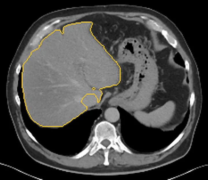
Normal karaciğer portal fazda homojen atenüasyon ve kontrastlanma gösterir; parankim boyunca yaygın ince nodüler görünüm, splenomegali, kollateraller veya asit bulunmaz. Kıyaslamada kapsüler yüzey ve lob hacimlerinin sirotik yeniden dağılım göstermediği halde portal hipertansiyon işaretlerinin var olup olmadığına odaklanın.
Normal BT ile kıyasla
Aynı bölgenin, kaynak veri setinde patoloji içermediği belirtilen bir BT incelemesini kesit kesit kaydırın; organ sınırlarını açarak anatomiyi hasta görüntüsüyle karşılaştırın. Küçük rastlantısal bulgular tümüyle dışlanmamıştır.
Kaynak: TotalSegmentator v2.01 veri seti (Wasserthal ve ark., Radiology: Artificial Intelligence 2023), CC BY 4.0, vaka s0541; organ sınırları veri setinin otomatik segmentasyonundan, birleştirilerek sadeleştirildi. Görüntüler 2,5 mm kesit aralığına yeniden örneklendi.
Ayırıcı tanı
- Siroz
- nodüler yüzey ve fibrotik lobar yeniden dağılım daha belirgindir; NRH'de portal hipertansiyon siroz morfolojisi olmadan ortaya çıkabilir.
- Fokal nodüler hiperplazi
- PSVD'de FNH benzeri lezyonlar görülebilir; bu nedenle FNH benzeri görünüm NRH ile ilişkili vasküler hastalığı dışlamaz.
- Hepatik adenom
- tipik olarak hipervasküler bir hepatoselüler lezyondur; NRH ise karaciğer parankiminin yaygın mikronodüler dönüşümüyle karakterizedir.
- Portal ven trombozu
- portal hipertansiyona yol açabilir ve PSVD ile birlikte bulunabilir; bu nedenle portal ven açıklığı ayrıca değerlendirilmelidir.
- Sinüzoidal obstrüksiyon sendromu
- kemoterapiyle ilişkili karaciğer hasarı örüntüleri arasında yer alabilir; vasküler karaciğer hastalıklarında damar yapıları ve parankim kontrastlanma örüntüleri görüntülemeyle değerlendirilir.
Tuzaklar
- Görüntülemede değişikliklerin ince ve kolay gözden kaçırılabilen yama yerleşimli olması nedeniyle, tek başına negatif görüntüleme NRH'yi kesin dışlamaz; tanı için biyopsi gereklidir.
- Splenomegali ve varisleri tek başına siroz kanıtı saymayın; yüzey, lob hacimleri ve karaciğer hastalığı bağlamını birlikte değerlendirin.
- NRH'yi yalnız BT ile kesinleştirmeyin; görüntüleme bulguları özgül değildir ve histolojik tanı için nodüler dönüşüm ile fibrotik septaların yokluğu aranır.
Kendini dene
Belirgin siroz morfolojisi olmayan hastada splenomegali ve varisler görülüyor; karaciğer parankimi ince heterojen. Hangi tanı düşünülmelidir?
Cevabı göster
Nodüler rejeneratif hiperplazi. NRH, siroz morfolojisi belirgin olmadan portal hipertansiyon yapabilen diffüz mikronodüler dönüşümdür. FNH ve adenom gibi lezyonlar genellikle sınırlı alanlarda bulunur ve NRH'deki gibi diffüz mikronodüler yapıya sahip değildirler.
BT'de NRH lehine portal hipertansiyon bulguları var ancak nodüller seçilemiyor. En doğru yorum hangidir?
Cevabı göster
BT bulguları özgül değildir. NRH'nin BT bulguları özgül değildir ve tanı için histolojik doğrulama gereklidir.
İlgili konular
Kaynaklar
Bu sayfadaki 36 cümle ve 2 quiz sorusu aşağıdaki kaynaklardan alıntılarla tek tek karşılaştırıldı; 5 cümle bu karşılaştırmada düzeltildi (2026-10-09).
- Nodular Regenerative Hyperplasia of the Liver in Rheumatic Disease: Cases and Review of the Literaturepmc.ncbi.nlm.nih.gov
- Trastuzumab Emtansine–Associated Porto-Sinusoidal Vascular Disorder: Clinical Features and Outcomes from Published Casespmc.ncbi.nlm.nih.gov
- Rituximab and improved nodular regenerative hyperplasia-associated non-cirrhotic liver disease in common variable immunodeficiency: a case report and literature studypmc.ncbi.nlm.nih.gov
- Vascular tumors of the liver: A brief reviewpmc.ncbi.nlm.nih.gov
- EASL postgraduate course report: Vascular biology in chronic liver disease and clinical management implications ☆pmc.ncbi.nlm.nih.gov
- Hepatic manifestations of telomere biology disorders.pubmed.ncbi.nlm.nih.gov
- Benign liver tumors in pediatric patients - Review with emphasis on imaging features.pubmed.ncbi.nlm.nih.gov
- Portal Vein Thrombosis: Imaging the Spectrum of Disease With an Emphasis on MRI Features.pubmed.ncbi.nlm.nih.gov
- Space Occupying Lesions in the Liver.pubmed.ncbi.nlm.nih.gov
- Rare benign tumors of the liver: still rare?pubmed.ncbi.nlm.nih.gov
- A pictorial review of benign hepatocellular nodular lesions: comprehensive radiological assessment incorporating the concept of anomalous portal tract syndrome.pubmed.ncbi.nlm.nih.gov
- Nodular Regenerative Hyperplasia Induced by Trastuzumab Emtansine: Role of Emtansine?pubmed.ncbi.nlm.nih.gov
- Benign Hepatocellular Nodules: Hepatobiliary Phase of Gadoxetic Acid-enhanced MR Imaging Based on Molecular Background.pubmed.ncbi.nlm.nih.gov
- Congenital Extrahepatic Portosystemic Shunts: Spectrum of Findings on Ultrasound, Computed Tomography, and Magnetic Resonance Imaging.pubmed.ncbi.nlm.nih.gov
- Treatment of a non-typical hepatic pseudolesion complicated by greatly elevated alpha fetoprotein: case report and literature review.pubmed.ncbi.nlm.nih.gov
- Hyperintense liver lesions on hepatobiliary phase of gadoxetic acid-enhanced magnetic resonance imaging: clinical insights and significance.pubmed.ncbi.nlm.nih.gov
- Porto-sinusoidal vascular disorder.pubmed.ncbi.nlm.nih.gov
- Non-cirrhotic portal hypertension - diagnosis and management.pubmed.ncbi.nlm.nih.gov
- Colorectal liver metastases.pubmed.ncbi.nlm.nih.gov
- Imaging features of porto-sinusoidal vascular disorder in a case-control study: diagnostic value for differentiation from liver cirrhosis.pubmed.ncbi.nlm.nih.gov
- Current approach to diagnosis and treatment of focal nodular hyperplasia in children with a management algorithm.pubmed.ncbi.nlm.nih.gov
- Role of Vascular Liver Diseases in Hepatocellular Carcinoma Development.pubmed.ncbi.nlm.nih.gov
- A Real World Analysis of Imaging Compared to Biopsy for Diagnosis of Pediatric Liver Tumors.pubmed.ncbi.nlm.nih.gov
- Primary Hepatic Masses in Ten Dogs: A Retrospective Surgical Case Series.pubmed.ncbi.nlm.nih.gov
- Suspected Paramalignant Pleural Effusion as the Primary Presentation of Disseminated Cholangiocarcinoma in a Cat.pubmed.ncbi.nlm.nih.gov
- Trastuzumab Emtansine-Associated Porto-Sinusoidal Vascular Disorder: Clinical Features and Outcomes from Published Cases.pubmed.ncbi.nlm.nih.gov
- Life-Threatening Variceal Bleeding in Antisynthetase Syndrome: Nodular Regenerative Hyperplasia and Porto-Sinusoidal Vascular Disorder.pubmed.ncbi.nlm.nih.gov
- Overview of Non-Cirrhotic Portal Hypertension in Pediatric Patients.pubmed.ncbi.nlm.nih.gov
- Case Report: Primary myelofibrosis presenting with non-cirrhotic portal hypertension and initially misdiagnosed as cirrhosis.pubmed.ncbi.nlm.nih.gov
- POEMS syndrome misdiagnosed as liver cirrhosis: a case report.pubmed.ncbi.nlm.nih.gov
- Imaging and pathological features of pediatric sclerosing angiomatoid nodular transformation of the spleen: a case series and hypothesis-generating perspective on non-operative management.pubmed.ncbi.nlm.nih.gov
- Segmental atrophy of the liver: Review of a rare pseudotumor.pubmed.ncbi.nlm.nih.gov
- The role of ceus in the characterization of indeterminate focal liver lesions at second-level imaging methods (INFOLIL STUDY).pubmed.ncbi.nlm.nih.gov
- Surgical treatment and prognosis of type II congenital extrahepatic portosystemic shunts: A single-center experience of 31 cases.pubmed.ncbi.nlm.nih.gov
- Portal Vein Thrombosis in Patients Without Cirrhosis: Current Practical Approaches and Treatment Strategies.pubmed.ncbi.nlm.nih.gov
- A case of hepatic segmental atrophy mimicking spontaneously necrotized hepatocellular carcinoma.pubmed.ncbi.nlm.nih.gov
- Focal Liver Lesions in Budd-Chiari Syndrome: Spectrum of Imaging Findings.pubmed.ncbi.nlm.nih.gov
- Contrast-Enhanced Low-Mechanical-Index Endoscopic Ultrasound for the Evaluation of Focal Liver Lesions.pubmed.ncbi.nlm.nih.gov
- Case Report: Contrast-enhanced US and CT imaging features of nodule-in-nodule hepatocellular carcinoma in a dog.pubmed.ncbi.nlm.nih.gov
- Case Report: Rare spontaneous hepatic rupture hemorrhage complicated by autoimmune liver disease.pubmed.ncbi.nlm.nih.gov
- Histological and Molecular Evaluation of Liver Biopsies: A Practical and Updated Review.pubmed.ncbi.nlm.nih.gov
- Imaging of a glomus tumor of the liver in a child.pubmed.ncbi.nlm.nih.gov
- Noncirrhotic portal hypertension.pubmed.ncbi.nlm.nih.gov
- Spontaneous bacteremia and spontaneous bacterial peritonitis share similar prognosis in patients with cirrhosis: a cohort study.pubmed.ncbi.nlm.nih.gov
- Nodular Regenerative Hyperplasia Suggested by Noninvasive Findings during Adjuvant Trastuzumab Emtansine Therapy for Human Epidermal Growth Factor Receptor 2-Positive Breast Cancer.pubmed.ncbi.nlm.nih.gov
- [Postoperative Imaging Findings of Biliary Atresia].pubmed.ncbi.nlm.nih.gov
- Transjugular intrahepatic portosystemic shunt as a feasible option for portal hypertension due to Porto-sinusoidal vascular disorder: a case report.pubmed.ncbi.nlm.nih.gov
- China Liver Cancer Guidelines for the Diagnosis and Treatment of Hepatocellular Carcinoma (2024 Edition).pubmed.ncbi.nlm.nih.gov
- Pulmonary vascular manifestations of hereditary haemorrhagic telangiectasia.pubmed.ncbi.nlm.nih.gov
- Triple-Phase Computed Tomography for Differentiating Canine Cutaneous and Subcutaneous Mast Cell Tumors From Benign Lesions: A Size-Based Evaluation.pubmed.ncbi.nlm.nih.gov
- Clinical and CT/MRI features of hepatic AL amyloidosis: preliminary experience in 10 cases.pubmed.ncbi.nlm.nih.gov
- Case Report: Multimodality imaging features and serial metabolic changes on <sup>18</sup> F-FDG PET/CT in a dog with granulomatous hepatitis.pubmed.ncbi.nlm.nih.gov
- Acute portal vein thrombosis postimage-guided liver biopsy in common variable immunodeficiency.pubmed.ncbi.nlm.nih.gov
- Advances in Precise Assessment of Portal Pressure in Porto-sinusoidal Vascular Disease.pubmed.ncbi.nlm.nih.gov
- Imaging features facilitate diagnosis of porto-sinusoidal vascular disorder.pubmed.ncbi.nlm.nih.gov
- Chinese Multidisciplinary Expert Consensus on Orphan/Anticopper Drugs and Other Non-drug Management of Hepatolenticular Degeneration.pubmed.ncbi.nlm.nih.gov
- Magnetic Resonance Elastography of Liver: Current Update.pubmed.ncbi.nlm.nih.gov
- Case Report: Indocyanine green fluorescence imaging in complex focal nodular hyperplasia resection: report of two cases.pubmed.ncbi.nlm.nih.gov
- Idiopathic portal hypertension with transfusion-associated hemolytic anemia: a case report.pubmed.ncbi.nlm.nih.gov
- Type Ib Abernethy Malformation Complicated by Hepatopulmonary Syndrome: A Case Report of a Rare Entity in a Pediatric Patient.pubmed.ncbi.nlm.nih.gov
- Fontan-associated liver disease: the importance of multidisciplinary teamwork in its management.pubmed.ncbi.nlm.nih.gov
- Imaging of the chemotherapy-induced hepatic damage: Yellow liver, blue liver, and pseudocirrhosis.pubmed.ncbi.nlm.nih.gov
- Diagnostic challenges in non-cirrhotic portal hypertension - porto sinusoidal vascular disease.pubmed.ncbi.nlm.nih.gov
- Hypervascular liver lesions.pubmed.ncbi.nlm.nih.gov
- Focal nodular hyperplasia-like nodules arising in the setting of hepatic vascular disorders with portosystemic shunting show β-catenin activation.pubmed.ncbi.nlm.nih.gov
- Ultrasound Evaluation of Fontan-Associated Liver Disease: A State-of-the-Art Review.pubmed.ncbi.nlm.nih.gov
- All about portal vein: a pictorial display to anatomy, variants and physiopathology.pubmed.ncbi.nlm.nih.gov
- The Impact of Genetics on Pediatric Interstitial Lung Diseases: A Narrative Literature Review and Clinical Implications.pubmed.ncbi.nlm.nih.gov
- Pediatric Cholestasis: A Practical Approach to Histological Diagnosis.pubmed.ncbi.nlm.nih.gov
- Interobserver study on histologic features of idiopathic non-cirrhotic portal hypertension.pubmed.ncbi.nlm.nih.gov
- Imaging in Vascular Liver Diseases.pubmed.ncbi.nlm.nih.gov
- Preoperative imaging of colorectal liver metastases: what the radiologist and the multidisciplinary team need to know.pubmed.ncbi.nlm.nih.gov
- Correlation of pretreatment imaging features with lung shunt fraction in patients with large hepatocellular carcinoma (maximum diameter >8 cm) planned for <sup>90</sup>Y-SIRT.pubmed.ncbi.nlm.nih.gov
- Assessment of Three-Phasic CT Scan Findings of Cirrhosis Due to Primary Sclerosing Cholangitis Versus Cryptogenic Cirrhosis.pubmed.ncbi.nlm.nih.gov
- Genicular Artery Embolization for Osteoarthritis Related Knee Pain: A Systematic Review and Qualitative Analysis of Clinical Outcomes.pubmed.ncbi.nlm.nih.gov
- Massive bleeding from a Bricker ileal conduit during FOLFIRINOX therapy in noncirrhotic portal hypertension: suspected oxaliplatin-associated sinusoidal injury and presumed irinotecan-associated conduit mucositis: a case report.pubmed.ncbi.nlm.nih.gov
- Hepatic encephalopathy due to non-cirrhotic portal hypertension associated with non-alcoholic steatohepatitis and chronic hepatitis B: a Case Report.pubmed.ncbi.nlm.nih.gov
- Noninvasive imaging of hepatic dysfunction: A state-of-the-art review.pubmed.ncbi.nlm.nih.gov
- Imaging features, diagnosis, and clinical management of hepatic involvement in hereditary hemorrhagic telangiectasia: a case series report and literature review.pubmed.ncbi.nlm.nih.gov
- Hepatic Vascular Involvement in Adenosine Deaminase 2 Deficiency (DADA2): Case Reports and Literature Review.pubmed.ncbi.nlm.nih.gov
- The pseudocirrhotic liver: Hepar lobatum carcinomatosum in a patient with triple-negative breast cancer.pubmed.ncbi.nlm.nih.gov
- Functional liver imaging score derived from hepatobiliary-specific contrast-enhanced MRI: a study on agreement and correlation.pubmed.ncbi.nlm.nih.gov
- Focal Nodular Hyperplasia Presenting With Biliary Obstruction and Intrahepatic Gallstones: A Case Report and Literature Review.pubmed.ncbi.nlm.nih.gov
- Vascular tumors of the liver: A brief review.pubmed.ncbi.nlm.nih.gov
- Update on the Pathology of Pediatric Liver Tumors: A Pictorial Review.pubmed.ncbi.nlm.nih.gov
- Liver tumors in children with chronic liver diseases.pubmed.ncbi.nlm.nih.gov
- Technical overview of pediatric liver transplantation for congenital absence of the portal vein: a collective review of six cases at a single center.pubmed.ncbi.nlm.nih.gov
- Severe Bleeding From Portal Hypertension in a Young Patient With Common Variable Immunodeficiency-Associated Nodular Regenerative Hyperplasia.pubmed.ncbi.nlm.nih.gov
- Segmental Atrophy of the Liver: An Increasingly Recognized Pseudotumor With Distinct Clinicopathologic and Imaging Features.pubmed.ncbi.nlm.nih.gov
- Long-term outcome of sinusoidal obstruction syndrome secondary to hematopoietic stem cell transplantation.pubmed.ncbi.nlm.nih.gov
- Diagnostic Yield of Liver Biopsy in Porto-Sinusoidal Vascular Disorder: Are Conventional Adequacy Criteria Always Indispensable?pubmed.ncbi.nlm.nih.gov
- Reliable diagnosis of liver focal nodular hyperplasia by a combination of T2 weighted signal and three diffusion magnetic resonance metrics of diffusion-derived 'vessel density', slow diffusion coefficient, and apparent diffusion coefficient: a validation study of two centers' data.pubmed.ncbi.nlm.nih.gov
- Gadoxetic acid-enhanced MRI in hepatocellular carcinoma: a comprehensive review of diagnostic, surveillance, and treatment response prediction and assessment.pubmed.ncbi.nlm.nih.gov
- Case Report: Hepatic sinusoidal obstruction syndrome caused by <i>Gynura <b>s</b>egetum</i>.pubmed.ncbi.nlm.nih.gov
- Tegafur/Uracil-Induced Sinusoidal Obstruction Syndrome as a Suspected Cause of Liver Cirrhosis during Adjuvant Chemotherapy for Lung Adenocarcinoma: A Case Report.pubmed.ncbi.nlm.nih.gov
- Hepatic Angiosarcoma Mimicking Sinusoidal Obstruction Syndrome: A Case Report.pubmed.ncbi.nlm.nih.gov
Eğitim amaçlıdır; hasta başında tanı ve tedavi kararının yerine geçmez. İçerik yapay zekâ destekli bir süreçle güncel literatüre göre cümle cümle kaynaklarla karşılaştırıldı, ancak uzman radyolog incelemesinden geçmedi. Klinik kararlarda güncel kılavuzları, kurum protokollerini ve radyoloji raporunu esas alın.