Özet
Portal ven anevrizması, ana portal ven, bifurkasyon veya dallarında gelişebilen nadir sakküler ya da fuziform genişlemedir; konjenital olabilir veya portal hipertansiyon ve kronik karaciğer hastalığıyla ilişkili gelişebilir. Çoğu olgu tesadüfen saptanır, ancak lümen trombozu, safra yollarına bası ve komşu yapılarda etkilenme önem taşır. Kontrastlı BT, genişlemenin portal venle bağlantısını ve tüm portal venöz ağ içindeki yerini gösterirken trombüs ve kalsifikasyonları da ortaya koyar. Damarla ilişkisi kurulmadan kistik veya solid kitle sanılması yanlış tanıya, trombüsün gözden kaçması ise portal akım komplikasyonlarının eksik değerlendirilmesine yol açabilir.
Faz ve pencere
- Portal tanısal
- İyi opaklaşmış portal venle aynı yoğunlukta kontrastlanan, portal venle kesintisiz devamlılığı görülen sınırlı sakküler çıkıntı veya fuziform genişleme aranır; kısmi trombüs kontrastlanmayan eksantrik dolum defekti, duvar kalsifikasyonu ise yüksek atenüasyonlu ince çizgiler şeklindedir.
Önerilen pencereler: Karaciğer (G 150 / M 30), Batın (G 400 / M 50), Anjiyo (G 600 / M 150).
BT bulguları
- Ana portal ven, bifurkasyon, intrahepatik dal veya splenomezenterik konfluens düzeyinde sakküler/fuziform lümen genişlemesi
- Portal ven lümeniyle doğrudan devamlılık ve portal fazda damar havuzuna benzer opaklaşma
- Anevrizma kesesi içinde kontrastlanmayan mural ya da santral trombüs
- Kronik trombüs veya anevrizma duvarında ince kalsifikasyon odakları
- Ana portal venin ve sağ-sol dalların açık olup olmadığının, trombüsün splenik ven veya üst mezenterik vene uzanımının gösterilmesi
- Safra kanalına, duodenuma veya inferior vena kavaya bası varsa yer değiştirme ya da kanal genişlemesi
- Siroz, splenomegali, kollateral damarlar ve asit gibi eşlik eden portal hipertansiyon bulguları
Ölçütler ve sınıflamalar
- Ekstrahepatik ana portal ven anevrizması için kullanılan çap eşiği
- Ekstrahepatik portal ven anevrizması tanısında >20 mm çap yaygın kullanılan eşiktir; kaynaklarda sirozlu hastalar için >19 mm, normal karaciğer için >15 mm değerleri de tanımlanmıştır.
Normalde
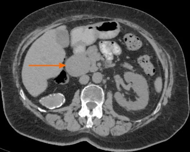
Normal ana portal venle karşılaştırmada, fokal sakküler anomali veya fuziform genişleme portal ven anevrizmasını düşündüren morfolojilerdir. Portal ven anevrizması kistik veya solid kitleleri taklit edebileceğinden, kistik görünümlü odağın portal venöz sistemle devamlılığını çok düzlemli görüntülerde değerlendir.
Normal BT ile kıyasla
Aynı bölgenin, kaynak veri setinde patoloji içermediği belirtilen bir BT incelemesini kesit kesit kaydırın; organ sınırlarını açarak anatomiyi hasta görüntüsüyle karşılaştırın. Küçük rastlantısal bulgular tümüyle dışlanmamıştır.
Kaynak: TotalSegmentator v2.01 veri seti (Wasserthal ve ark., Radiology: Artificial Intelligence 2023), CC BY 4.0, vaka s0541; organ sınırları veri setinin otomatik segmentasyonundan, birleştirilerek sadeleştirildi. Görüntüler 2,5 mm kesit aralığına yeniden örneklendi.
Ayırıcı tanı
- Portal ven trombozu
- lümen genişlemesi eşlik edebilse de kontrastlanmayan intraluminal materyal damar boyunca uzanır; kronikte porta hepatis çevresinde kavernöz kollateraller gelişebilir.
- Portal ven psödoanevrizması
- çoğunlukla cerrahi veya travma sonrası ortaya çıkar; eşlik eden doku hasarı ve düzensiz damar duvarı, gerçek duvarlı anevrizmadan ayrımda yardımcıdır.
- Hepatik arter anevrizması
- arteriyel fazda arterle aynı anda ve yoğunlukta dolar, portal venöz sisteme değil hepatik arter dallarına bağlanır.
- Koledok kisti
- safra yollarıyla devamlı sıvı yoğunluklu yapı görünür ve damar lümeni gibi portal faz kontrastı almaz.
- Basit karaciğer kisti
- ultrasonografide sakküler portal ven anevrizmasıyla karışabilir; her ikisi de iyi sınırlı ve anekoik görünebilir.
Tuzaklar
- Portal ven anevrizması bifurkasyon düzeyinde yerleşebilir; portal venöz faz BT'de fokal sakküler anomali veya fuziform genişleme olarak görünür.
- Kısmi trombüs kesenin tümü kontrastlanmıyormuş izlenimi verebilir; anevrizma içindeki opaklaşan akım kanalını ve trombüsün portal ven boyunca uzanımını ayrı ayrı tanımla.
- Eşik değer kaynaklarda farklı normal karaciğer ve siroz grupları için tartışılmıştır; çapı ölçerken düzlemi damara dik seç ve klinik bağlamdan bağımsız tek bir sayı ile küçük intrahepatik dalı sınıflandırma.
Kendini dene
Portal faz BT'de porta hepatis'teki yuvarlak oluşum ana portal venle kesintisiz devam ediyor ve damar gibi opaklaşıyor. En olası tanı nedir?
Cevabı göster
Portal ven anevrizması. Portal ven anevrizması portal venöz sistemin sakküler veya fuziform genişlemesidir; BT'de portal venöz fazda kontrastlanan bir odak olarak görülebilir. Ana portal venle kesintisiz devamlılık, odağın portal ven kökenini destekler.
Genişlemiş portal ven kesesinde kontrastlanmayan eksantrik materyal izleniyor. Bu alan en olası nedir?
Cevabı göster
Mural trombüs. Portal venle devamlı genişlemiş lümen içindeki kontrastlanmayan eksantrik dolum defekti mural trombüsle uyumludur. Safra çamuru safra kesesi veya kanalı içinde olur; hepatoselüler karsinom parankimal kitle oluşturur; akım artefaktı gerçek mural materyal görünümünü ve kesitler arası sürekliliği açıklamaz.
İlgili konular
Kaynaklar
Bu sayfadaki 36 cümle ve 2 quiz sorusu aşağıdaki kaynaklardan alıntılarla otomatik olarak karşılaştırıldı; 5 cümle bu karşılaştırmada düzeltildi (2026-10-09).
- Portal vein aneurysm-etiology, multimodal imaging and current managementpmc.ncbi.nlm.nih.gov
- Asymptomatic saccular portal vein aneurysm: a case report and review of the literaturepmc.ncbi.nlm.nih.gov
- Giant extra-hepatic thrombosed portal vein aneurysm: a case report and review of the literaturepmc.ncbi.nlm.nih.gov
- Portal vein aneurysm mimicking a pancreatic mass: A case report with an emphasis on imaging pitfallspmc.ncbi.nlm.nih.gov
- Thrombosis of extrahepatic portal vein aneurysm immediately after laparoscopic distal gastrectomy: A case reportpmc.ncbi.nlm.nih.gov
- Portal vein aneurysm: What to know.pubmed.ncbi.nlm.nih.gov
- Giant extra-hepatic thrombosed portal vein aneurysm: a case report and review of the literature.pubmed.ncbi.nlm.nih.gov
- [A case of extrahepatic portal vein aneurysm with massive thrombosis].pubmed.ncbi.nlm.nih.gov
- Asymptomatic thrombosis of extrahepatic portal vein aneurysm necessitating hybrid operative repair.pubmed.ncbi.nlm.nih.gov
- Extrahepatic portal vein aneurysm.pubmed.ncbi.nlm.nih.gov
- Portal vein aneurysm in thalassaemia.pubmed.ncbi.nlm.nih.gov
- Portal vein aneurysms: a case series with literature review.pubmed.ncbi.nlm.nih.gov
- Extrahepatic Portal Vein Aneurysm.pubmed.ncbi.nlm.nih.gov
- Calcified wall portal venous aneurysm: a case report.pubmed.ncbi.nlm.nih.gov
- Asymptomatic portal vein aneurysm: Three case reports.pubmed.ncbi.nlm.nih.gov
- Portal vein aneurysm and portal biliopathy.pubmed.ncbi.nlm.nih.gov
- Visceral artery aneurysms: a literature review of etiology, natural history, diagnosis and treatment paradigms.pubmed.ncbi.nlm.nih.gov
- Hypertrophic Cardiomyopathy as a Key Feature of MRAS-Related Noonan Syndrome: New Case and Comprehensive Literature Review.pubmed.ncbi.nlm.nih.gov
- Extrahepatic Portal Vein Occlusion and Collateral Formation Following Anticoagulant-Induced Thrombosis Regression in a Portal Vein Aneurysm: Potential for Misdiagnosis As Primary Extrahepatic Portal Vein Occlusion.pubmed.ncbi.nlm.nih.gov
- Vascular complications in percutaneous biliary interventions.pubmed.ncbi.nlm.nih.gov
- Ruptured Pseudoaneurysm of Right Hepatic Artery as Immediate Complication of Lumen-Apposing Metal Stent Placement for Choledochoduodenostomy: A Case Report.pubmed.ncbi.nlm.nih.gov
- Arc of Buhler pseudoaneurysm complicated by an arterioportal fistula: a combined endovascular and open surgical approach.pubmed.ncbi.nlm.nih.gov
- A Silent Mimic: Biopsy-Proven Focal Nodular Hyperplasia Masquerading as Chronic Intrahepatic Biliary Obstruction.pubmed.ncbi.nlm.nih.gov
- Giant Azygos Vein Aneurysm Mimicking a Mediastinal Mass With Segmental Partial Anomalous Pulmonary Venous Return.pubmed.ncbi.nlm.nih.gov
- Diagnostic and management dilemmas in pulmonary hypertension.pubmed.ncbi.nlm.nih.gov
- Emphysematous conditions of the abdomen and pelvis: pearls and pitfalls.pubmed.ncbi.nlm.nih.gov
- The inferior vena cava: anatomical variants and acquired pathologies.pubmed.ncbi.nlm.nih.gov
- Pitfall in necrotizing pancreatitis: Organized hematoma with arteriovenous fistula mimicking a splenic artery pseudoaneurysm.pubmed.ncbi.nlm.nih.gov
- Cinematic rendering in CT angiography: a pictorial review of clinical applications.pubmed.ncbi.nlm.nih.gov
- Cystic lesions and their mimics involving the intrahepatic bile ducts and peribiliary space: diagnosis, complications, and management.pubmed.ncbi.nlm.nih.gov
- Portal vein aneurysm-etiology, multimodal imaging and current management.pubmed.ncbi.nlm.nih.gov
- Asymptomatic saccular portal vein aneurysm: a case report and review of the literature.pubmed.ncbi.nlm.nih.gov
- Technical considerations of endovascular management of true visceral artery aneurysms.pubmed.ncbi.nlm.nih.gov
- Portal vein aneurysm mimicking a pancreatic mass: A case report with an emphasis on imaging pitfalls.pubmed.ncbi.nlm.nih.gov
- Computed tomography of pediatric abdominal trauma: optimizing utilization and enhancing diagnostic interpretation.pubmed.ncbi.nlm.nih.gov
- Pancreatic Pseudocyst Masquerading as Right Atrial Thrombus: An Unusual Intracardiac Extension.pubmed.ncbi.nlm.nih.gov
- Calcified left ventricular aneurysm: Delayed thrombus formation leading to multiorgan thrombosis.pubmed.ncbi.nlm.nih.gov
- Gut Dysbiosis, Sex, and Vascular Smooth Muscle Cells in Abdominal Aortic Aneurysms.pubmed.ncbi.nlm.nih.gov
- Open surgical resection of giant common hepatic artery aneurysm after failed endovascular repair.pubmed.ncbi.nlm.nih.gov
- Celiac artery stenosis threshold for stratifying management in spontaneous isolated dissection.pubmed.ncbi.nlm.nih.gov
- Widespread pylephlebitis and infective thromboembolism due to <i>Streptococcus anginosus</i>-related appendiceal rupture: A case report.pubmed.ncbi.nlm.nih.gov
- Hemosuccus Pancreaticus from Large Inferior Pancreaticoduodenal Artery Pseudoaneurysm Complicating Chronic Pancreatitis.pubmed.ncbi.nlm.nih.gov
- Retrospective Three-Dimensional CT Reconstruction in Complex Emergency Hepato-Pancreato-Biliary Surgery: A Proof-of-Concept Case Series with Anatomical Concordance to Intraoperative Findings and Narrative Review.pubmed.ncbi.nlm.nih.gov
- Clinical applications of 4D flow magnetic resonance imaging in hepatic vascular disorders: practical biomarkers, protocol design, and translational implementation.pubmed.ncbi.nlm.nih.gov
- The challenging diagnosis of sinistral portal hypertension in a patient with liver cirrhosis: a case report and literature review.pubmed.ncbi.nlm.nih.gov
- A rare case of splenic arteriovenous fistula causing portal hypertension, treated by embolisation.pubmed.ncbi.nlm.nih.gov
- Cardiovascular Computed Tomography Angiographic Assessment of Simple Cardiac Shunts in Adults.pubmed.ncbi.nlm.nih.gov
- A Protocol for Body MRI/CT and Extraction of Imaging-Derived Phenotypes (IDPs) from the China Phenobank Project.pubmed.ncbi.nlm.nih.gov
- Case Report: Delayed intra-abdominal hemorrhage in a patient with a duodenal stump tumor after Billroth II gastrectomy.pubmed.ncbi.nlm.nih.gov
- Detecting biliary complications following liver transplantation with contrast-enhanced ultrasound.pubmed.ncbi.nlm.nih.gov
- An Accessory Right Hepatic and Cystic Arteries Derived from the Superior Mesenteric Artery: A Cadaveric Case Report.pubmed.ncbi.nlm.nih.gov
- Navigating the biliary maze: a retrospective cohort study of three-dimensional navigation technology improving outcomes in congenital biliary dilatation resection.pubmed.ncbi.nlm.nih.gov
- Narrative review of the current management of ectopic varices: an update.pubmed.ncbi.nlm.nih.gov
- Case Report: Making room for radicality: balancing liver reserve and oncologic clearance in hilar cholangiocarcinoma through portal vein embolisation.pubmed.ncbi.nlm.nih.gov
- Peribiliary Inflammatory Phlegmon Masquerading as Cholangiocarcinoma in Janus Kinase 2 V617F-positive Primary Myelofibrosis: A Multimodality Imaging Diagnosis.pubmed.ncbi.nlm.nih.gov
- Massive Esophagogastric Variceal Bleeding from Chronic Portal Vein Thrombosis in a Non-Cirrhotic Young Man: A Resource-Limited Endoscopic Management Dilemma.pubmed.ncbi.nlm.nih.gov
- An unusual presentation of portal biliopathy manifesting as chronic abdominal pain as a delayed post-splenectomy complication: a case report.pubmed.ncbi.nlm.nih.gov
- Gastrointestinal bleeding from an arterial-enteric fistula postcoil embolisation of GDA pseudoaneurysm after pancreatico-duodenectomy.pubmed.ncbi.nlm.nih.gov
- Traumatic superior mesenteric artery pseudoaneurysm complicated by high-output arteriovenous fistula.pubmed.ncbi.nlm.nih.gov
- A safe seal: controlled flow arrest with dual balloon protection for embolisation of a large spontaneous splenic arteriovenous fistula-a case report.pubmed.ncbi.nlm.nih.gov
- An unusual superior mesenteric arteriovenous malformation causing obscure gastrointestinal bleeding: a case report.pubmed.ncbi.nlm.nih.gov
- Adult Hepatic Infarction for Internists: A Practical Diagnostic and Management Pathway to Avoid Misdiagnosis, Unnecessary Drainage, and Delayed Vascular Recognition.pubmed.ncbi.nlm.nih.gov
- Hepatoduodenal ligament teratoma presenting with fever in a young adult.pubmed.ncbi.nlm.nih.gov
- Imaging insights into pediatric liver masses: A comprehensive minireview for hepatology practice.pubmed.ncbi.nlm.nih.gov
- Fistulating Intraductal Papillary Mucinous Neoplasms (IPMNs): Case Series and Discussion of a Rare Complication.pubmed.ncbi.nlm.nih.gov
- Dual-Energy CT Images: Pearls and Pitfalls.pubmed.ncbi.nlm.nih.gov
- The Role of CT Imaging in Characterization of Small Renal Masses.pubmed.ncbi.nlm.nih.gov
- Technical and clinical outcomes of endovascular treatment for splenic artery aneurysms.pubmed.ncbi.nlm.nih.gov
- Multiparametric Abdominal Ultrasound as a Source of Imaging Biomarkers in Predictive and Personalized Medicine: A Narrative Review.pubmed.ncbi.nlm.nih.gov
- Multimodal clinical parameters-based immune status associated with the prognosis in patients with hepatocellular carcinoma.pubmed.ncbi.nlm.nih.gov
- Liver Transplantation for Biliary Atresia in Adulthood: Single-Centre Surgical Experience.pubmed.ncbi.nlm.nih.gov
- Portal vein recanalization and transjugular intrahepatic portosystemic shunt in chronic portal vein thrombosis: a narrative review.pubmed.ncbi.nlm.nih.gov
- Preoperative CT and inflammatory index-based risk scores for predicting short-term recurrence following resection of pancreatic ductal adenocarcinoma.pubmed.ncbi.nlm.nih.gov
- CT-Based Reference Values for Splenic Artery and Vein Diameters in Individuals Aged 1-80 Years.pubmed.ncbi.nlm.nih.gov
- Transjugular Intrahepatic Portosystemic Shunt for Portal Vein Thrombosis in Cirrhotic Patients: 18-Year Experience in a Tertiary Referral Hospital.pubmed.ncbi.nlm.nih.gov
- Endovascular Treatment of Congenital Portosystemic Shunt: A Single-Center Prospective Study.pubmed.ncbi.nlm.nih.gov
- Endovascular flow reduction after portal vein arterialization: a technical note.pubmed.ncbi.nlm.nih.gov
- Pulmonary artery aneurysm: computed tomography (CT) imaging findings and diagnosis.pubmed.ncbi.nlm.nih.gov
- Role of imaging in penetrating vascular injuries of the craniocervical region.pubmed.ncbi.nlm.nih.gov
- The filling defect of pulmonary artery, an imaging finding what we should know.pubmed.ncbi.nlm.nih.gov
- Demonstration of Hepatic Vein Abnormalities Using Contrast-Enhanced Sonography in Liver Diseases.pubmed.ncbi.nlm.nih.gov
- Behçet's Disease Revealed by a Thrombosed Aneurysm of the Superior Mesenteric Vein: A Case Report.pubmed.ncbi.nlm.nih.gov
- Pathologically confirmed complete remission after neoadjuvant chemotherapy in patients with advanced intrapancreatic cholangiocarcinoma following robot-assisted excision of choledochal cyst: A case report.pubmed.ncbi.nlm.nih.gov
- Surgical Anatomy of the Liver-Significance in Ovarian Cancer Surgery.pubmed.ncbi.nlm.nih.gov
- Endovascular closure of a large intrahepatic portosystemic shunt in a three-month-old infant: a case report and literature review.pubmed.ncbi.nlm.nih.gov
- Patient-specific computational fluid dynamics analysis of portal vein hemodynamics before and after balloon angioplasty following living donor liver transplantation: a proof-of-concept study.pubmed.ncbi.nlm.nih.gov
- From congenital variants to critical clues: A radiologic review of inferior vena cava pathologies.pubmed.ncbi.nlm.nih.gov
- The Role of Imaging in Portal Vein Thrombosis: From the Diagnosis to the Interventional Radiological Management.pubmed.ncbi.nlm.nih.gov
- The British technique of purely robotic Whipple's pancreatoduodenectomy a systematic and stepwise ('clockwise') approach.pubmed.ncbi.nlm.nih.gov
- Clinical anatomy of hepatic vessels by computed tomography angiography: A minireview.pubmed.ncbi.nlm.nih.gov
- Endovascular treatment of a pancreatic pseudoaneurysm associated with massive pancreatic arteriovenous malformation using flow modification technique.pubmed.ncbi.nlm.nih.gov
- Mapping the vascular complications of acute pancreatitis: focus on splanchnic vein thrombosis.pubmed.ncbi.nlm.nih.gov
- Percutaneous left renal vein stenting in posterior nutcracker syndrome: technical and antithrombotic nuances - a case report.pubmed.ncbi.nlm.nih.gov
- Non-contrast microvascular flow imaging in abdominal ultrasound: a pictorial essay.pubmed.ncbi.nlm.nih.gov
- Hepatic portal venous gas in severe neurocritical illness after intracranial surgery: Two case reports and literature review.pubmed.ncbi.nlm.nih.gov
- Beyond embryological remnants: imaging of ligamentum teres hepatis and falciform ligament pathologies.pubmed.ncbi.nlm.nih.gov
- Ablative stereotactic MR-guided radiotherapy for >10 cm solid pseudopapillary tumor of the pancreas with liver oligometastases: a case report and literature review.pubmed.ncbi.nlm.nih.gov
- The application of computational fluid dynamics in hepatic portal vein haemodynamics research: a narrative review.pubmed.ncbi.nlm.nih.gov
- Long-term safety and effectiveness of the "OptEase" vena cava filter.pubmed.ncbi.nlm.nih.gov
- The Blood Supply of the Stomach: Anatomical and Surgical Considerations.pubmed.ncbi.nlm.nih.gov
- Role of Grey Scale and Color Doppler in the Diagnosis of Portal Hypertension: A Comprehensive Review.pubmed.ncbi.nlm.nih.gov
- Missed Opportunities in the Prevention of First Esophageal Variceal Bleeding: A Real-World Clinical Audit of Undiagnosed and Under-Treated Patients.pubmed.ncbi.nlm.nih.gov
- Case Report: Primary myelofibrosis presenting with non-cirrhotic portal hypertension and initially misdiagnosed as cirrhosis.pubmed.ncbi.nlm.nih.gov
- Asymmetric Sensitivity of Hepatic Venous Pressure Gradient to Portal and Sinusoidal Resistance: An In Vitro Experimental Study.pubmed.ncbi.nlm.nih.gov
- Hughes-Stovin Syndrome: A Rare Cause of Thrombosis and Pulmonary Artery Aneurysm.pubmed.ncbi.nlm.nih.gov
- Splenic infarction: an uncommon yet significant complication in COVID-19 patients - a case series report and literature review.pubmed.ncbi.nlm.nih.gov
- Thoracic Complications in Behçet's Disease: Imaging Findings.pubmed.ncbi.nlm.nih.gov
- Clinical, Radiological, and Endoscopic Features of Pancreatic Pseudocyst and Walled-Off Necrosis: How to Diagnose and How to Drain Them.pubmed.ncbi.nlm.nih.gov
- Adrenal hemorrhage and hemorrhagic masses; diagnostic workup and imaging findings.pubmed.ncbi.nlm.nih.gov
- Macroscopic and Histopathologic Findings From a Mass Stranding of Rough-Toothed Dolphins ( Steno bredanensis ) in 2005 on Marathon Key, Florida, USA.pubmed.ncbi.nlm.nih.gov
- The Arc of Bühler: A Cadaveric Case Report of a Novel Variant.pubmed.ncbi.nlm.nih.gov
Eğitim amaçlıdır; hasta başında tanı ve tedavi kararının yerine geçmez, tıbbi tavsiye değildir. Metinler yapay zekâ desteğiyle yazıldı ve otomatik bir adımda kaynak alıntılarıyla karşılaştırıldı; bu bir tıbbi doğrulama değildir ve içerik uzman radyolog incelemesinden geçmedi. Klinik kararlarda güncel kılavuzları, kurum protokollerini ve radyoloji raporunu esas alın.