
Özet
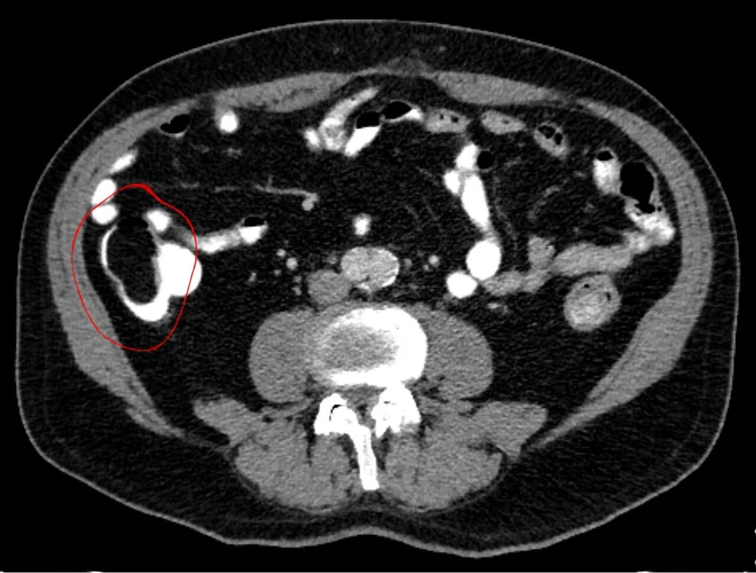
Kolon lipomu, submukozadan kaynaklanan ve çoğu kez tesadüfen saptanan benign yağ dokusu kitlesidir. Kontrastsız BT'de düzgün sınırlı, homojen yağ atenüasyonlu intralüminal lezyon olarak tanınır; büyük lezyonlarda invajinasyon veya tıkanma eşlik edebilir. BT, yağ içeriğini ve bağırsak segmentleri arasındaki invajinasyonu göstererek olası öncü odağı ortaya koyar. Yağ atenüasyonunun kaybolduğu infarkte invajinasyon veya eşlik eden obstrüksiyon yanlışlıkla malignite olarak değerlendirilebilir; duvar ve çevre bağırsak da incelenmelidir.
Faz ve pencere
- Kontrastsız tanısal
- Kolon lümenine doğru uzanan, düzgün sınırlı homojen kitle içinde makroskopik yağ atenüasyonu görülür; yağ dansitesi tanı için kontrastsız seride gösterilebilir ve intravenöz kontrast gerekli değildir. İntussusepsiyonda BT’de hedef veya sosis biçimli görünüm oluşabilir; mezenterik yağ ve damarlar invajine bağırsak segmentleriyle birlikte görülebilir. İnfarkte uğramış lipom heterojen görünüm alabilir.
Önerilen pencereler: Batın (G 400 / M 50), Yumuşak doku (G 400 / M 40).
BT bulguları
- Homojen yağ atenüasyonlu intralüminal kitle — kolon lümeni içinde, çevre yumuşak dokudan belirgin düşük atenüasyonlu odak.
- Düzgün ve keskin sınır — submukozal yerleşimli lezyon kolon lümenine doğru yuvarlak ya da oval kabarıklık yapar.
- Yağ yoğunluğu — saf lipom tipik olarak makroskopik yağ değerleri taşır; eşik aralığı ölçüm ölçütlerinde verilmiştir.
- Submukozal büyüme biçimi — kitle bağırsak dış yüzünden değil lümen iç yüzünden kaynaklanan yumuşak, düzgün konturlu kabarıklıktır.
- Kolokolik invajinasyon — önde gelen yağ kitlesi iç içe geçmiş kolon duvarlarının başında görülebilir.
- Mezenterik çekilme — invajinasyon içinde eşlik eden mezenterik yağ ve damarlar hedef veya tübüler yapı içine taşınır.
- İnfarkt/yağ nekrozu — invajine lipomda tipik homojen yağ atenüasyonu yerini heterojen içeriğe bırakabilir.
- Mekanik obstrüksiyon eşlik ettiğinde kolonda belirgin dilatasyon görülebilir.
Ölçütler ve sınıflamalar
- Tipik lipom BT atenüasyonu
- −40 ile −120 HU
Normalde
Normal kolon lümeninde dışkı ve gaz bulunabilir, ancak bağırsak duvarına bağlı düzgün sınırlı makroskopik yağ kitlesi ve iç içe geçmiş kolon segmentleri görülmez. Şüpheli kitleyi çevre perikolik yağla HU açısından kıyaslayın ve lezyonun lümen içindeki konumunu, submukozal tabanını, lümen açıklığını ve proksimal kolon çapını değerlendirin.
Normal BT ile kıyasla
Aynı bölgenin, kaynak veri setinde patoloji içermediği belirtilen bir BT incelemesini kesit kesit kaydırın; organ sınırlarını açarak anatomiyi hasta görüntüsüyle karşılaştırın. Küçük rastlantısal bulgular tümüyle dışlanmamıştır.
Kaynak: TotalSegmentator v2.01 veri seti (Wasserthal ve ark., Radiology: Artificial Intelligence 2023), CC BY 4.0, vaka s0119; organ sınırları veri setinin otomatik segmentasyonundan, birleştirilerek sadeleştirildi. Görüntüler 2,5 mm kesit aralığına yeniden örneklendi.
Ayırıcı tanı
- Kolon adenomu
- yumuşak doku atenüasyonlu mukozal kitle oluşturur; homojen makroskopik yağ yoğunluğu beklenmez.
- Kolon adenokarsinomu
- BT’de kolon duvarı kalınlaşması malignite şüphesi doğurabilir; düzgün sınırlı, homojen yağ atenüasyonlu görünüm lipom lehinedir.
- Kolokolik invajinasyon
- hedef biçimli iç içe bağırsak duvarları bir bulgudur; öncü kitle lipom olmayabilir ve özellikle erişkinde başka tümör odağı aranmalıdır.
- Liposarkom veya başka yağlı kitle
- düzgün sınırlı yağ atenüasyonlu görünüm lipomu düşündürse de BT’de benign ve malign lezyonları ayırt etmek mümkün olmayabilir.
Tuzaklar
- İnvajinasyon içindeki lipomun yağ yoğunluğunu korumayabileceğini unutmayın; ödem, iskemi ve yağ nekrozu kitleyi heterojen yumuşak doku gibi gösterebilir.
- Kolon içindeki dışkı parçalarını duvara bağlı kitle sanmayın; kolon lipomu BT’de düzgün sınırlı, homojen yağ atenüasyonlu intralüminal kitle olarak görülebilir.
- İnvajinasyonu gördüğünüzde yalnız öncü yağlı lezyona odaklanmayın; tıkanma, mezenterik damarların çekilmesi ve bağırsak duvarında iskemi bulgularını da arayın.
Kendini dene
Transvers kolon içinde düzgün sınırlı, homojen −80 HU ölçülen submukozal kitle saptanıyor. En olası tanı?
Cevabı göster
Kolon lipomu. Düzgün sınırlı submukozal kitlenin makroskopik yağ atenüasyonu lipom lehinedir. Adenom ve adenokarsinom yumuşak doku dansitesinde olur; fekalom ise duvara bağlı düzenli kitle değildir ve gaz/dışkı karışımı görünür.
BT’de intussusepsiyon için tanımlanan karakteristik görünüm hangisidir?
Cevabı göster
Hedef veya sosis biçimli görünüm. BT’de intussusepsiyon hedef veya sosis biçimli görünüm oluşturabilir. Diğer seçenekler lipom için endoskopik veya baryumlu incelemelerde tarif edilen işaretlerdir.
Kaynaklar
Bu sayfadaki 38 cümle ve 2 quiz sorusu aşağıdaki kaynaklardan alıntılarla tek tek karşılaştırıldı; 4 cümle bu karşılaştırmada düzeltildi (2026-10-09).
- Colonic lipoma, a rare cause of intestinal intussusception: A narrative review and how to diagnose itpmc.ncbi.nlm.nih.gov
- Colonic Lipoma Causing Bowel Intussusception: An Up-to-Date Systematic Reviewpmc.ncbi.nlm.nih.gov
- Giant Colonic Lipoma Remains a Surgeon's Domain: A Report of Two Casespmc.ncbi.nlm.nih.gov
- Giant lipoma of the colon presenting as a case of adult intussusception: A case seriespmc.ncbi.nlm.nih.gov
- Minimally invasive management of colonic lipoma causing intussusception with endoloop in an adultpmc.ncbi.nlm.nih.gov
- Colonic lipomas revisited on CT colonography.pubmed.ncbi.nlm.nih.gov
- Colonic Lipoma Causing Bowel Intussusception: An Up-to-Date Systematic Review.pubmed.ncbi.nlm.nih.gov
- Giant submucosal lipoma cause colo-colonic intussusception. A case report and review of literature.pubmed.ncbi.nlm.nih.gov
- Colonic lipoma, a rare cause of intestinal intussusception: A narrative review and how to diagnose it.pubmed.ncbi.nlm.nih.gov
- Splenic flexure colonic lipoma causing intussusception.pubmed.ncbi.nlm.nih.gov
- Pedunculated sigmoid lipoma causing colo-colonic intussusception.pubmed.ncbi.nlm.nih.gov
- Submucosal lipoma of the large intestine masquerading as a colonic malignancy.pubmed.ncbi.nlm.nih.gov
- Laparoscopic assisted submucosal excision of an intussuscepting colonic lipoma.pubmed.ncbi.nlm.nih.gov
- Pedunculated colonic lipoma prolapsing through the anus.pubmed.ncbi.nlm.nih.gov
- Ileocolic intussusception caused by giant submucosal colonic lipoma: A rare case report.pubmed.ncbi.nlm.nih.gov
- Unroofing technique for endoscopic resection of a large colonic lipoma.pubmed.ncbi.nlm.nih.gov
- Large benign submucosal lipoma presented with descending colonic intussusception in an adult.pubmed.ncbi.nlm.nih.gov
- Large pedunculated colonic lipoma: a rare cause of colorectal intussusception in adults.pubmed.ncbi.nlm.nih.gov
- Colonic lipoma of the hepatic flexure causing invagination in an adult.pubmed.ncbi.nlm.nih.gov
- Colocolic intussusception caused by a descending colon lipoma in an adult: a rare case report.pubmed.ncbi.nlm.nih.gov
- Giant Sigmoid Lipoma With Necrosis Mimicking Colorectal Cancer and Intussusception: A Case Report.pubmed.ncbi.nlm.nih.gov
- Gastrointestinal Tract Polyp Detection, Tracking, and Surveillance and the Role of Artificial Intelligence.pubmed.ncbi.nlm.nih.gov
- Minimally invasive management of colonic lipoma causing intussusception with endoloop in an adult.pubmed.ncbi.nlm.nih.gov
- Colonic lipoma presenting as colo-colic intussusception in an adult.pubmed.ncbi.nlm.nih.gov
- Rare Adult Ileo-Ileo-Colic Intussusception Caused by an Ileal Lipoma with Spontaneous Ileal Rupture and Rectal Hemorrhage: A Case Report with a Hypothesis-Generating Discussion of Mesenteric Anatomical Variability.pubmed.ncbi.nlm.nih.gov
- International Society of Paediatric Surgical Oncology (IPSO) Surgical Practice Guidelines.pubmed.ncbi.nlm.nih.gov
- Does laparoscopic surgery decrease the risk of atrial fibrillation after foregut surgery?pubmed.ncbi.nlm.nih.gov
- Descending Colon Intussusception Presenting With Bleeding per Rectum.pubmed.ncbi.nlm.nih.gov
- Adult Colonic Intussusception: A Focused Review of Diagnostic and Management Strategies.pubmed.ncbi.nlm.nih.gov
- Small Intestine Tumors: Diagnostic Role of Multiparametric Ultrasound.pubmed.ncbi.nlm.nih.gov
- Advances in gastrointestinal endoscopy: A comprehensive review of innovations in cancer diagnosis and management.pubmed.ncbi.nlm.nih.gov
- International Harmonization of Nomenclature and Diagnostic Criteria (INHAND): Non-proliferative and Proliferative Lesions of the Non-human Primate ( M. fascicularis ).pubmed.ncbi.nlm.nih.gov
- A colonic lipoma presenting as intussusception in an elderly patient.pubmed.ncbi.nlm.nih.gov
- Giant lipoma of the colon presenting as a case of adult intussusception: A case series.pubmed.ncbi.nlm.nih.gov
- Ileocolic intussusception due to intestinal lipoma in an adult patient.pubmed.ncbi.nlm.nih.gov
- Beyond the abdominal and pelvic cavity: abdominal wall and spinal "Aunt Minnies".pubmed.ncbi.nlm.nih.gov
- Ileocecal thickening: Clinical approach to a common problem.pubmed.ncbi.nlm.nih.gov
- Sigmoid volvulus revealing large bowel cancer: A case report with a literature review.pubmed.ncbi.nlm.nih.gov
- Colorectal intussusception with rectal prolapse related to cecal lipoma in adult: A rare case report and review of the literature.pubmed.ncbi.nlm.nih.gov
- Usefulness of ultrasound in diagnosing constipation in children: a narrative review.pubmed.ncbi.nlm.nih.gov
- International Harmonization of Nomenclature and Diagnostic Criteria (INHAND): Nonproliferative and Proliferative Lesions of the Rabbit.pubmed.ncbi.nlm.nih.gov
- Case Report: Sphincter-preserving surgery after conversion therapy and longitudinal recovery of bowel function in ultra-low rectal cancer.pubmed.ncbi.nlm.nih.gov
- Pelvic schwannoma misdiagnosed as colorectal cancer metastasis and the corresponding diagnostic lessons: A report of two cases.pubmed.ncbi.nlm.nih.gov
- Spectral CT imaging in colorectal cancer: current applications, limitations, and future perspectives.pubmed.ncbi.nlm.nih.gov
- Intussusception as the Primary Presentation of a Cecal Tumor.pubmed.ncbi.nlm.nih.gov
- Magnetic resonance imaging findings of degenerating uterine fibroids: A pictorial review.pubmed.ncbi.nlm.nih.gov
- Multimodal Imaging of Oncocytic Lipoadenoma Arising from the Parotid Deep Lobe with Medial Extension into the Parapharyngeal Space: A Case Report with Histopathologic Findings and Literature Review.pubmed.ncbi.nlm.nih.gov
- An unusual presentation of abdominopelvic inflammatory myofibroblastic tumor in an adolescent male: a rare case report.pubmed.ncbi.nlm.nih.gov
- The SLC-ome of membrane transport: From molecular discovery to physiology and clinical applications.pubmed.ncbi.nlm.nih.gov
- Commentary: an eye on PET quantification.pubmed.ncbi.nlm.nih.gov
- Computed tomography colonography for the practicing radiologist: A review of current recommendations on methodology and clinical indications.pubmed.ncbi.nlm.nih.gov
- Radiologist's perspective for the Meckel's diverticulum and its complications.pubmed.ncbi.nlm.nih.gov
- Role of magnetic resonance in bowel pathologies: Beyond inflammatory bowel disease.pubmed.ncbi.nlm.nih.gov
- Spontaneous expulsion of a duodenal lipoma after endoscopic biopsy: A case report.pubmed.ncbi.nlm.nih.gov
- Role of CT Colonography in Colonic Lesions and Its Correlation with Conventional Colonoscopic Findings.pubmed.ncbi.nlm.nih.gov
- MDCT of small bowel tumours.pubmed.ncbi.nlm.nih.gov
- Patterns of injury and violence in Yaoundé Cameroon: an analysis of hospital data.pubmed.ncbi.nlm.nih.gov
- Adenocarcinoma Developing Overlying a Colonic Lipoma: A Case Report.pubmed.ncbi.nlm.nih.gov
- Ileocecal Lipoma as a Lead Point for Adult Ileocolic Intussusception: A Diagnostic Challenge.pubmed.ncbi.nlm.nih.gov
- Giant pedunculated lipoma of the colon causing colo-colic intussusception in a young adult woman: a case report and review of the literature.pubmed.ncbi.nlm.nih.gov
- Is adrenal adenoma to carcinoma transformation possible?-Illustrative cases and literature review.pubmed.ncbi.nlm.nih.gov
- Ancillary Diagnostic Utility of Glycoprotein Nonmetastatic Melanoma Protein B (GPNMB) in Diagnostically Challenging Hepatic Angiomyolipoma.pubmed.ncbi.nlm.nih.gov
- Dual-layer spectral detector computed tomography for adrenal adenoma characterization: radiation dose reduction and quantitative agreement of multiphase virtual noncontrast with true noncontrast imaging.pubmed.ncbi.nlm.nih.gov
- Diagnostic value of dual-source CT dual-energy technology for assessing differentiation degree and serosal invasion in colorectal cancer: A retrospective study.pubmed.ncbi.nlm.nih.gov
- Obstructing colonic involvement by gastric-origin adenocarcinoma during adjuvant chemotherapy after curative-intent gastrectomy: a case report with literature review.pubmed.ncbi.nlm.nih.gov
- Adenocarcinoma of the Duodenojejunal Flexure in a Patient With Intestinal Nonrotation.pubmed.ncbi.nlm.nih.gov
- Colonic pleomorphic undifferentiated sarcoma with a leukemoid reaction: case report.pubmed.ncbi.nlm.nih.gov
- Primary Chondroblastic Osteosarcoma of the Kidney: A Case Report and Review of Literature.pubmed.ncbi.nlm.nih.gov
- Bridging Imaging and Pathohistology in Pancreatic Hamartoma: A Systematic Review of the Literature with an Integrated Case Report.pubmed.ncbi.nlm.nih.gov
- EACR 2025 Congress: Innovative Cancer Science, 16-19 June 2025.pubmed.ncbi.nlm.nih.gov
- The CT colonography radiology and data system: history, updates, and future directions.pubmed.ncbi.nlm.nih.gov
- Computed tomography and magnetic resonance imaging features of struma ovarii: A retrospective case series.pubmed.ncbi.nlm.nih.gov
- A case report of multiple primary malignant tumors: breast cancer and colorectal cancer.pubmed.ncbi.nlm.nih.gov
- Inguinal Lymph Node Metastasis from a Sigmoid Stomal Cancer: A Case Report.pubmed.ncbi.nlm.nih.gov
- Challenges in the initial diagnosis of colon carcinoma from symptoms of a bladder and colon fistula: a case report.pubmed.ncbi.nlm.nih.gov
- Robot-assisted nephrectomy for retroperitoneal liposarcoma compressing the kidney: a case report with urological management perspective.pubmed.ncbi.nlm.nih.gov
- Myxoid liposarcoma of sigmoid mesocolon: a case report and review of the literature.pubmed.ncbi.nlm.nih.gov
- The Clinical Significance of Extrapancreatic Incidental Findings Detected During Pancreas Surveillance.pubmed.ncbi.nlm.nih.gov
- Large colonic lipomas presenting as a rare cause of adult bowel obstruction and intussusception: A two-case surgical series.pubmed.ncbi.nlm.nih.gov
- Faecalibacterium prausnitzii EXL01 for the Maintenance of Steroid-induced Clinical Response or Remission in Patients with Crohn's Disease: a first in human trial.pubmed.ncbi.nlm.nih.gov
- Colocolic intussusception causing by a large lipoma of the ascending colon: A rare case report.pubmed.ncbi.nlm.nih.gov
- Pedunculated colonic lipoma causing adult colo-colic intussusception: A case report and literature review.pubmed.ncbi.nlm.nih.gov
- Large Bowel Obstruction in the Emergency Department: Imaging Spectrum of Common and Uncommon Causes.pubmed.ncbi.nlm.nih.gov
- Recurrent intestinal intussusception due to ileal angiolipoma: an uncommon, rare, and potentially life-threatening entity: a case report.pubmed.ncbi.nlm.nih.gov
- CT imaging features of fat stranding in cats and dogs with abdominal disorder.pubmed.ncbi.nlm.nih.gov
- Large-bowel obstruction secondary to Enterobius vermicularis pseudotumour.pubmed.ncbi.nlm.nih.gov
- Duodenal tumors on cross-sectional imaging with emphasis on multidetector computed tomography: a pictorial review.pubmed.ncbi.nlm.nih.gov
- Gastrointestinal Ultrasound: Measurements and Normal Findings - What Do You Need to Know?pubmed.ncbi.nlm.nih.gov
- Desmoid tumor suggesting recurrence of retroperitoneal liposarcoma: case report and review of the literature.pubmed.ncbi.nlm.nih.gov
- Exploring Experimental Models of Colorectal Cancer: A Critical Appraisal from 2D Cell Systems to Organoids, Humanized Mouse Avatars, Organ-on-Chip, CRISPR Engineering, and AI-Driven Platforms-Challenges and Opportunities for Translational Precision Oncology.pubmed.ncbi.nlm.nih.gov
- Invited review: Recommendations for reporting intervention studies on reproductive performance in dairy cattle: Improving design, analysis, and interpretation of research on reproduction.pubmed.ncbi.nlm.nih.gov
- Incidental giant mesenteric lipoma: A case report.pubmed.ncbi.nlm.nih.gov
- Dedifferentiated liposarcoma with low-grade fibromyxoid sarcoma and inflammatory myofibroblastic tumor: A case report and literature review.pubmed.ncbi.nlm.nih.gov
- Imaging Approach to Diverticular Disease of the Small Bowel.pubmed.ncbi.nlm.nih.gov
- A Cecal Lipoma Causing Intussusception, Detected on Routine Outpatient Abdominal Ultrasound.pubmed.ncbi.nlm.nih.gov
- A case of late-diagnosed colovesical fistula leading to complete colonic obstruction and urethral fecal diversion.pubmed.ncbi.nlm.nih.gov
- CT of acute abdomen in the elderly.pubmed.ncbi.nlm.nih.gov
Eğitim amaçlıdır; hasta başında tanı ve tedavi kararının yerine geçmez. İçerik yapay zekâ destekli bir süreçle güncel literatüre göre cümle cümle kaynaklarla karşılaştırıldı, ancak uzman radyolog incelemesinden geçmedi. Klinik kararlarda güncel kılavuzları, kurum protokollerini ve radyoloji raporunu esas alın.