Özet
Kapsülleyici peritoneal skleroz, en çok uzun süreli periton diyaliziyle ilişkili, ince barsak anslarını fibrotik bir zar içinde hapseden ağır peritoneal hastalıktır; diyaliz dışı nedenlerle de gelişebilir. Kontrastlı batın BT, bağırsakların merkezde kümelenmesini, kalınlaşmış veya kalsifiye peritonu, loküle sıvıyı ve mekanik obstrüksiyonu aynı incelemede gösterir. Peritoneal kalınlaşma ve ansların zar içinde toplanması özellikle klinik obstrüksiyonla birlikte değerlendirilir; tek bir BT bulgusu hastalığı tek başına doğrulamaz. Tanının gecikmesi tekrarlayan obstrüksiyon, beslenme bozukluğu ve sepsis gibi ciddi sonuçlarla ilişkili olabilir.
Faz ve pencere
- Portal tanısal
- Kontrastlanan kalınlaşmış periton veya kapsül, merkezde kümelenip birbirine yapışmış ince barsak ansları, loküle asit/septalı sıvı ve proksimal ans genişlemesi aranır. Kalsifikasyonlar eşlik edebilir; ancak ince peritoneal kalsifikasyonlar düz grafide normal görünümlü olabileceği için dikkatli izlenmelidir.
Önerilen pencereler: Batın (G 400 / M 50), Yumuşak doku (G 400 / M 40), Kemik (G 1800 / M 400).
BT bulguları
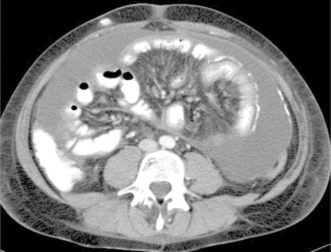
- İnce barsak ansları orta hatta veya karın merkezinde kümelenir ve çevresel fibrotik zar/kapsül içinde toplanmış görünür.
- Visseral ya da parietal peritonda kalınlaşma ve kontrastlanma izlenebilir.
- Peritoneal yüzey boyunca ince veya kaba kalsifikasyonlar görülebilir; dağılımı kapsül paternini tamamlar.
- Loküle veya septalı asit, bağırsak ansları arasında sınırlanmış sıvı cepleri oluşturabilir.
- Anslar birbirine yapışık, çekilmiş veya angüle olabilir; lümen daralması ve geçiş noktası mekanik obstrüksiyonu düşündürür.
- Kapsül içindeki ince barsak duvarında kalınlaşma ve kontrastlanma artışı inflamasyon ya da ileri fibrozisle ilişkili olabilir.
- Obstrüksiyon varsa kapsül proksimalindeki ince barsak ansları genişleyebilir ve hava-sıvı seviyeleri eşlik edebilir.
Ölçütler ve sınıflamalar
- Yayınlanmış CT bileşik ölçütü
- Küçük kohortta (15 EPS/16 kontrol) şu altı BT bulgusundan en az üçü aranır: peritoneal kontrastlanma, peritoneal kalınlaşma, peritoneal kalsifikasyon, ans yapışıklığı, obstrüksiyon ve loküle/septalı sıvı. Eşik tek başına veya evrensel tanı ölçütü değildir.
Normalde
Normal peritonda ince bağırsak ansları tek bir zarla çevrelenmez; anslar ayrı izlenir, mezenterik yağ korunur ve anslar arasında az miktarda serbest sıvı bulunabilir. Normal ince bağırsak duvarı belirgin kalınlaşma göstermez. Aynı düzeyde ansların kümelenmesini, peritoneal çizginin kalınlığını ve kalsifikasyonunu, sıvının loküle olup olmadığını ve kapsül proksimalindeki ans çapını karşılaştır.
Normal BT ile kıyasla
Aynı bölgenin, kaynak veri setinde patoloji içermediği belirtilen bir BT incelemesini kesit kesit kaydırın; organ sınırlarını açarak anatomiyi hasta görüntüsüyle karşılaştırın. Küçük rastlantısal bulgular tümüyle dışlanmamıştır.
Kaynak: TotalSegmentator v2.01 veri seti (Wasserthal ve ark., Radiology: Artificial Intelligence 2023), CC BY 4.0, vaka s0119; organ sınırları veri setinin otomatik segmentasyonundan, birleştirilerek sadeleştirildi. Görüntüler 2,5 mm kesit aralığına yeniden örneklendi.
Ayırıcı tanı
- Konjenital peritoneal kapsül
- ince ve çoğunlukla kalsifiye olmayan zar bağırsakları örtebilir; peritoneal inflamasyon, kalınlaşma ve diyaliz öyküsü EPS lehine olur.
- Peritoneal tüberküloz
- asit, peritoneal kalınlaşma ve yapışık anslar yapabilir; lenf nodu nekrozu ve eşlik eden enfeksiyon bulguları ayrımı destekler.
- Peritoneal karsinomatozis
- nodüler implantlar, omental kitle ve asit öne çıkar; tek tip fibrotik kapsül içinde ans kümelenmesi daha az tipiktir.
- Adezyon obstrüksiyonu
- geçiş noktası ve proksimal dilatasyon oluşturur ancak çevresel kalınlaşmış/kalsifiye peritoneal kapsül beklenmez.
- İnternal herni
- ansların kapalı kese içinde kümelenmesi ve mezenter damarlarında girdap görülebilir; peritoneal kalsifik fibrotik örtü EPS'yi destekler.
Tuzaklar
- Yalnız peritoneal kalsifikasyon EPS tanısı koydurmaz; ansların kümelenmesi, peritoneal kalınlaşma, loküle sıvı ve obstrüksiyon bulgularını birlikte ara.
- Hafif BT değişiklikleri klinik tanıdan aylar önce belirgin olmayabilir; normal ya da silik tek BT görünümü güçlü klinik kuşkuyu sonlandırmaz.
- Yapışık ansları tek başına internal herni veya adezyon obstrüksiyonu olarak yorumlama; bağırsakların kapsülle çevrelenip çevrelenmediğini tüm kesitlerde izle.
- Loküle sıvı ve asit, bakteriyel peritonit veya ilerlemiş vakalarda perforasyon ile ilişkilendirilebilir.
Kendini dene
Uzun süre periton diyalizi alan hastada anslar merkezde kümeli, kalın kalsifiye zarla çevrili ve loküle sıvı var. En olası tanı?
Cevabı göster
Kapsülleyici peritoneal skleroz. Diyaliz öyküsüyle kümelenmiş ansların kalın/kalsifiye peritoneal örtü içinde hapsolması EPS'yi destekler. Konjenital kapsül çoğunlukla ince ve kalsifikasyonsuzdur; tüberküloz ve internal herni bu tam kapsül paternini tipik olarak oluşturmaz.
Kapsül proksimalinde dilate anslar ve keskin geçiş noktası görülüyor. Bu BT bulgusu hangi komplikasyonu gösterir?
Cevabı göster
Mekanik ince barsak obstrüksiyonu. Proksimal ans dilatasyonu ve geçiş noktası mekanik obstrüksiyon örüntüsüdür. Diğer seçeneklerden peritoneal lenfomatozis tipik olarak asit, diffüz peritoneal kalınlaşma ve omental tutulumla seyreder; keskin geçiş noktası ve proksimal ans dilatasyonu ise bu tabloda değil, mekanik obstrüksiyonda beklenen bulgulardır.
Kaynaklar
Bu sayfadaki 40 cümle ve 2 quiz sorusu aşağıdaki kaynaklardan alıntılarla tek tek karşılaştırıldı; 7 cümle bu karşılaştırmada düzeltildi (2026-10-08).
- Imaging in encapsulating peritoneal sclerosispmc.ncbi.nlm.nih.gov
- Idiopathic Encapsulating Peritoneal Sclerosis Mimicking Acute Appendicitis: A Case Report and Systematic Literature Reviewpmc.ncbi.nlm.nih.gov
- Non-infectious complications of peritoneal dialysis in childrenpmc.ncbi.nlm.nih.gov
- Encapsulating Peritoneal Sclerosis: A Case Report and Literature Reviewpmc.ncbi.nlm.nih.gov
- Surgical management of Encapsulating Peritoneal Sclerosis (EPS) in children: international case series and literature reviewpmc.ncbi.nlm.nih.gov
- Encapsulating peritoneal sclerosis.pubmed.ncbi.nlm.nih.gov
- Encapsulating Peritoneal Sclerosis: The Abdominal Cocoon.pubmed.ncbi.nlm.nih.gov
- Encapsulating Peritoneal Sclerosis: A Case Report and Literature Review.pubmed.ncbi.nlm.nih.gov
- Encapsulating peritoneal sclerosis: the state of affairs.pubmed.ncbi.nlm.nih.gov
- Encapsulating peritoneal sclerosis--correlation of radiological findings at CT with underlying pathogenesis.pubmed.ncbi.nlm.nih.gov
- Congenital peritoneal encapsulation: A review and novel classification system.pubmed.ncbi.nlm.nih.gov
- Encapsulating peritoneal sclerosis: what have we learned?pubmed.ncbi.nlm.nih.gov
- Imaging features of encapsulating peritoneal sclerosis in continuous ambulatory peritoneal dialysis patients.pubmed.ncbi.nlm.nih.gov
- Abdominal cocoon in children: A case report and review of literature.pubmed.ncbi.nlm.nih.gov
- Encapsulating peritoneal sclerosis with favorable outcome in hemodialysis: an unusual case with a literature review.pubmed.ncbi.nlm.nih.gov
- A New Experience With Encapsulating Peritoneal Sclerosis: Role of Early Intervention.pubmed.ncbi.nlm.nih.gov
- Imaging pearls and differential diagnosis of encapsulating peritoneal sclerosis: Emphasis on computed tomography.pubmed.ncbi.nlm.nih.gov
- Successful surgical approach for a patient with encapsulating peritoneal sclerosis after hyperthermic intraperitoneal chemotherapy: a case report and literature review.pubmed.ncbi.nlm.nih.gov
- Case report: abdominal cocoon syndrome following peritoneal dialysis presenting as closed-loop small bowel obstruction.pubmed.ncbi.nlm.nih.gov
- Case Report: A case of refractory tuberculous peritonitis mimicking and complicating suspected encapsulating peritoneal sclerosis in a long-term peritoneal dialysis patient.pubmed.ncbi.nlm.nih.gov
- Idiopathic Encapsulating Peritoneal Sclerosis Mimicking Acute Appendicitis: A Case Report and Systematic Literature Review.pubmed.ncbi.nlm.nih.gov
- CT in predicting abdominal cocoon in patients on peritoneal dialysis.pubmed.ncbi.nlm.nih.gov
- Research progress on abdominal cocoon syndrome.pubmed.ncbi.nlm.nih.gov
- Case Report: Suspected <i>Mycobacterium abscessus</i>-associated polyserositis following coronary stenting, diagnosed with the aid of metagenomic next-generation sequencing.pubmed.ncbi.nlm.nih.gov
- Current Approaches and Emerging Strategies in the Treatment of Peritoneal Carcinomatosis.pubmed.ncbi.nlm.nih.gov
- Caught in a Cocoon: Operative Release of Primary Sclerosing Encapsulating Peritonitis With Endoscopic Management of Early Postoperative Duodenojejunal Intussusception.pubmed.ncbi.nlm.nih.gov
- Ascitic Fluid Drainage before Dialysis Catheter Removal for Peritoneal Dialysis-Related Peritonitis.pubmed.ncbi.nlm.nih.gov
- Fibrous membrane stripping combined with nasointestinal ileus tube for abdominal cocoon: A case report and literature review.pubmed.ncbi.nlm.nih.gov
- Role of PET imaging in peritoneal involvement of subcutaneous panniculitis-like T-cell lymphoma.pubmed.ncbi.nlm.nih.gov
- Nanoparticle-enabled molecular imaging diagnosis of osteoarthritis.pubmed.ncbi.nlm.nih.gov
- Advancements in nanotechnology for diagnostics: a literature review, part II: advanced techniques in nuclear and optical imaging.pubmed.ncbi.nlm.nih.gov
- Advances in immune cell-based therapeutic agents for the treatment of inflammation-related diseases.pubmed.ncbi.nlm.nih.gov
- Peritoneal carcinomatosis with desmoplasia and osseous metaplasia mimicking encapsulating peritoneal sclerosis in a cat: case report.pubmed.ncbi.nlm.nih.gov
- Case report: Sclerosing encapsulating peritonitis in a cat with disseminated pancreatic adenocarcinoma of presumed ductal origin.pubmed.ncbi.nlm.nih.gov
- A case report of idiopathic sclerosing encapsulating peritonitis causing an acute bowel occlusion in adult.pubmed.ncbi.nlm.nih.gov
- A Case Series and Brief Review of Literature on Encapsulating Peritoneal Sclerosis: Unveiling the Cocoon.pubmed.ncbi.nlm.nih.gov
- Calcified encapsulating peritoneal sclerosis associated with peritoneal dialysis: A case report.pubmed.ncbi.nlm.nih.gov
- An intra-abdominal abscess or "rind" as a consequence of peritoneal dialysis-associated pseudomonas peritonitis.pubmed.ncbi.nlm.nih.gov
- Encapsulating peritoneal sclerosis outcomes: a retrospective study on the significance of nutritional parameter variations.pubmed.ncbi.nlm.nih.gov
- Encapsulating Peritoneal Sclerosis Leading to Small Bowel Obstruction in a Young Woman with End-Stage Renal Disease: A Case Report.pubmed.ncbi.nlm.nih.gov
- A Case Report and Clinical Insights on Encapsulating Peritoneal Sclerosis: A Rare yet Critical Complication of Peritoneal Dialysis.pubmed.ncbi.nlm.nih.gov
- Imaging and treatment of idiopathic abdominal cocoon in 9 patients.pubmed.ncbi.nlm.nih.gov
- C-reactive protein levels in combination with abdominal CT scans is a useful tool to predict the macroscopic appearance in late-stage EPS patients prior to surgery.pubmed.ncbi.nlm.nih.gov
- Human biomarker navigator.pubmed.ncbi.nlm.nih.gov
- Mechanical bowel displacement to enable safe microwave ablation of a peripheral hepatic tumor in Chilaiditi anatomy.pubmed.ncbi.nlm.nih.gov
- Situs inversus totalis with bilateral broad ligament defects identified during laparoscopic surgery: a case report.pubmed.ncbi.nlm.nih.gov
- Diagnostic performance of an artificial intelligence algorithm for detecting pneumoperitoneum on abdominal CT scans.pubmed.ncbi.nlm.nih.gov
- Relative colon wall attenuation - distinguishing ischemia from other colon wall changes.pubmed.ncbi.nlm.nih.gov
- Usefulness of ultrasound in diagnosing constipation in children: a narrative review.pubmed.ncbi.nlm.nih.gov
- CT-Based Reference Ranges for Canine Pyloric Thickness: Influence of Body Size and Normalization With Aortic Diameter.pubmed.ncbi.nlm.nih.gov
- Heterogeneity of primary and metastatic CAFs: From differential treatment outcomes to treatment opportunities (Review).pubmed.ncbi.nlm.nih.gov
- Special Issue: EACR 2026 Congress: Innovative Cancer Science, 8-11 June 2026.pubmed.ncbi.nlm.nih.gov
- EACR 2025 Congress: Innovative Cancer Science, 16-19 June 2025.pubmed.ncbi.nlm.nih.gov
- Cocoon Syndrome: A Clinical Case of Intestinal Obstruction Highlighting the Diagnostic and Therapeutic Importance of Exploratory Laparoscopy in the Face of Ambiguous Imaging.pubmed.ncbi.nlm.nih.gov
- A Case of Congenital Peritoneal Encapsulation Complicated with Left Paraduodenal Hernia Presenting as Recurrent Small Bowel Obstruction.pubmed.ncbi.nlm.nih.gov
- Abdominal Cocoon Syndrome Masquerading as Intussusception on Computed Tomography: A Case Report.pubmed.ncbi.nlm.nih.gov
- Encapsulating peritoneal sclerosis: a three-case series highlighting diagnostic challenges and surgical managements.pubmed.ncbi.nlm.nih.gov
- A Rare Case of Recurrent Intestinal Obstruction in a Young Female: Abdominal Cocoon.pubmed.ncbi.nlm.nih.gov
- Peritoneal lymphomatosis mimicking complicated hydatid cyst on imaging: A rare presentation of diffuse large B-cell lymphoma.pubmed.ncbi.nlm.nih.gov
- Peritoneal Lymphomatosis Mimicking Advanced Ovarian Malignancy: A Diagnostic Challenge in Diffuse Large B-Cell Lymphoma.pubmed.ncbi.nlm.nih.gov
- Coccidioidal peritonitis: A rare manifestation of disseminated coccidioidomycosis.pubmed.ncbi.nlm.nih.gov
- A Focus on Inflammatory and Bacterial Biomarkers in Secondary Peritonitis.pubmed.ncbi.nlm.nih.gov
- Validity of the American Association for the Surgery of Trauma Intestinal Obstruction Grading System.pubmed.ncbi.nlm.nih.gov
- Outcomes of the surgical management of encapsulating peritoneal sclerosis: A case series from a single center in Korea.pubmed.ncbi.nlm.nih.gov
- Case Report: Intestinal necrosis secondary to internal abdominal hernia in a young female.pubmed.ncbi.nlm.nih.gov
- Radiology insights into Petersen's hernia complication of post Roux-en-Y gastric bypass.pubmed.ncbi.nlm.nih.gov
- The role of computed tomography in acute bowel obstruction due to a supravesical hernia: a case report from Nepal.pubmed.ncbi.nlm.nih.gov
- Successful treatment of non-occlusive mesenteric ischemia in a patient undergoing combined therapy with peritoneal dialysis and hemodialysis: a case report.pubmed.ncbi.nlm.nih.gov
- Advances in nanotechnology for the diagnosis and management of autoimmune diseases.pubmed.ncbi.nlm.nih.gov
- Understanding the Mechanisms Driving Fibrosis Following Cochlear Implantation-Lessons from Other Tissues.pubmed.ncbi.nlm.nih.gov
- Abdominal Cocoon Syndrome With Greater Omental Hypoplasia: A Rare Anatomical Insight and Diagnostic Challenge.pubmed.ncbi.nlm.nih.gov
- Acute presentation of cocoon abdomen as intestinal obstruction mimicking with strangulated eventration: A case report.pubmed.ncbi.nlm.nih.gov
- Periostin: a matricellular protein involved in peritoneal injury during peritoneal dialysis.pubmed.ncbi.nlm.nih.gov
- Abdominal cocoon syndrome: Rare cause of intestinal obstruction-Case report and systematic review of literature.pubmed.ncbi.nlm.nih.gov
- Non-neoplastic conditions mimicking peritoneal carcinomatosis at CT imaging.pubmed.ncbi.nlm.nih.gov
- Unraveling the cocoon: A case report on preoperative ambiguity and definitive surgical management of encapsulating peritoneal sclerosis.pubmed.ncbi.nlm.nih.gov
- Medical hyperspectral imaging: an updated review of technology advancements and biomedical applications.pubmed.ncbi.nlm.nih.gov
- Commentary: an eye on PET quantification.pubmed.ncbi.nlm.nih.gov
- An Idiopathic Case of Sclerosing Encapsulating Peritonitis: A Case Report.pubmed.ncbi.nlm.nih.gov
- Benign Sclerosing and Fibrosing Conditions of the Abdomen and Their Potential Mimics.pubmed.ncbi.nlm.nih.gov
- Sclerosing Encapsulating Peritonitis Is More Common Following Repeat Hyperthermic Intraperitoneal Chemotherapy.pubmed.ncbi.nlm.nih.gov
- Primary sclerosing encapsulating peritonitis: a case report.pubmed.ncbi.nlm.nih.gov
- Mitochondrial neurogastrointestinal encephalomyopathy: approaches to diagnosis and treatment.pubmed.ncbi.nlm.nih.gov
- Recurrent Intestinal Obstruction Secondary to Crohn's Disease in Dextrocardia With Situs Inversus Totalis: A Case Report.pubmed.ncbi.nlm.nih.gov
- Urgent da Vinci-Assisted Laparoscopic Repair of an Incarcerated Adult Bochdalek Hernia Presenting With Large-Bowel Obstruction: A Case Report and Literature Review.pubmed.ncbi.nlm.nih.gov
- Comprehensive Imaging Evaluation and Staging of Crohn's Disease: When and Why to Use Intestinal Ultrasound, MRE, or CTE: Current Guidelines and Future Directions.pubmed.ncbi.nlm.nih.gov
- A clinical evaluation of HyperSight CBCT enhanced offline adaptive breast radiotherapy.pubmed.ncbi.nlm.nih.gov
- Standardized processing of explanted hearts after stereotactic arrhythmia radiotherapy (Bio-STAR): a STOPSTORM framework and first human findings.pubmed.ncbi.nlm.nih.gov
- Conduction Abnormalities After Transcatheter Aortic Valve Replacement: Comprehensive Review of Current Literature, Guidelines, and Clinical Practices.pubmed.ncbi.nlm.nih.gov
- Rare Adult Ileo-Ileo-Colic Intussusception Caused by an Ileal Lipoma with Spontaneous Ileal Rupture and Rectal Hemorrhage: A Case Report with a Hypothesis-Generating Discussion of Mesenteric Anatomical Variability.pubmed.ncbi.nlm.nih.gov
- Case Report: Lumbar herniation of a low-grade appendiceal mucinous neoplasm mimicking a chronic abscess.pubmed.ncbi.nlm.nih.gov
- Case Report: Strangulated intestinal obstruction due to chronic migration of an intrauterine device (IUD): a 30-year latent complication.pubmed.ncbi.nlm.nih.gov
- Acute Emergent Biliary Pathologies Identified Using Point-of-Care Ultrasound.pubmed.ncbi.nlm.nih.gov
- Benign Pneumoperitoneum: A Case Report and Literature Review.pubmed.ncbi.nlm.nih.gov
- Early-onset colorectal cancer in patients under 30: a case series from Tanzania highlighting an emerging epidemiological shift.pubmed.ncbi.nlm.nih.gov
- Entero-Appendiceal Fistula Presenting with Overt Gastrointestinal Bleeding: A Case Report.pubmed.ncbi.nlm.nih.gov
- Characteristic CT Imaging Features of Atraumatic Peritoneal and Subperitoneal Pathologies in Emergency Settings: A Pictorial Review.pubmed.ncbi.nlm.nih.gov
- Primary dermoid cysts of the cecum: A systematic review and case report.pubmed.ncbi.nlm.nih.gov
- Splenic torsion mistaken for an ovarian cyst: a case report.pubmed.ncbi.nlm.nih.gov
- Encapsulating Peritoneal Sclerosis Improvement Following Conservative Treatment in a Child Undergoing Peritoneal Dialysis.pubmed.ncbi.nlm.nih.gov
- Postmortem diagnosis of exocrine pancreatic adenocarcinoma mimicking encapsulating peritoneal sclerosis in a cat: Clinical and histopathological insights.pubmed.ncbi.nlm.nih.gov
- Unraveling the enigma of sclerosing encapsulating peritonitis: a comprehensive review.pubmed.ncbi.nlm.nih.gov
- Intraoperative Diagnosis and Surgical Management of Encapsulating Peritoneal Sclerosis: A Case Report.pubmed.ncbi.nlm.nih.gov
- Successful treatment of sclerosing encapsulating peritonitis in a cat using bioresorbable hyaluronate-carboxymethylcellulose membrane after surgical adhesiolysis and long-term prednisolone.pubmed.ncbi.nlm.nih.gov
Eğitim amaçlıdır; hasta başında tanı ve tedavi kararının yerine geçmez. İçerik yapay zekâ destekli bir süreçle güncel literatüre göre cümle cümle kaynaklarla karşılaştırıldı, ancak uzman radyolog incelemesinden geçmedi. Klinik kararlarda güncel kılavuzları, kurum protokollerini ve radyoloji raporunu esas alın.