Özet
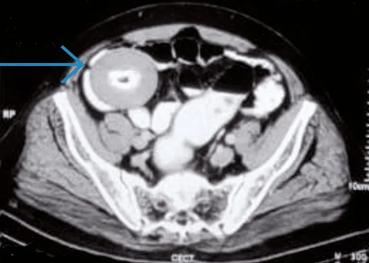
Peritoneal serbest cisim, sıklıkla torsiyone epiploik apendiks yağ dokusunun zamanla fibrotikleşip kalsifiye olmasıyla oluşan, periton boşluğunda serbest hareket edebilen nadir bir oluşumdur. Kontrastsız BT, yuvarlak veya oval yumuşak doku dansiteli cismin merkezindeki kalsifikasyonu ve çevresindeki ince yumuşak doku halkasını gösterebilir. Önceki ya da pozisyon değiştirilerek yapılan görüntülemede yer değiştirmesi tanı için güçlü bir ipucudur. Pelvik tümör veya yabancı cisim sanılması gereksiz cerrahiye yol açabilir; sabit, invaziv ya da çevre reaksiyonu oluşturan kitleler ise farklı nedenleri düşündürür.
Faz ve pencere
- Kontrastsız tanısal
- Peritoneal serbest cisim için kontrastsız kesit yeterli morfolojik değerlendirmeyi sağlar: iyi sınırlı yuvarlak/oval yumuşak doku dansiteli cisim içinde merkezî, tabakalı veya düzensiz kalsifikasyon ve çevresinde ince yumuşak doku halkası görülebilir. Kontrast fazı tanı için zorunlu değildir.
Önerilen pencereler G genişlik, M merkez; değerler Hounsfield birimi (HU)
- BatınG 400 / M +50
- Yumuşak dokuG 400 / M +40
- KemikG 1800 / M +400
BT bulguları
- Pelviste veya başka bir peritoneal reseste yuvarlak ya da oval, düzgün sınırlı solid cisim izlenir.
- Cismin merkezinde küçük kalsifik odak veya konsantrik tabakalı kalsifikasyon bulunabilir.
- Tipik olarak düzgün sınırlı yuvarlak/oval yumuşak doku dansiteli cisim içinde merkezi veya konsantrik kalsifikasyon ve çevresinde yumuşak doku halkası izlenir; hipodens merkez ya da tabakalı dış görünüm değişken olabilir.
- Komşu organlardan ince yağ planıyla ayrılması ve çevre dokuda belirgin inflamasyon oluşturmaması serbest yerleşimi destekler.
- Pozisyon değişikliği veya seri incelemede cismin farklı peritoneal konumda görülmesi mobiliteyi doğrular.
- Büyük cisimler nadiren mesane, rektum veya ince barsağa bası yapabilir; komşu lümen ve organ konturları incelenmelidir.
- Önceki cerrahi varsa yabancı cisim/gossypiboma ayırıcı tanıya girer; gaz kabarcıkları, tekstil işaretleyici veya çevresel reaksiyon lehine bulgular aranır.
Normalde
Normal peritoneal boşlukta ayrı, yuvarlak kalsifikasyonlu yumuşak doku cismi bulunmaz; mesane ve rektum çevresi yağ planları serbesttir. Kıyaslamada lezyonun merkezî kalsifikasyonunu, ince dış yumuşak doku tabakasını, çevre organlara yapışık olmamasını ve eski görüntülerdeki konumunu değerlendir.
Normal BT ile kıyasla
Aynı bölgenin, kaynak veri setinde patoloji içermediği belirtilen bir BT incelemesini kesit kesit kaydırın; organ sınırlarını açarak anatomiyi hasta görüntüsüyle karşılaştırın. Küçük rastlantısal bulgular tümüyle dışlanmamıştır.
Kaynak: TotalSegmentator v2.01 veri seti (Wasserthal ve ark., Radiology: Artificial Intelligence 2023), CC BY 4.0, vaka s0119; organ sınırları veri setinin otomatik segmentasyonundan, birleştirilerek sadeleştirildi. Görüntüler 2,5 mm kesit aralığına yeniden örneklendi.
Ayırıcı tanı
- Gossypiboma
- cerrahi öyküyle ilişkili sabit kitle, tekstil materyali, gaz odakları veya çevresel granülomatöz reaksiyon gösterebilir; serbestçe göç etmesi beklenmez.
- Kalsifiye lenf düğümleri de peritoneal serbest cismin ayırıcı tanısında yer alır.
- Dermoid/teratom, merkezî kalsifikasyonlu pelvik peritoneal serbest cisim için ayırıcı tanıda düşünülmüştür.
- Uterin leiomyom pelvik kitle ayırıcı tanısında yer alır; kadınlarda bazı peritoneal serbest cisimlerin otoampute uterin leiomyomdan kaynaklanabileceği bildirilmiştir.
- Düşmüş safra taşları, özellikle laparoskopik kolesistektomi sonrasında peritoneal boşlukta kalabilir ve peritoneal serbest cismi taklit edebilir.
Tuzaklar
- Tek bir BT'de mesane arkasındaki serbest cisim over veya rektosigmoid kitle gibi görünebilir; organla devamlılık olup olmadığını çok düzlemde ara.
- Önceki görüntüleme yoksa farklı pozisyonda ek tarama veya takip incelemesi cismin mobilitesini gösterebilir ve tanıya yardımcı olabilir.
- Ameliyat öyküsünde kalsifik kitleyi otomatik olarak serbest cisim sayma; gossypiboma genellikle sabit kalır ve çevresel reaksiyon veya karakteristik tekstil işaretleri gösterebilir.
- Merkezî kalsifikasyon boyutu ve düzeni değişkenlik gösterir; kesit incelemelerinde hipodens merkezli yapılar dikkatle izlenmelidir.
Kendini dene
Kontrastsız BT'de kalsifikasyon içeren iyi sınırlı bir kitle görülüyor. Önceki BT'de aynı kitlenin yeri farklı. Bu bulgu neyi gösterir?
Cevabı göster
Kitlenin hareketli olduğunu. Seri görüntülerde kitlenin yer değiştirmesi, periton içi serbest cismin hareketli olduğunu gösterir ve tanıya yardımcı olabilir.
Önceki cerrahi öyküsü olan bir hastada BT'de çevre yapılara veya cerrahi skara yapışık, düzensiz kenarlı bir kitle görülüyor. Hangi ayırıcı tanı bu bulgularla uyumludur?
Cevabı göster
Unutulmuş cerrahi materyal (gossypiboma). Gossypiboma BT'de düzensiz kenarlı olabilir ve çevre yapılara veya önceki cerrahi skarlara yapışıklık gösterebilir.
Kaynaklar
Bu sayfadaki 36 cümle ve 2 quiz sorusu aşağıdaki kaynaklardan alıntılarla otomatik olarak karşılaştırıldı; 5 cümle bu karşılaştırmada düzeltildi (2026-10-09).
- Rolling Egg-Shaped Peritoneal Loose Body (PLB): A Diagnostic Dilemma for Surgeons and Radiologistspmc.ncbi.nlm.nih.gov
- Deciding to Remove or Leave a Peritoneal Loose Body: A Case Report and Review of Literaturepmc.ncbi.nlm.nih.gov
- Pancreatic cancer complicated with a giant peritoneal loose body: case report and literature reviewpmc.ncbi.nlm.nih.gov
- A giant peritoneal loose body: A case report and updated literature review and data synthesispmc.ncbi.nlm.nih.gov
- Giant peritoneal loose body in the pelvic cavity confirmed by laparoscopic exploration: a case report and review of the literaturepmc.ncbi.nlm.nih.gov
- Giant peritoneal loose body : a case report and review of the literature.pubmed.ncbi.nlm.nih.gov
- Giant peritoneal loose body in the pelvic cavity confirmed by laparoscopic exploration: a case report and review of the literature.pubmed.ncbi.nlm.nih.gov
- CT diagnosis of a large peritoneal loose body: a case report and review of the literature.pubmed.ncbi.nlm.nih.gov
- Deciding to Remove or Leave a Peritoneal Loose Body: A Case Report and Review of Literature.pubmed.ncbi.nlm.nih.gov
- A giant peritoneal loose body: A case report and updated literature review and data synthesis.pubmed.ncbi.nlm.nih.gov
- Imaging Manifestation and Pathological Comparison of the Peritoneal Loose Body: A Case Report and Review of the Literature.pubmed.ncbi.nlm.nih.gov
- Pancreatic cancer complicated with a giant peritoneal loose body: case report and literature review.pubmed.ncbi.nlm.nih.gov
- Giant peritoneal loose body: A case report.pubmed.ncbi.nlm.nih.gov
- Peritoneal Mouse as Detected on (18)F-FDG PET-CT.pubmed.ncbi.nlm.nih.gov
- Dropped Gallstone Presenting as Recurrent Abdominal Wall Abscess.pubmed.ncbi.nlm.nih.gov
- Giant peritoneal loose body - case report.pubmed.ncbi.nlm.nih.gov
- Symptomatic giant peritoneal loose body in the pelvic cavity: A case report.pubmed.ncbi.nlm.nih.gov
- Giant peritoneal loose body: a case report and review of literature.pubmed.ncbi.nlm.nih.gov
- Revisiting the enigma of a large symptomatic peritoneal loose body: a case report and critical literature review.pubmed.ncbi.nlm.nih.gov
- Coexisting adhesive small-bowel obstruction and giant peritoneal loose body in a surgically naïve patient: a case report.pubmed.ncbi.nlm.nih.gov
- Unexpected Metal Ion Elevation and Adverse Tissue Reactions in Trident II Dual Mobility Constructs With Ceramic Heads.pubmed.ncbi.nlm.nih.gov
- Long-term recovery of cauda equina syndrome and sexual dysfunction following delayed decompression of a giant lumbar disc herniation: illustrative case.pubmed.ncbi.nlm.nih.gov
- Rectal diffuse large B-cell lymphoma misdiagnosed as bleeding cancer in an elderly patient.pubmed.ncbi.nlm.nih.gov
- Hemostatic Cellulose Masquerading as a Postoperative Abscess Following Elective Colon Surgery: A Unique Case and Literature Review.pubmed.ncbi.nlm.nih.gov
- Case report: Reactive nodular fibrous pseudotumor: report of 4 new cases including a rare pulmonary presentation and a literature review covering 40 cases.pubmed.ncbi.nlm.nih.gov
- Computed Tomography and Sonography of Spleen-related Complications in Sickle Cell Anemia.pubmed.ncbi.nlm.nih.gov
- Congenital pulmonary airway malformation mimicking lung cancer in adults: a pitfall in oncologic diagnosis.pubmed.ncbi.nlm.nih.gov
- Evaluation of benign and malignant pulmonary nodules based on clinical and contrast-enhanced computed tomography data: a multicenter study.pubmed.ncbi.nlm.nih.gov
- Retromammary Schwannoma Mimicking a Breast Tumor in a Patient with Neurofibromatosis Type 1: A Case Report Demonstrating Surgical Avoidance Using Contrast-Enhanced Ultrasound-Informed Biopsy.pubmed.ncbi.nlm.nih.gov
- Intrapancreatic accessory spleen mimicking pancreatic malignancy: Diagnostic role of Tc-99m heat-damaged red blood cell scintigraphy.pubmed.ncbi.nlm.nih.gov
- Moyamoya disease masquerading as CNS vasculitis: Diagnostic challenges and imaging insights.pubmed.ncbi.nlm.nih.gov
- MRI insights into endometrial stromal tumors.pubmed.ncbi.nlm.nih.gov
- Pure Sertoli cell tumor of the right ovary managed with fertility-sparing surgery: a case report.pubmed.ncbi.nlm.nih.gov
- Swyer syndrome complicated by rapidly progressive invasive ovarian dysgerminoma with bladder and rectal direct invasion: a rare case report.pubmed.ncbi.nlm.nih.gov
- Isolated Cardiac Hydatid Disease Presenting as Ventricular Remodeling and Tamponade.pubmed.ncbi.nlm.nih.gov
- Ministromatolite Modular Reef Framework, ~1.5 Ga, Yesanpo, North China.pubmed.ncbi.nlm.nih.gov
- Hydrodynamic control of fluvial oncoid formation in a modern high-energy tropical cascade system.pubmed.ncbi.nlm.nih.gov
- Diagnostic Musculoskeletal Ultrasound in the Evaluation of the Glenoid Labrum.pubmed.ncbi.nlm.nih.gov
- Synovial Chondromatosis of the Elbow: A Case Report and Discussion of Feared Complications.pubmed.ncbi.nlm.nih.gov
- Disseminated hepatic epithelioid hemangioendothelioma with multiorgan metastases achieving long-term stability with Pazopanib: a case report.pubmed.ncbi.nlm.nih.gov
- Technique-Driven and Patient-Specific Variations in Complications of Mastectomy with Immediate Breast Reconstruction: An Evidence-Based Review.pubmed.ncbi.nlm.nih.gov
- Beyond Palmer: a topographic classification of non-Palmer triangular fibrocartilage complex injuries.pubmed.ncbi.nlm.nih.gov
- Metabolism, Morphology and Function of the Anterior Abdominal Wall Musculature in Ventral Hernias and After Repair: A Narrative Review and Research Agenda.pubmed.ncbi.nlm.nih.gov
- Three-Grade Neurological Recovery (ASIA Impairment Scale A to D) Following Surgical Stabilization of Traumatic D11-D12 Meyerding Grade IV Spondylolisthesis: A Case Report.pubmed.ncbi.nlm.nih.gov
- Spontaneous Reduction of an Incarcerated Gravid Uterus With a Giant Uterine Fibroid in Late Pregnancy: A Case Report.pubmed.ncbi.nlm.nih.gov
- Dynamic Changes in Peripheral Nerve Stiffness After Regional Anesthesia: Implications of Shear Wave Elastography in Adductor Canal Block.pubmed.ncbi.nlm.nih.gov
- Progressive Pelvic and Gluteal Desmoid Tumor in a 16-Year-Old Female: A Five-Year Multimodal Imaging Follow-Up.pubmed.ncbi.nlm.nih.gov
- Imaging and Histopathological Findings of Phyllodes Tumor of the Prostate in a Cat.pubmed.ncbi.nlm.nih.gov
- Primary Synovial Sarcoma of the Prostate: A Case Report and Review of the Literature.pubmed.ncbi.nlm.nih.gov
- Incidental Diagnosis of a Gossypiboma With Complete Transmural Migration Into the Jejunum After a Hysterectomy: A Case Report.pubmed.ncbi.nlm.nih.gov
- A rare cause of late benign biliary stricture after laparoscopic cholecystectomy: gossypiboma.pubmed.ncbi.nlm.nih.gov
- Gossypiboma Mimicking Ovarian Mass: A Rare Case Report from Rwanda.pubmed.ncbi.nlm.nih.gov
- Navigating pelvic anatomy for exenteration: a clinical guide for radiologists.pubmed.ncbi.nlm.nih.gov
- Giant uterine leiomyoma successfully managed by surgical excision: a case report.pubmed.ncbi.nlm.nih.gov
- Gynecologic imaging errors and pitfalls: how to avoid misdiagnosis on pelvic MRI.pubmed.ncbi.nlm.nih.gov
- A case report of synovial sarcoma arising in the abdominal wall muscles.pubmed.ncbi.nlm.nih.gov
- Compliance for Resistance: The Intrinsic Extraordinary Isotropic Anti-Fatigue Performance of Fish Bladder.pubmed.ncbi.nlm.nih.gov
- Gossypiboma Following Cesarean Section Presenting as Bilateral Abdominal Abscesses: A Case Report.pubmed.ncbi.nlm.nih.gov
- The Enigma of Gossypiboma: An Entity to Explore.pubmed.ncbi.nlm.nih.gov
- Asymptomatic intrahepatic gossypiboma incidentally discovered: A case report.pubmed.ncbi.nlm.nih.gov
- Primary chondrosarcoma arising from the L5 transverse process: A rare cause of chronic low back pain.pubmed.ncbi.nlm.nih.gov
- AWMF S2k Guideline: Diagnosis and Assessment of Lung Disease Caused by Occupational Silica Dust Exposure (Silicosis) - Disease No. 4101 as per Occupational Diseases Ordinance (BKV) Annex 1.pubmed.ncbi.nlm.nih.gov
- Uniportal robotic-assisted thoracoscopic lung resection with the da Vinci Xi surgical system: technical modifications and practical guidance.pubmed.ncbi.nlm.nih.gov
- A Para-Ovarian Cyst Infected With Salmonella: A Case Report.pubmed.ncbi.nlm.nih.gov
- Complete adnexal torsion of a large dermoid ovarian cyst with ovarian and tubal preservation in a 17-year-old patient: a case report.pubmed.ncbi.nlm.nih.gov
- Collision Tumour of the Ovary: Mature Cystic Teratoma and Benign Mucinous Cystadenoma.pubmed.ncbi.nlm.nih.gov
- Spontaneous Expulsion of a Large Submucosal Fibroid in the Postpartum Period: A Report of a Rare Case.pubmed.ncbi.nlm.nih.gov
- Ultrasound diagnosis of intrauterine device embedment complicated by bladder stone formation: a case report.pubmed.ncbi.nlm.nih.gov
- Laparoscopic resection of a small intestinal venous malformation presenting as acute bowel obstruction without hemorrhage in a child: a case report.pubmed.ncbi.nlm.nih.gov
- Imaging of Venolymphatic Malformation in a Child Extending from Inguinoscrotal Region to Thoracic Region: Case Report with Review of Literature.pubmed.ncbi.nlm.nih.gov
- Quantitative Susceptibility Mapping of Kidney Stones: An Ex Vivo MRI Phantom Study.pubmed.ncbi.nlm.nih.gov
- Heterotrimeric G proteins containing Gβ1γ2 subunits exhibit subtype-specific mobility differences in live cells.pubmed.ncbi.nlm.nih.gov
- Efficacy of Myofascial Release in Improving the Flexibility of Erector Spinae and Thoracolumbar Fascia in Subjects with Chronic Low Back Pain-A Sonoelastographic Analysis.pubmed.ncbi.nlm.nih.gov
- Inflammation vs. multiple AVN sites: a nuclear medicine evaluation in systemic allergic-immune syndrome - a case report.pubmed.ncbi.nlm.nih.gov
- Case Report: Bezoar-mimicking intraluminal gossypiboma causing small-bowel obstruction and chronic malabsorption 15 years after hysterectomy.pubmed.ncbi.nlm.nih.gov
- Late intestinal obstruction due to intraluminal textiloma: a case report.pubmed.ncbi.nlm.nih.gov
- Delayed Intrathoracic Textile Foreign Body Following Penetrating Shrapnel Injury Presenting with Expectoration of Fabric-Like Material and Chronic Middle-Lobe Destruction: A Case Report.pubmed.ncbi.nlm.nih.gov
- Metastatic Retroperitoneal Rhabdomyosarcoma in a Child: A Case Report Highlighting the Role of Imaging in Diagnosis, Staging, and Follow-Up.pubmed.ncbi.nlm.nih.gov
- A rare case of thyroid cartilage chondroma: diagnostic challenges and the role of multimodal imaging and histopathology.pubmed.ncbi.nlm.nih.gov
- Sphenoid Temporal Meningioma With Granular Cell Features and Extracranial Infratemporal Extension.pubmed.ncbi.nlm.nih.gov
- Multifocal small bowel gastrointestinal stromal tumor causing perforation peritonitis: A rare cause of acute abdomen.pubmed.ncbi.nlm.nih.gov
- AI-Based Approaches for the Identification and Quantification of Traumatic Brain Injury in Computed Tomography Imaging: Systematic Review.pubmed.ncbi.nlm.nih.gov
- Pilocytic astrocytoma: A rare case report.pubmed.ncbi.nlm.nih.gov
- Radiological Tumor Calcification in Sinonasal Adenocarcinoma: Prevalence and Prognostic Implications.pubmed.ncbi.nlm.nih.gov
- Pulmonary nodules with explosive calcification were finally diagnosed as lung adenocarcinom: A case report.pubmed.ncbi.nlm.nih.gov
- Rare anatomical localizations of hydatid cysts (2020-2025): Narrative review of presentation, imaging, and management.pubmed.ncbi.nlm.nih.gov
- Staged management of a compound combined pelvi-acetabular injury with perineal degloving in a young female: a case report and literature review.pubmed.ncbi.nlm.nih.gov
- Skull Base Metastases: A Pictorial Review.pubmed.ncbi.nlm.nih.gov
- MRI visualization of maternal pelvic and perineal anatomical changes during the second stage of labor: a feasibility study.pubmed.ncbi.nlm.nih.gov
- Ribitoborate Synergism with Histone Deacetylase Inhibitor Romidepsin as a Potential Treatment for Breast Cancer.pubmed.ncbi.nlm.nih.gov
- Hemodynamic remnant drift after coil embolization: longitudinal silent MRA and computational fluid dynamics analysis of a migratory neck remnant. Illustrative case.pubmed.ncbi.nlm.nih.gov
- Conservative Management of a Traumatic Deep Palmar Arch Pseudoaneurysm with Delayed Ulnar Motor Dysfunction: A Case Report.pubmed.ncbi.nlm.nih.gov
- When Meckel's diverticulum mimics malignancy: a case report of massive enterolithiasis in an adult patient.pubmed.ncbi.nlm.nih.gov
- Capsaicin-Triggered Vaginal Burning Due to Obstructed Rectovaginal Fistula: A Rare Case of Perineal Coproliths.pubmed.ncbi.nlm.nih.gov
- A rare case of epiploic appendagitis of the vermiform appendix.pubmed.ncbi.nlm.nih.gov
- Pelvic osseous foreign body identified during laparoscopic appendectomy in a child with acute appendicitis: a case report.pubmed.ncbi.nlm.nih.gov
- An unusual presentation of cystic lymphangioma in the mesoappendix: a case report.pubmed.ncbi.nlm.nih.gov
- Peritoneal loose body presenting as a hepatic mass: A case report and review of the literature.pubmed.ncbi.nlm.nih.gov
- Tuberculosis revisted: classic imaging findings in childhood.pubmed.ncbi.nlm.nih.gov
- The space of Retzius: anatomy, imaging features, and patterns of disease spread.pubmed.ncbi.nlm.nih.gov
- Ovarian Germ Cell Tumors: Pictorial Essay.pubmed.ncbi.nlm.nih.gov
- Bilateral primary ovarian Ewing sarcoma recurring as left submandibular lymphadenopathy diagnosed on cytology.pubmed.ncbi.nlm.nih.gov
- Rolling Egg-Shaped Peritoneal Loose Body (PLB): A Diagnostic Dilemma for Surgeons and Radiologists.pubmed.ncbi.nlm.nih.gov
- Mucocoele of the appendix.pubmed.ncbi.nlm.nih.gov
- A rare peritoneal egg: Case report with literature review.pubmed.ncbi.nlm.nih.gov
- Recent advances in nanomaterial-based precision medicine for orthotopic tumor therapy.pubmed.ncbi.nlm.nih.gov
- International Society of Paediatric Surgical Oncology (IPSO) Surgical Practice Guidelines.pubmed.ncbi.nlm.nih.gov
- International Harmonization of Nomenclature and Diagnostic Criteria (INHAND): Non-proliferative and Proliferative Lesions of the Non-human Primate ( M. fascicularis ).pubmed.ncbi.nlm.nih.gov
- Long-term safety and effectiveness of the "OptEase" vena cava filter.pubmed.ncbi.nlm.nih.gov
- Solid malignant retroperitoneal masses-a pictorial review.pubmed.ncbi.nlm.nih.gov
- Non-Epithelial Ovarian Cancers: How Much Do We Really Know?pubmed.ncbi.nlm.nih.gov
- Gastrointestinal Stromal Tumors (GISTs) Mimicking Primary Ovarian Tumors or Metastasizing to the Ovaries: A Systematic Literature Review.pubmed.ncbi.nlm.nih.gov
- Assessing the Hepatic Safety of Epigallocatechin Gallate (EGCG) in Reproductive-Aged Women.pubmed.ncbi.nlm.nih.gov
- Inflammatory myofibroblastic tumor from molecular diagnostics to current treatment.pubmed.ncbi.nlm.nih.gov
- Epiploic appendagitis: An overlooked cause of acute abdominal pain.pubmed.ncbi.nlm.nih.gov
Eğitim amaçlıdır; hasta başında tanı ve tedavi kararının yerine geçmez, tıbbi tavsiye değildir. Metinler yapay zekâ desteğiyle yazıldı ve otomatik bir adımda kaynak alıntılarıyla karşılaştırıldı; bu bir tıbbi doğrulama değildir ve içerik uzman radyolog incelemesinden geçmedi. Klinik kararlarda güncel kılavuzları, kurum protokollerini ve radyoloji raporunu esas alın.