
Özet
Malign peritoneal mezotelyoma, mezotelyal yüzeyden gelişen ve karın ağrısı, karında büyüme ya da asitle ortaya çıkabilen nadir bir primer periton tümörüdür. İntravenöz kontrastlı batın-pelvis BT, peritoneal yayılımı, omental yükü, mezenter ve ince barsak ilişkisini, asiti ve olası komşu invazyonu haritalamak için ilk basamak görüntülemedir. Yaygın nodüler periton kalınlaşması ve omental kekleşme görülebilir; kesin tanı için genellikle biyopsi gerekir. İnce barsak veya mezenter tutulumunun atlanması hastalık yaygınlığının ve obstrüksiyon riskinin eksik değerlendirilmesine yol açabilir.
Faz ve pencere
- Portal tanısal
- İntravenöz kontrast sonrası heterojen kontrastlanan, düzensiz sınırlı peritoneal yumuşak doku nodülleri ve plakları; omental kalınlaşma veya kekleşme, ince barsak çevresinde serozal yumuşak doku halkası, mezenterik nodüller ve asit birlikte haritalanır.
Önerilen pencereler G genişlik, M merkez; değerler Hounsfield birimi (HU)
- BatınG 400 / M +50
- Yumuşak dokuG 400 / M +40
BT bulguları
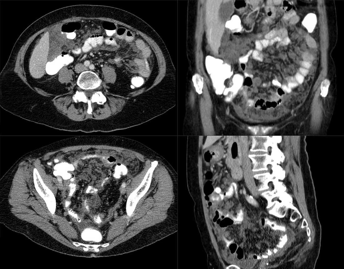
- Sheet-like peritoneal tutulum — parietal yüzeyde yaygın, plak biçimli kalınlaşma ve düzensiz kontrastlanan yumuşak doku alanları gelişebilir.
- Omental tümör yükü — büyük omentum kalınlaşır; nodüler infiltrasyon birleşerek omental cake görünümüne ulaşabilir.
- Mezenterik tutulum — malign peritoneal mezotelyomada ince barsak mezenteri içinde implantlar görülebilir.
- Malign peritoneal mezotelyomada yaygın nodüler periton kalınlaşmasına bağırsaklarda anatomik distorsiyon eşlik edebilir.
- Peritoneal kitleler — tek ya da çok sayıda, çoğunlukla heterojen kontrastlanan solid kitle ve düzensiz sınırlı implantlar görülebilir.
- Asit eşlik edebilir; bazı olgularda yaygın asit, minimal yumuşak doku kitlesiyle baskın bulgu olabilir.
- Primer odağın görünmemesi — peritoneal hastalık yaygınken gastrointestinal ya da jinekolojik bir primer kitle saptanmaması mezotelyomayı akla getirebilir, ancak özgül değildir.
- Komşu yayılım — diyafragma, abdominal duvar veya retroperitoneuma uzanım; barsak anslarında çekilme, distorsiyon ya da obstrüksiyon bulguları araştırılır.
- Toraks görüntülerinde plevral plak veya plevral efüzyon gibi eşlik eden bulguları araştırın; malign peritoneal mezotelyoma olgularında plevral plak bildirilmiştir, ancak BT bulguları tek başına tanıyı doğrulamaz.
Ölçütler ve sınıflamalar
- Peritoneal Cancer Index (PCI), bölgesel en büyük implant boyutu
- LS-0: görünür hastalık yok; LS-1: <0,5 cm; LS-2: ≥0,5 cm ve ≤5 cm; LS-3: >5 cm veya konfluent hastalık
- PCI toplam bölge sayısı ve puan aralığı
- 13 bölge; toplam 0–39 puan
- Peritoneal Cancer Index (PCI)
- Dokuz abdominopelvik alan ve dört ince barsak alanı ayrı puanlanır; her alandaki en büyük hastalık odağı LS-0 ile LS-3 arasında sınıflanır ve puanlar toplanır. BT ile ameliyat öncesi tahmin edilebilir, fakat en doğru hastalık yükü değerlendirmesi cerrahi incelemedir.
Normalde
Periton, karın boşluğunu döşeyen ve karın organlarını örten ince bir membrandır. Aynı aksiyel düzeyde karaciğer-dalak yüzeylerini, kolon önündeki omentumu ve mezenter kökünden anslara uzanan peritoneal yüzeyleri karşılaştırın.
Normal BT ile kıyasla
Aynı bölgenin, kaynak veri setinde patoloji içermediği belirtilen bir BT incelemesini kesit kesit kaydırın; organ sınırlarını açarak anatomiyi hasta görüntüsüyle karşılaştırın. Küçük rastlantısal bulgular tümüyle dışlanmamıştır.
Kaynak: TotalSegmentator v2.01 veri seti (Wasserthal ve ark., Radiology: Artificial Intelligence 2023), CC BY 4.0, vaka s0119; organ sınırları veri setinin otomatik segmentasyonundan, birleştirilerek sadeleştirildi. Görüntüler 2,5 mm kesit aralığına yeniden örneklendi.
Ayırıcı tanı
- Peritoneal karsinomatozis
- gastrointestinal veya over primeri, organ yüzeyinde düzensiz implantlar ve benzer omental kekleşme görülebilir; BT'de mezotelyomadan kesin ayrım çoğu zaman mümkün değildir.
- Peritoneal lenfomatozis, malign peritoneal mezotelyomanın daha nadir ayırıcı tanılarındandır.
- Tüberküloz peritoniti, malign peritoneal mezotelyomanın daha nadir ayırıcı tanılarındandır.
- Primer peritoneal seröz karsinom
- asit ve yaygın peritoneal-omental implantlar mezotelyomayla örtüşür; pelvik/over kitlesi, kalsifiye odaklar ve klinik bağlam ayırıcı olabilir.
- Desmoplastik küçük yuvarlak hücreli tümör
- özellikle gençlerde görülen bu tümör abdomen veya pelviste kitle şeklinde ortaya çıkabilir ve peritoneal tutulum gösterebilir.
Tuzaklar
- Omental cake ve yaygın peritoneal kalınlaşma mezotelyomaya özgü değildir; primer gastrointestinal ve jinekolojik tümörleri aramadan görüntüye dayanarak kesin tanı koymayın.
- Lenfadenopati veya uzak metastaz görülmemesi mezotelyomayı destekleyebilir, fakat bu bulguların yokluğu tek başına tanı ölçütü değildir.
- Asit varlığı peritoneal implantların tespitini kolaylaştırır; ince barsak duvarı ve mezenter yaprakları üzerindeki küçük nodüller için yumuşak doku penceresi ve ince kesitli görüntüler kritik öneme sahiptir.
- İnce barsak serozasındaki yumuşak doku kılıfı, yalnız duvar kalınlaşması olarak okunabilir; ansın dış konturunu ve mezenter damarlarının doğrusal seyrini birlikte takip edin.
- Asbest maruziyeti malign peritoneal mezotelyoma için tanımlanmış bir risk etmenidir, ancak bu hastalıkta maruziyet öyküsü her zaman bulunmaz; BT görünümü tek başına tanı koydurmaz.
Kendini dene
Asbest maruziyeti öyküsü olan hastada uygun incelemelere rağmen gastrointestinal ya da jinekolojik primer bulunmuyor. BT’de yaygın peritoneal kalınlaşma ve omental kekleşme izleniyor. Hangi primer peritoneal malignite özellikle akla gelmelidir?
Cevabı göster
Malign peritoneal mezotelyoma. Asbest maruziyeti MPM için bilinen bir risk etmenidir, ancak MPM’de asbestle ilişki plevral mezotelyomaya göre daha zayıftır; primer lezyon saptanmaması ve yaygın kitle dağılımı, omental kekleşme veya periton kalınlaşması MPM ile uyumlu olabilir, ancak bu bulgular özgül değildir. BT’de malign peritoneal mezotelyoma ile gastrointestinal veya over kaynaklı peritoneal karsinomatozis ayırıcı tanıdadır; kesin tanı genellikle BT eşliğinde kor biyopsi veya tanısal laparoskopiyle alınan biyopsiyle konur.
Cerrahi PCI değerlendirmesinde bir bölgede en büyük implant 3 cm ölçülüyor. Bu bölgenin lezyon boyutu (LS) puanı nedir?
Cevabı göster
LS-2. PCI’de en büyük implant çapı <0,5 cm ise LS-1, 0,5–5 cm arasındaysa LS-2, >5 cm ise veya lezyonlar konfluent ise LS-3 verilir. 3 cm’lik implant bu nedenle LS-2’dir.
İlgili konular
Kaynaklar
Bu sayfadaki 45 cümle ve 2 quiz sorusu aşağıdaki kaynaklardan alıntılarla otomatik olarak karşılaştırıldı; 20 cümle bu karşılaştırmada düzeltildi (2026-10-09).
- Peritoneal mesotheliomapmc.ncbi.nlm.nih.gov
- CT imaging of peritoneal carcinomatosis with surgical correlation: a pictorial reviewpmc.ncbi.nlm.nih.gov
- 胸膜间皮瘤相关小肠受累致回肠穿孔1例并文献复习pmc.ncbi.nlm.nih.gov
- The genetics of cancer heterogeneity and mesotheliomapmc.ncbi.nlm.nih.gov
- Diffuse malignant peritoneal mesothelioma: A reviewpmc.ncbi.nlm.nih.gov
- Treatment of Malignant Peritoneal Mesothelioma.pubmed.ncbi.nlm.nih.gov
- Advances in malignant peritoneal mesothelioma.pubmed.ncbi.nlm.nih.gov
- Malignant peritoneal mesothelioma: a review.pubmed.ncbi.nlm.nih.gov
- Intra-abdominal manifestations of pleural mesothelioma.pubmed.ncbi.nlm.nih.gov
- CT imaging of peritoneal carcinomatosis and its mimics.pubmed.ncbi.nlm.nih.gov
- Malignant pericardial mesothelioma : A systematic review of current practice.pubmed.ncbi.nlm.nih.gov
- Diffuse malignant peritoneal mesothelioma: A review.pubmed.ncbi.nlm.nih.gov
- Multicystic peritoneal mesothelioma: A short review.pubmed.ncbi.nlm.nih.gov
- Diffuse malignant peritoneal mesothelioma--an update on treatment.pubmed.ncbi.nlm.nih.gov
- Chinese expert consensus on cytoreductive surgery and hyperthermic intraperitoneal chemotherapy for peritoneal malignancies.pubmed.ncbi.nlm.nih.gov
- [Recurrent benign multicystic peritoneal mesothelioma: Approach to this rare condition].pubmed.ncbi.nlm.nih.gov
- Communicating imaging findings in peritoneal mesothelioma: the impact of 'PAUSE' on surgical decision-making.pubmed.ncbi.nlm.nih.gov
- Malignant peritoneal mesothelioma: clinical aspects, and therapeutic perspectives.pubmed.ncbi.nlm.nih.gov
- [A case of malignant peritoneal mesothelioma successfully treated with systemic chemothrapy].pubmed.ncbi.nlm.nih.gov
- From Ascites to Malignant Peritoneal Mesothelioma: The Diagnostic Workup of a Rare Tumor.pubmed.ncbi.nlm.nih.gov
- Case Report: Iparomlimab and Tuvonralimab-based therapy for epithelioid malignant peritoneal mesothelioma: a three-case series.pubmed.ncbi.nlm.nih.gov
- Pregnancy Deciduosis Mimicking Abdominal Tuberculosis and Peritoneal Carcinomatosis: A Case Report.pubmed.ncbi.nlm.nih.gov
- Diffuse peritoneal mesothelioma presenting with the appearance of mesenteric panniculitis: A case report.pubmed.ncbi.nlm.nih.gov
- Laparoscopy and Peritoneal Biopsy in the Diagnosis of Peritoneal Tuberculosis: A Case Report.pubmed.ncbi.nlm.nih.gov
- Cystic epithelioid malignant mesothelioma of the mesentery with rapid postoperative metastasis: an unusual case report.pubmed.ncbi.nlm.nih.gov
- Localized peritoneal epithelioid clear cell subtype mesothelioma: a case report and literature review.pubmed.ncbi.nlm.nih.gov
- Malignant pleural mesothelioma in a young adult without asbestos exposure: a case report highlighting diagnostic pitfalls and literature review.pubmed.ncbi.nlm.nih.gov
- Role of diagnostic laparoscopy in preoperative staging and resectability assessment for cytoreductive surgery combined with hyperthermic intraperitoneal chemotherapy in peritoneal metastasis.pubmed.ncbi.nlm.nih.gov
- Recurrent aseptic peritonitis in a dog diagnosed with peritoneal mesothelioma post-mortem.pubmed.ncbi.nlm.nih.gov
- CT and MR in peritoneal malignancies: pearls and pitfalls at preoperative examination.pubmed.ncbi.nlm.nih.gov
- Disseminated peritoneal leiomyomatosis post-laparoscopic myomectomy.pubmed.ncbi.nlm.nih.gov
- Computed tomography findings of peritoneal tumour in the absence of primary lesions: preliminary study.pubmed.ncbi.nlm.nih.gov
- Primary diffuse malignant epithelioid peritoneal mesothelioma of the greater omentum in an asbestos-naive patient: a case report.pubmed.ncbi.nlm.nih.gov
- Role of Computed Tomography in the Evaluation of Peritoneal Carcinomatosis.pubmed.ncbi.nlm.nih.gov
- Neoadjuvant Pressurized Intraperitoneal Aerosol Chemotherapy for Conversion to Cytoreductive Surgery in Unresectable Malignant Peritoneal Mesothelioma: A Report of 2 Cases.pubmed.ncbi.nlm.nih.gov
- Adenoid cystic carcinoma of the uterine cervix with metastasis 10 years postsurgery: Case report and literature review.pubmed.ncbi.nlm.nih.gov
- Postmortem diagnosis of exocrine pancreatic adenocarcinoma mimicking encapsulating peritoneal sclerosis in a cat: Clinical and histopathological insights.pubmed.ncbi.nlm.nih.gov
- Real-world risk factors for incomplete cytoreduction in peritoneal carcinomatosis patients scheduled for cytoreductive surgery and hyperthermic intraperitoneal chemotherapy.pubmed.ncbi.nlm.nih.gov
- Primary and secondary tumors of the peritoneum: key imaging features and differential diagnosis with surgical and pathological correlation.pubmed.ncbi.nlm.nih.gov
- Gynecologic imaging errors and pitfalls: how to avoid misdiagnosis on pelvic MRI.pubmed.ncbi.nlm.nih.gov
- The genetics of cancer heterogeneity and mesothelioma.pubmed.ncbi.nlm.nih.gov
- Primary malignant mesothelioma of the tunica vaginalis presenting as hydrocele: imaging pitfalls.pubmed.ncbi.nlm.nih.gov
- Occult Metastatic Invasive Lobular Breast Carcinoma Presenting as Gastric Outlet Obstruction and Peritoneal Carcinomatosis.pubmed.ncbi.nlm.nih.gov
- Malignant Mesothelioma Diagnosed From a Spermatic Cord Lesion After Inconclusive Thoracoscopy: A Case With Concomitant Pleural and Peritoneal Involvement.pubmed.ncbi.nlm.nih.gov
- Multicystic peritoneal mesothelioma mimicking recurrent pseudomyxoma peritonei.pubmed.ncbi.nlm.nih.gov
- Umbilical hernia as the initial manifestation of pseudomyxoma peritonei: a case report.pubmed.ncbi.nlm.nih.gov
- Computed Tomographic Findings of Primary Hepatic Extraskeletal Osteosarcoma With Peritoneal Metastasis via Periportal Space in a Dog.pubmed.ncbi.nlm.nih.gov
- Isolated Port-Site Recurrence after Laparoscopic Distal Pancreatectomy for Pancreatic Adenosquamous Carcinoma: A Case Report.pubmed.ncbi.nlm.nih.gov
- The umbilical canal revisited: historical anatomy and radiologic implications of a commonly misnamed CT finding.pubmed.ncbi.nlm.nih.gov
- Left-sided omental torsion secondary to recurrent inguinal hernia in a 24-year-old male: A case report and review of literature.pubmed.ncbi.nlm.nih.gov
- Torsion of the Falciform Ligament Fatty Appendage: An Overlooked Cause of Abdominal Pain in Adults.pubmed.ncbi.nlm.nih.gov
- A hybrid omental-parietal peritoneal fibrous sheath: a case report of an unreported cause of recurrent ventriculoperitoneal shunt failure.pubmed.ncbi.nlm.nih.gov
- Dual single-port laparoscopic surgery for metastatic low-grade serous ovarian carcinoma: a case report of an innovative minimally invasive approach adhering to no-touch isolation technique.pubmed.ncbi.nlm.nih.gov
- Gastric perforation repaired by hepatic round ligament: Case report and literature review.pubmed.ncbi.nlm.nih.gov
- [<sup>18</sup>F]AlF-FAPI-74 PET/CT for preoperative assessment of the peritoneal cancer index and comparison with MRI-based, surgical and pathological assessment in colorectal cancer patients eligible for CRS-HIPEC: study protocol for a prospective observational proof-of-concept study.pubmed.ncbi.nlm.nih.gov
- The use of MRI in the diagnosis of ovarian cancer.pubmed.ncbi.nlm.nih.gov
- The optimal SVM model from multi-model comparison outperforms conventional criteria in preoperative prediction of ovarian cancer residual disease.pubmed.ncbi.nlm.nih.gov
- Comparative Evaluation of [<sup>68</sup>Ga]Ga-FAPI-04 PET Versus Conventional Imaging for Peritoneal Carcinomatosis Detection in Ovarian Carcinoma: A Multimodal Imaging Study.pubmed.ncbi.nlm.nih.gov
- Fagotti Score (FS) Compared with Peritoneal Cancer Index (PCI) for Preoperative Assessment of Optimal Cytoreduction in Upfront Laparoscopy.pubmed.ncbi.nlm.nih.gov
- Diagnostic Differentiation Between Two Rare Entities-Metastatic Peritoneal Mesothelioma With Duodenal Involvement and Duodenal Synovial Sarcoma: A Case Report.pubmed.ncbi.nlm.nih.gov
- Malignant Peritoneal Mesothelioma Features Shown by FDG-PET/CT.pubmed.ncbi.nlm.nih.gov
- Establishment and Prospective Validation of a Classification System for Surface Microvessels in Peritoneal Lesions Under Magnifying Endoscopy.pubmed.ncbi.nlm.nih.gov
- Image-guided puncture for differentiating malignant from benign peritoneal lesions: a systematic review and meta-analysis.pubmed.ncbi.nlm.nih.gov
- The role of <sup>18</sup>F-FDG PET/CT in patients with serous cavity effusion of undetermined origin: a retrospective clinical study.pubmed.ncbi.nlm.nih.gov
- Diagnosis and management of pleural mesothelioma: three unique cases and review of the literature.pubmed.ncbi.nlm.nih.gov
- Distinct epidemiologic patterns of mesothelioma: evidence from a 16-year provincial cohort in China.pubmed.ncbi.nlm.nih.gov
- Targeting Fibroblast Activation Protein with [177Lu]Lu-FAP-2286 in Patients with Advanced Solid Tumors in the Phase I LuMIERE Trial.pubmed.ncbi.nlm.nih.gov
- The use of ROTEM in Detection of Coagulopathy and Altered Hemostasis in Patients Undergoing Cytoreductive Surgery and Hyperthermic Intraperitoneal Chemotherapy.pubmed.ncbi.nlm.nih.gov
- Ovarian Carcinosarcoma With Heterologous Chondrosarcomatous Differentiation: A Case Report and Review of Literature.pubmed.ncbi.nlm.nih.gov
- A case of omental endometriosis complicated by hemorrhagic ascites leading to pelvic encapsulated effusion.pubmed.ncbi.nlm.nih.gov
- Case Report: Diagnostic and therapeutic reflections on one case of greater omental adhesion misdiagnosed as peritoneal metastasis following rectal cancer surgery.pubmed.ncbi.nlm.nih.gov
- Primary Peritoneal High-Grade Serous Carcinoma Presenting With Diffuse Carcinomatosis and No Ovarian Mass: A Case of Exceptional Response.pubmed.ncbi.nlm.nih.gov
- Long Road to Identifying Peritoneal Mesothelioma: An Elusive Diagnosis.pubmed.ncbi.nlm.nih.gov
- Non-Asbestos-Related Malignant Peritoneal Mesothelioma: A Diagnostic Challenge in a Case of Unexplained Ascites.pubmed.ncbi.nlm.nih.gov
- Emerging Near-Infrared Targeted Imaging Pharmaceutics for Ovarian Cancer.pubmed.ncbi.nlm.nih.gov
- Imaging of Peritoneal Surface Malignancies.pubmed.ncbi.nlm.nih.gov
- The Many Faces of the Angry Peritoneum.pubmed.ncbi.nlm.nih.gov
- Unicentric or Multicentric Castleman disease? A case report of a pelvic intraperitoneal mass in a middle aged woman.pubmed.ncbi.nlm.nih.gov
- Adult abdominal Burkitt lymphoma with isolated peritoneal involvement.pubmed.ncbi.nlm.nih.gov
- Postpancreatitis extrapancreatic necrosis mimicking peritoneal carcinomatosis: A pediatric diagnostic pitfall.pubmed.ncbi.nlm.nih.gov
- Primary Omental Infarction Mimicking Acute Appendicitis: A Case Report.pubmed.ncbi.nlm.nih.gov
- Perigastric appendagitis mimicking biliary colic: Recognition of the central dot sign on CT.pubmed.ncbi.nlm.nih.gov
- Peritoneal lymphomatosis mimicking complicated hydatid cyst on imaging: A rare presentation of diffuse large B-cell lymphoma.pubmed.ncbi.nlm.nih.gov
- Characteristic CT Imaging Features of Atraumatic Peritoneal and Subperitoneal Pathologies in Emergency Settings: A Pictorial Review.pubmed.ncbi.nlm.nih.gov
- Ascites Decompensation in a Patient without Liver Disease: A Case Report.pubmed.ncbi.nlm.nih.gov
- Hidden in plain sight: Extrapulmonary manifestations of tuberculosis in Asia.pubmed.ncbi.nlm.nih.gov
- Executive Summary of the American Radium Society Appropriate Use Criteria for the Management of Peritoneal Carcinomatosis From Different Tumor Origins: Systematic Review and Guidelines.pubmed.ncbi.nlm.nih.gov
- Case Report: Refractory desmoplastic small round cell tumor: a report of a molecularly confirmed case with <i>EWSR1-WT1</i> fusion.pubmed.ncbi.nlm.nih.gov
- Aggressive Desmoplastic Small Round Cell Tumor in an Adult: A Case Report and Literature Review.pubmed.ncbi.nlm.nih.gov
- Extensive Intra-abdominal Desmoplastic Small Round Cell Tumor With Hepatic Metastases: A Report of a Rare Case.pubmed.ncbi.nlm.nih.gov
- Localised malignant mesothelioma presenting as an esophageal mass: An unusual manifestation.pubmed.ncbi.nlm.nih.gov
- Diagnostic Challenges in Pleural Mesothelioma.pubmed.ncbi.nlm.nih.gov
- A case of well-differentiated papillary mesothelial tumor concomitant with primary gastric cancer: case report and literature review.pubmed.ncbi.nlm.nih.gov
- Ultrasound and computed tomography in the evaluation of mesenteric lesions: A pictorial review.pubmed.ncbi.nlm.nih.gov
- Peritoneal Carcinosis: What the Radiologist Needs to Know.pubmed.ncbi.nlm.nih.gov
- Pseudomyxoma Peritonei Presenting With Progressive Abdominal Distension and Abdominal Hernia: A Case Report.pubmed.ncbi.nlm.nih.gov
- Metastatic appendiceal mucinous adenocarcinoma presenting as bilateral ovarian masses mimicking advanced ovarian cancer: a case report.pubmed.ncbi.nlm.nih.gov
- Carbohydrate antigen 72-4 and tumor-associated glycoprotein 72 - tumor marker: Past, present and future.pubmed.ncbi.nlm.nih.gov
- Peritoneal Inclusion Cyst in an Unusual Location in a "Virgin" Abdomen: A Diagnostic Dilemma Expanding the Clinical Spectrum.pubmed.ncbi.nlm.nih.gov
- A decision-support tool for management of advanced epithelial ovarian cancer in a single centre in India (CT PAUSE Nomogram): a prospective Study (2022-2024).pubmed.ncbi.nlm.nih.gov
- Morrison's Pouch: Anatomy and Radiological Appearance of Pathological Processes.pubmed.ncbi.nlm.nih.gov
- Radiologically Occult Desmoplastic Small Round Cell Tumor in a 36-Year-Old Man: A Case Report.pubmed.ncbi.nlm.nih.gov
- Abdominopelvic desmoplastic small round cell tumor with metastasis: A case report and literature review.pubmed.ncbi.nlm.nih.gov
- The Imaging of Primary Fallopian Tube Carcinoma: A Literature Review.pubmed.ncbi.nlm.nih.gov
- Diagnostic Imaging of Extrapulmonary Tuberculosis Across Organ Systems.pubmed.ncbi.nlm.nih.gov
- Multimodality imaging of mediastinal masses and mimics.pubmed.ncbi.nlm.nih.gov
- Spontaneous transomental internal hernia causing small bowel obstruction in an elderly patient with a virgin abdomen: a case report.pubmed.ncbi.nlm.nih.gov
- Reactive nodular fibrous pseudotumor of the omentum: a rare case report and literature review.pubmed.ncbi.nlm.nih.gov
- Case Report: Strangulated transverse colon inguinal hernia complicated by cecal perforation in a patient with sclerosing mesenteritis.pubmed.ncbi.nlm.nih.gov
- Robotic retroperitoneal partial nephrectomy: rediscovering a proven approach in the era of minimally invasive surgery.pubmed.ncbi.nlm.nih.gov
- Research progress on the application of dual energy CT in gastric diseases.pubmed.ncbi.nlm.nih.gov
- Suspended Microbubbles With Ultrasound Visualization and Sustained Drug Release Properties for Enhancing Intraperitoneal Perfusion Therapy.pubmed.ncbi.nlm.nih.gov
- Approach to Cystic Lesions in the Abdomen and Pelvis, with Radiologic-Pathologic Correlation.pubmed.ncbi.nlm.nih.gov
- CT, MRI and PET imaging in peritoneal malignancy.pubmed.ncbi.nlm.nih.gov
- Anthropometric measurements of the abdominal wall: toward standardization and difficulty grading in rTAPP inguinal.pubmed.ncbi.nlm.nih.gov
Eğitim amaçlıdır; hasta başında tanı ve tedavi kararının yerine geçmez, tıbbi tavsiye değildir. Metinler yapay zekâ desteğiyle yazıldı ve otomatik bir adımda kaynak alıntılarıyla karşılaştırıldı; bu bir tıbbi doğrulama değildir ve içerik uzman radyolog incelemesinden geçmedi. Klinik kararlarda güncel kılavuzları, kurum protokollerini ve radyoloji raporunu esas alın.