Özet
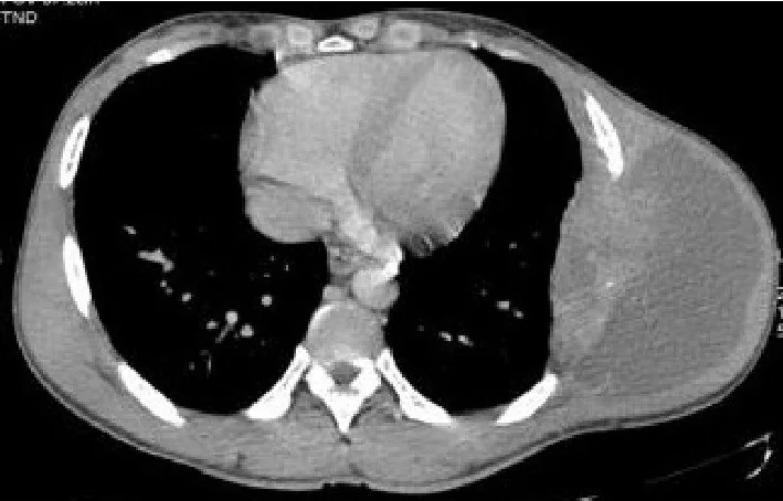
Göğüs duvarı tüberkülozu çoğunlukla kaburgaları, daha seyrek sternum veya sternoklaviküler eklemi tutan ekstrapulmoner enfeksiyondur. BT, kemik lezyonunun sınırını ve komşu koleksiyonun plevra, kas planları ve cilde uzanımını birlikte göstererek tanısal örnekleme için anatomik yol haritası sağlar. Akciğer ya da plevrada eşlik eden aktif hastalık görülmeyebilir; bu nedenle normal akciğer bulguları göğüs duvarındaki şüpheli odağı önemsizleştirmemelidir. Tümör veya piyojenik enfeksiyon olarak yorumlanması mikrobiyolojik ve patolojik doğrulamayı geciktirebilir.
Faz ve pencere
- Venöz tanısal
- Kontrastlı toraks BT’de koleksiyon içeriği çevre yumuşak dokudan düşük atenüasyonlu görünür; duvarı rim tarzında boyanabilir. Aynı kesitlerde kaburga ya da sternumdaki kemik kaybının koleksiyonla ilişkisi ve plevral uzanım değerlendirilir.
- Kemik algoritması tanısal
- İnce kemik rekonstrüksiyonlarında kaburga ve sternum korteksindeki erozyon veya osteoliz, eklem yüzü düzensizliği, ayrılmış kemik parçası ve eşlik eden skleroz araştırılır.
Önerilen pencereler: Mediasten (G 350 / M 50), Yumuşak doku (G 400 / M 40), Kemik (G 1800 / M 400), Akciğer (G 1500 / M -600).
BT bulguları
- Kaburga osteolizi — juxtakostal yumuşak doku kitlesine komşu kortikal kayıp görülebilir; kemik tutulumu olmaması da olasıdır.
- Sternal destrüksiyon — sternumda fokal litik alan, bazen çevresindeki yumuşak doku kitlesiyle devamlılık gösterir.
- Soğuk apse — içi sıvı dansitesinde, duvarı kontrastlanan koleksiyon; çevresel inflamasyon klinik şişlikle kıyaslandığında sınırlı olabilir.
- Kemik sekestrumu — destrüksiyon alanı içinde canlı korteksten ayrılmış yoğun kemik odağı seçilebilir.
- İnce yumuşak doku kalsifikasyonu — kronik koleksiyon çevresinde veya içinde noktasal kalsifik odaklar bulunabilir.
- Göğüs duvarı boyunca yayılım — koleksiyon kaburganın iç ve dış yüzleri arasında uzanabilir, interkostal kas planını aşabilir.
- Cilde açılan trakt — koleksiyondan deri altına ilerleyen ince yol ve varsa cilt açıklığı takip edilebilir.
- Plevra-akciğer eşlikçisi — plevral kalınlaşma, efüzyon veya parankimal TB bulguları aranır; bunların yokluğu izole duvar tutulumunu dışlamaz.
Normalde
Normal kaburga korteksi düzgün ve kesintisiz izlenir; sternumun kortikal konturu korunur ve sternoklaviküler eklem yüzlerinde erozyon bulunmaz. Karşı taraftaki aynı seviyeli kaburgayı ve eklemi kıyaslayarak fokal kemik kaybını, komşu interkostal kası ve plevra boyunca gelişen sıvı ya da kalınlaşmayı ayırt edin.
Normal BT ile kıyasla
Aynı bölgenin, kaynak veri setinde patoloji içermediği belirtilen bir BT incelemesini kesit kesit kaydırın; organ sınırlarını açarak anatomiyi hasta görüntüsüyle karşılaştırın. Küçük rastlantısal bulgular tümüyle dışlanmamıştır.
Kaynak: TotalSegmentator v2.01 veri seti (Wasserthal ve ark., Radiology: Artificial Intelligence 2023), CC BY 4.0, vaka s1278; organ sınırları veri setinin otomatik segmentasyonundan, birleştirilerek sadeleştirildi. Görüntüler 2,5 mm kesit aralığına yeniden örneklendi.
Ayırıcı tanı
- Piyojenik osteomiyelit
- akut klinik başlangıç, belirgin çevre ödemi ve bazen gaz eşlik edebilir; BT tek başına mikroorganizmayı ayıramaz.
- Metastatik kemik tutulumu
- litik kaburga odağı ve ek kitle oluşturabilir; başka kemik veya organ odaklarının dağılımı neoplastik olasılığı destekler.
- Primer göğüs duvarı sarkomu
- kemik kökenli geniş, çoğunlukla solid yumuşak doku bileşeni beklenir; nekroz görüntüde apseyi taklit edebilir.
- Sternoklaviküler septik artrit
- değişiklikler eklem aralığı ve komşu eklem yüzlerinde merkezlenir; çevresel koleksiyonun tek başına bu eklemden kaynaklandığı varsayılmamalıdır.
- Lenfoma
- kemik iliği ve yumuşak dokuda kitle yapabilir; doku tanısı olmadan enfeksiyon ile güvenilir biçimde ayrılamayabilir.
Tuzaklar
- Akciğer parankiminde kavite ya da plevral hastalık görülmemesi göğüs duvarı tutulumunu dışlamaz; lezyonun kendi kemik-yumuşak doku örüntüsünü değerlendirin.
- Çevresel boyanma enfeksiyonu düşündürse de nekrotik tümör benzer görünür; BT etkeni kanıtlamaz ve örnekleme ihtiyacını ortadan kaldırmaz.
- Göğüs duvarı tüberkülozunda cilde açılan sinüs traktı gelişebilir.
- Yalnız yumuşak doku penceresiyle sınırlı kalmak küçük kortikal odağı ve sekestrumu gözden kaçırabilir; aynı seviyeyi kemik rekonstrüksiyonunda yeniden inceleyin.
Kendini dene
Kaburga litik odağına komşu çevresel boyanan göğüs duvarı koleksiyonu var, akciğer BT’sinde aktif tüberküloz bulgusu yok. Hangi yorum en doğrudur?
Cevabı göster
İzole göğüs duvarı tüberkülozu olasılığı sürer; normal akciğer bulguları dışlamaz.. Göğüs duvarı tüberkülozu akciğer veya plevrada aktif hastalık olmadan da görülebilir; ancak çevresel boyanan koleksiyon ve litik odak özgül değildir. BT yayılımı gösterir, etkeni doğrulamaz.
Göğüs duvarında rim tarzı boyanan koleksiyon ve kaburga destrüksiyonu var; görüntüleme tüberküloz, piyojenik enfeksiyon ve nekrotik tümör arasında kesin ayrım yapmıyor. Etiyolojiyi doğrulamak için en uygun yaklaşım hangisidir?
Cevabı göster
Mikrobiyolojik ve histopatolojik örnekleme. Rim boyanması ve kemik destrüksiyonu farklı enfeksiyonlarda ve nekrotik tümörlerde görülebilir. BT anatomik yayılımı tarif eder; etken veya histolojiyi güvenilir biçimde belirlemek için örnekleme gerekir.
İlgili konular
Kaynaklar
Bu sayfadaki 40 cümle ve 2 quiz sorusu aşağıdaki kaynaklardan alıntılarla otomatik olarak karşılaştırıldı (2026-10-09).
- Imaging of thoracic tuberculosis: pulmonary and extrapulmonarypmc.ncbi.nlm.nih.gov
- Chest wall tuberculosis - A clinical and imaging experiencepmc.ncbi.nlm.nih.gov
- Atypical imaging of spinal tuberculosis: a case report and review of literaturepmc.ncbi.nlm.nih.gov
- Unusual manifestations of chest wall tuberculosis: diagnostic challenges in two cases from a low-incidence tuberculosis countrypmc.ncbi.nlm.nih.gov
- Diagnosis of Chest Wall Tuberculosis Using Fine Needle Aspiration: A Single-Center Experiencepmc.ncbi.nlm.nih.gov
- Imaging of thoracic tuberculosis: pulmonary and extrapulmonary.pubmed.ncbi.nlm.nih.gov
- Atypical imaging of spinal tuberculosis: a case report and review of literature.pubmed.ncbi.nlm.nih.gov
- Chest wall tuberculosis - A clinical and imaging experience.pubmed.ncbi.nlm.nih.gov
- Primary chest wall tuberculosis mimicking gynecomastia: A case report.pubmed.ncbi.nlm.nih.gov
- Isolated Chest Wall Necrotizing Fasciitis: An Unusual Fatal Manifestation of Extrapulmonary Tuberculosis.pubmed.ncbi.nlm.nih.gov
- Chest wall subcutaneous mass leading to the diagnosis of pericostal tuberculosis.pubmed.ncbi.nlm.nih.gov
- Role of Cross Sectional Imaging in Isolated Chest Wall Tuberculosis.pubmed.ncbi.nlm.nih.gov
- Breast Lump: A Rare Presentation of Tuberculosis in an Elderly Man.pubmed.ncbi.nlm.nih.gov
- 18F-FDG PET/CT Images in a Patient With Primary Chest Wall Tuberculosis Mimicking Malignant Tumor.pubmed.ncbi.nlm.nih.gov
- Solitary Chest Wall Tuberculosis Mimicking a Malignancy Demonstrated on FDG PET/CT.pubmed.ncbi.nlm.nih.gov
- Unusual manifestations of chest wall tuberculosis: diagnostic challenges in two cases from a low-incidence tuberculosis country.pubmed.ncbi.nlm.nih.gov
- Primary sternocostal tuberculosis in a young healthcare worker: a case report.pubmed.ncbi.nlm.nih.gov
- Case Report: Metagenomic next-generation sequencing diagnosed a rare case of sternal tuberculosis mimicking a malignant tumour.pubmed.ncbi.nlm.nih.gov
- Pulmonary Abscess With Contiguous Extension to the Chest Wall in an Immunocompetent Young Woman: A Case Report.pubmed.ncbi.nlm.nih.gov
- The role of thoracoplasty in modern thoracic surgery: a narrative review with three illustrative cases.pubmed.ncbi.nlm.nih.gov
- Perforated acute appendicitis on computed tomography revealing underlying tuberculosis.pubmed.ncbi.nlm.nih.gov
- Isolated Ovarian Tuberculosis Mimicking Ovarian Malignancy in a Young Female with Primary Infertility: A Case Report.pubmed.ncbi.nlm.nih.gov
- From misdiagnosis to definitive diagnosis of SAPHO syndrome during a routine health check-up: a case report and literature review.pubmed.ncbi.nlm.nih.gov
- Epidemiological Features, Etiopathogenesis, Diagnostic Evaluation, and Management Protocol of Splenic Tuberculosis: A Systematic Review.pubmed.ncbi.nlm.nih.gov
- Atypical Presentation of Pancreatic Adenocarcinoma as a Fever of Unknown Origin Mimicking Multiple Liver Abscesses: A Case Report.pubmed.ncbi.nlm.nih.gov
- A Rare Case of Sterile Retrostyloid Abscess in a Patient With Crohn's Disease: Diagnostic and Therapeutic Insights.pubmed.ncbi.nlm.nih.gov
- Case report: Tumor-like mediastinal tuberculous lymphadenitis with paravertebral cold abscess under cumulative immunosuppression: a case highlighting altered host-pathogen interactions.pubmed.ncbi.nlm.nih.gov
- Askin Tumor Presents with Ewing Sarcoma of the Femur in Young Female Patient: Case Report and Literature Review.pubmed.ncbi.nlm.nih.gov
- Spinal Cord Compression due to Pyothorax-Associated Lymphoma: A Rare Presentation of EBV-Positive Diffuse Large B-Cell Lymphoma.pubmed.ncbi.nlm.nih.gov
- Common and Rare Radiological Findings of Pulmonary and Thoracic Hydatid Cysts.pubmed.ncbi.nlm.nih.gov
- Purely osteolytic bone lesions in the early osteoarticular phase of synovitis, acne, pustulosis, hyperostosis and osteitis syndrome.pubmed.ncbi.nlm.nih.gov
- Tuberculous Otomastoiditis: A Diagnostic Dilemma.pubmed.ncbi.nlm.nih.gov
- Chronic Recurrent Osteomyelitis of Ribs Due to Bamboo Stick Injury in an Adult Man: A Case Report.pubmed.ncbi.nlm.nih.gov
- Multifocal Skeletal Tuberculosis Mimicking Metastatic Malignancy: A Case Report.pubmed.ncbi.nlm.nih.gov
- A pictorial essay of thoracic wall diseases: multiple pathologies in the same anatomical site.pubmed.ncbi.nlm.nih.gov
- Extensive thoracic vertebral and chest wall metastases as the initial presentation of breast cancer: a case report and literature review.pubmed.ncbi.nlm.nih.gov
- Hidden in plain sight: Extrapulmonary manifestations of tuberculosis in Asia.pubmed.ncbi.nlm.nih.gov
- <i>Salmonella</i> Osteomyelitis of the Rib.pubmed.ncbi.nlm.nih.gov
- Spontaneous appendicocutaneous fistula in a child: a case report.pubmed.ncbi.nlm.nih.gov
- Pediatric pulmonary tuberculosis masquerading as chest neoplasms.pubmed.ncbi.nlm.nih.gov
- A Rare Case of an Intrapulmonary Teratoma Masquerading as a Pulmonary Hamartoma.pubmed.ncbi.nlm.nih.gov
- Multifocal Necrotizing Fasciitis of the Bilateral Chest Wall and Contralateral Lower Limb in a Patient With Diabetes: A Case Report.pubmed.ncbi.nlm.nih.gov
- A Rare Case of Empyema Necessitans Due to Streptococcus Anginosus with Necrotizing Fasciitis and Retroperitoneal Abscesses.pubmed.ncbi.nlm.nih.gov
- Thoracic Actinomycosis: A Rare Cause of Pleural Thickening.pubmed.ncbi.nlm.nih.gov
- Chronic draining xiphisternal fistula associated with fractured right coronary artery stent and transdiaphragmatic inflammatory extension: a multimodality imaging and surgical correlation.pubmed.ncbi.nlm.nih.gov
- Hybrid eclipse: a case of isolated renal tuberculosis with an unusual infectious-inflammatory-neoplastic triad.pubmed.ncbi.nlm.nih.gov
- Primary Chest Wall Malignant Lymphoma in a Patient Without a History of Tuberculosis or Chronic Pyothorax: A Case Report With a Review of the Literature.pubmed.ncbi.nlm.nih.gov
- When Tuberculosis Left the Heart in a Cage: Multimodality Imaging Revealing Constrictive Pericarditis in a Young Adult.pubmed.ncbi.nlm.nih.gov
- An integrated digital-to-surgical framework for patient-specific chest wall resection and reconstruction using 3D-printed titanium implants: proof-of-concept study.pubmed.ncbi.nlm.nih.gov
- Single-stage repair of congenital complete sternal agenesis with PFO-type interatrial communication and aortic arch variant complicated by an unexpected ascending aortic pseudoaneurysm: a case report.pubmed.ncbi.nlm.nih.gov
- Anatomic variability of the ribs and costal cartilage: A pilot study.pubmed.ncbi.nlm.nih.gov
- Standardized bilateral thoracic ultrasound image comparison as a tool for the diagnosis of pneumothorax: a pilot exploratory study.pubmed.ncbi.nlm.nih.gov
- Tonsillar Leishmaniasis: A Rare Clinical Entity Mimicking Malignancy in the Oropharynx - A Case Series from Northeastern Italy.pubmed.ncbi.nlm.nih.gov
- Effects of Muscle Energy Technique and Joint Manipulation on Pulmonary Functions, Mobility, Disease Exacerbations, and Health-Related Quality of Life in Chronic Obstructive Pulmonary Disease Patients: A Quasiexperimental Study.pubmed.ncbi.nlm.nih.gov
- Blastomycosis of the Central Nervous System.pubmed.ncbi.nlm.nih.gov
- Isolated cryptococcal osteomyelitis of the sacrum in an immunocompetent patient: a case report and literature review.pubmed.ncbi.nlm.nih.gov
- Differentiation between infectious spondylodiscitis versus inflammatory or degenerative spinal changes: How can magnetic resonance imaging help the clinician?pubmed.ncbi.nlm.nih.gov
- Chronic <i>Mycobacterium kansasii</i> Pleural Infection Mimicking Metastatic Breast Cancer: A Seven-Year Diagnostic Odyssey and the Critical Role of Metagenomic Sequencing.pubmed.ncbi.nlm.nih.gov
- Xanthogranulomatous Osteomyelitis as a Bone Tumor Mimic: A Systematic Review of Pre-biopsy Diagnostic Clues and Management Decision Points.pubmed.ncbi.nlm.nih.gov
- Computed tomography-guided precision biopsy combined with metagenomic next-generation sequencing for etiological diagnosis in patients with blood culture-negative systemic infections.pubmed.ncbi.nlm.nih.gov
- Approach to mediastinal cytopathology: a diagnostic overview.pubmed.ncbi.nlm.nih.gov
- Incidental Lung Lesions on the CT Component of Oncologic FDG PET/CT: A Pictorial Review of Interpretive Pitfalls and Diagnostic Clues.pubmed.ncbi.nlm.nih.gov
- A Rare Case of Disseminated Bacteroides fragilis Sternoclavicular Osteomyelitis Following Cardiopulmonary Resuscitation (CPR) in an Immunocompromised Patient: Is CPR the Missing Link?pubmed.ncbi.nlm.nih.gov
- Complicated septic arthritis of the sternoclavicular joint due to <i>Bacteroides fragilis</i> in a healthy middle-aged woman; A case report and literature review.pubmed.ncbi.nlm.nih.gov
- Evaluation of sternoclavicular joint infection in adults, 2020 to 2025.pubmed.ncbi.nlm.nih.gov
- Prolonged fever in ESBL-producing <i>Salmonella Typhi</i> in Lebanon: Diagnostic challenges and the possible role of occult low-level carbapenemase activity.pubmed.ncbi.nlm.nih.gov
- Tuberculous brain abscess mimicking a neoplastic lesion in a patient with well-controlled human immunodeficiency virus infection: illustrative case.pubmed.ncbi.nlm.nih.gov
- The first CT-based classification system for pulmonary actinomycosis: correlating imaging patterns with therapeutic strategies and prognosis in a highly misdiagnosed disease.pubmed.ncbi.nlm.nih.gov
- Infiltrative and inflammatory systemic disorders involving the pituitary gland.pubmed.ncbi.nlm.nih.gov
- Anatomic Pulmonary Lobectomy via Video-Assisted Thoracoscopic Surgery in an Adult Patient With Congenital Lobar Emphysema: A Case Report.pubmed.ncbi.nlm.nih.gov
- Cavitary Pulmonary Adenocarcinoma in a Young Adult With a History of Treated Hodgkin Lymphoma: A Case Report.pubmed.ncbi.nlm.nih.gov
- Multimodal ultrasound in the diagnosis and treatment of caudate lobe Klebsiella pneumoniae liver abscess complicated by rare gas-containing inferior vena cava thrombosis: a case report.pubmed.ncbi.nlm.nih.gov
- Tuberculosis revisted: classic imaging findings in childhood.pubmed.ncbi.nlm.nih.gov
- Tuberculosis, a great masquerader: A case series unveiling rare sites of musculoskeletal involvement through imaging.pubmed.ncbi.nlm.nih.gov
- Efficacy and safety of an all-oral delamanid-containing regimen in the treatment of multidrug-resistant pulmonary tuberculosis complicated by extrapulmonary tuberculosis: Four case reports and review of the literature.pubmed.ncbi.nlm.nih.gov
- Mycobacterium bovis Bacillus Calmette-Guérin (BCG)-Related Sternal Osteomyelitis.pubmed.ncbi.nlm.nih.gov
- Synchronous Occurrence of Gastric Signet Ring Cell Carcinoma and Pulmonary Fibrosarcoma in 23-Year-Old Male Youth.pubmed.ncbi.nlm.nih.gov
- EACR 2025 Congress: Innovative Cancer Science, 16-19 June 2025.pubmed.ncbi.nlm.nih.gov
- Commentary: an eye on PET quantification.pubmed.ncbi.nlm.nih.gov
- High-affinity alphavbeta3 integrin targeted optical probe as a new imaging biomarker for early atherosclerosis: initial studies in Watanabe rabbits.pubmed.ncbi.nlm.nih.gov
- High-resolution ultrasound in the evaluation of musculoskeletal infections.pubmed.ncbi.nlm.nih.gov
- Diagnosis of Osteoarticular Tuberculosis: Perceptions, Protocols, Practices, and Priorities in the Endemic and Non-Endemic Areas of the World-A WAIOT View.pubmed.ncbi.nlm.nih.gov
- Post-Tuberculosis Lung Disease: Clinicopathologic Insights, Diagnosis, Prevention, and Management.pubmed.ncbi.nlm.nih.gov
- Diagnostic tools in respiratory medicine (Review).pubmed.ncbi.nlm.nih.gov
- CT Imaging Findings of Pulmonary Artery Stenosis: A Pictorial Review.pubmed.ncbi.nlm.nih.gov
- Lung squamous cell carcinoma complicated with immune checkpoint inhibitor-associated active tuberculosis: a case report and literature review.pubmed.ncbi.nlm.nih.gov
- Malignancy Presenting As Recurrent Empyema: A Report of Two Cases and a Systematic Review.pubmed.ncbi.nlm.nih.gov
- Distinct Presentations of Langerhans Cell Histiocytosis in Children: A Case Series.pubmed.ncbi.nlm.nih.gov
- Tuberculosis of Rib: Diagnosis and Treatment.pubmed.ncbi.nlm.nih.gov
- Case Report: delayed recognition of pulmonary tuberculosis during immunosuppression for suspected connective tissue disease.pubmed.ncbi.nlm.nih.gov
- Migratory Pulmonary Infiltrates Mimicking Malignancy, Tuberculosis, and Sarcoidosis in an Apparently Immunocompetent Adult: A Case of Tissue-Proven Invasive Fungal Disease Due to a Non-<i>marneffei</i><i>Penicillium</i> Species.pubmed.ncbi.nlm.nih.gov
- Acute mediastinitis, mediastinal granuloma, and fibrosing mediastinitis: a narrative review.pubmed.ncbi.nlm.nih.gov
- A Review of Posteromedial Lesions of the Chest Wall: What Should a Chest Radiologist Know?pubmed.ncbi.nlm.nih.gov
- Coexistence of Autoantibodies against the Golgi Complex and Ro52 Antigen in a Patient with Nonspecific Interstitial Pneumonia.pubmed.ncbi.nlm.nih.gov
- 2.5-mm articulated endoluminal forceps using a compliant mechanism.pubmed.ncbi.nlm.nih.gov
- Trans-thoracic Extension of a Lung Abscess With a Pleurocutaneous Fistula: A Report of a Rare Case.pubmed.ncbi.nlm.nih.gov
- Case Report: A rare case of oesophageal cellulitis caused by <i>Klebsiella pneumoniae</i>: diagnosis, management, and literature review.pubmed.ncbi.nlm.nih.gov
- Sigmoid colonic tuberculosis presenting as a colovesical fistula mimicking colorectal malignancy: a case report.pubmed.ncbi.nlm.nih.gov
- Cutaneous Tuberculous Sinus Tract: A Case Report.pubmed.ncbi.nlm.nih.gov
Eğitim amaçlıdır; hasta başında tanı ve tedavi kararının yerine geçmez, tıbbi tavsiye değildir. Metinler yapay zekâ desteğiyle yazıldı ve otomatik bir adımda kaynak alıntılarıyla karşılaştırıldı; bu bir tıbbi doğrulama değildir ve içerik uzman radyolog incelemesinden geçmedi. Klinik kararlarda güncel kılavuzları, kurum protokollerini ve radyoloji raporunu esas alın.