Özet
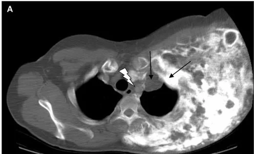
Göğüs duvarındaki primer kemik ve kıkırdak tümörleri benign veya malign olabilir; kondrosarkom göğüs duvarının önemli primer malign kemik tümörlerinden biridir ve sıklıkla kaburga-kostal kıkırdak bileşkesinde görülür. BT, lezyonun kemik merkezini, kondroid matriks mineralizasyonunu, korteks ve endosteal yüzeyi, ayrıca yumuşak doku bileşenini gösterir. Lobüle kalsifiye matriks tek başına benignlik ya da tümör derecesi anlamına gelmez; agresif kemik değişikliği ve ekstraosseöz uzanım ayrıca aranmalıdır. Göğüs duvarı kondrosarkomlarında yumuşak doku kitlesi sık görülebildiğinden, tümörün BT'de yayılımı değerlendirilirken kemik dışındaki bileşen de incelenmelidir.
Faz ve pencere
- Kontrastsız tanısal
- BT'de lezyonun yeri, litik ya da mikst görünümü ve kondroid matriks kalsifikasyonu; ayrıca yumuşak doku kitlesi ve patolojik kırık varlığı değerlendirilip kaydedilebilir. Kontrast fazı matriks ve korteks değerlendirmesi için gerekmez; ekstraosseöz kitlenin ayrıntılı yayılımı gerektiğinde yumuşak doku incelemesi ayrıca planlanabilir.
- Kemik algoritması tanısal
- BT, tümörün kondroid yapısını ve yayılımını değerlendirmeye; kemik dışına uzanan yumuşak doku dansitesindeki bileşeni göstermeye yardımcı olur.
Önerilen pencereler: Kemik (G 1800 / M 400), Yumuşak doku (G 400 / M 40), Mediasten (G 350 / M 50).
BT bulguları
- Kostokondral merkezli lobüle kitle — kaburga veya kıkırdak bileşkesinden gelişen ekspansil lezyon olarak izlenir.
- Halka-yay kondroid matriksi — kıkırdak matriksi içindeki kavisli mineralizasyon odakları lezyon karakterizasyonuna katkı sağlar.
- Endosteal çentiklenme — göğüs duvarı kondrosarkomlarında bildirilen radyolojik bulgulardandır.
- Kortikal destrüksiyon — kemik lezyonlarında agresif görüntüleme özelliklerinden biri olarak değerlendirilir.
- Ekstraosseöz yumuşak doku komponenti — kaburga/kıkırdak konturunu aşan lobüle doku kitlesi malignite açısından önem taşır.
- Matriks mineralizasyonunda değişkenlik — iyi organize kavisli kalsifikasyon düşük dereceli lezyonlarda görülebilir; daha az mineralize litik alanlar da olabilir.
- Patolojik kırık — bir göğüs duvarı kondrosarkomu serisinde 26 olgunun 2'sinde (%8) bildirilmiştir.
Normalde
Karşı taraftaki aynı seviyenin korteksini ve kostal kıkırdak konturunu kıyaslayın; kalsifikasyonun kitle içinde lobüle ve halka-yay biçiminde dağılıp dağılmadığını izleyin.
Normal BT ile kıyasla
Aynı bölgenin, kaynak veri setinde patoloji içermediği belirtilen bir BT incelemesini kesit kesit kaydırın; organ sınırlarını açarak anatomiyi hasta görüntüsüyle karşılaştırın. Küçük rastlantısal bulgular tümüyle dışlanmamıştır.
Kaynak: TotalSegmentator v2.01 veri seti (Wasserthal ve ark., Radiology: Artificial Intelligence 2023), CC BY 4.0, vaka s1278; organ sınırları veri setinin otomatik segmentasyonundan, birleştirilerek sadeleştirildi. Görüntüler 2,5 mm kesit aralığına yeniden örneklendi.
Ayırıcı tanı
- Enkondrom
- halka-yay matriksi olabilir; kortikal destrüksiyon ve yumuşak doku kitlesi yoksa agresif kondrosarkomdan daha az kuşkuludur, ancak göğüs duvarında klinik/radyolojik bağlam önemlidir.
- Osteokondrom
- kemik yüzeyinden gelişen, üzerini kıkırdak dokunun örttüğü benign bir tümördür.
- Osteosarkom
- osteoid matriks üretimiyle karakterize primer malign kemik tümörüdür; mevcut kaynaklar mineralizasyon biçimini veya periostal reaksiyonunu tarif etmiyor.
- Kaburga metastazı
- prostat kanserinde kaburga metastazları bildirilmiştir; verilen kaynaklar bunların litik, sklerotik veya mikst BT görünümünü ya da kondroid matriksle karşılaştırmasını açıklamıyor.
Tuzaklar
- Halka-yay kalsifikasyonunu tek başına kondrosarkom tanısı saymayın; benign kıkırdak lezyonlarında da görülebilir ve korteks/yumuşak doku davranışıyla birlikte yorumlanmalıdır.
- Uzun kemikler için tanımlanmış endosteal çentiklenme eşiğini kaburgaya otomatik taşımayın; bu sayfada göğüs duvarına özgü doğrulanmış sayısal eşik kullanılmıyor.
- Kısmi hacim etkisi kostal kıkırdak ve kortekste yalancı kesinti yaratabilir; ince kemik rekonstrüksiyonları ile ardışık düzlemlerde doğrulayın.
- BT'deki mineralizasyon miktarından histolojik derece çıkarmayın; düşük ve orta dereceli kondrosarkomların özellikleri örtüşebilir.
Kendini dene
Kostokondral bileşkede lobüle kitle, halka-yay matriksi, kortikal destrüksiyon ve kaburga dışına taşan doku var. Hangi tanı önceliklidir?
Cevabı göster
Kondrosarkom. Kondroid matriksle birlikte kortikal yıkım ve ekstraosseöz kitle agresif kondrosarkom lehine özelliklerdir. Enkondromda agresif destrüksiyon beklenmez; osteokondrom ana kemikle kortikomedüller devamlılık gösterir; kallus kırık hattı çevresinde gelişir.
BT'de pedinküllü kemik çıkıntısının korteksi ve medullası ana kemikle devamlılık gösteriyor. Bu bulgu en çok hangi lezyonu destekler?
Cevabı göster
Osteokondrom. Korteks ve medullanın ana kemikle devamlılığı, kaynakta osteokondrom olgusunda tarif edilen görüntüleme bulgusudur.
İlgili konular
Kaynaklar
Bu sayfadaki 35 cümle ve 2 quiz sorusu aşağıdaki kaynaklardan alıntılarla otomatik olarak karşılaştırıldı; 10 cümle bu karşılaştırmada düzeltildi (2026-10-09).
- Grade 1 and 2 Chondrosarcomas of the Chest Wall: CT Imaging Features and Review of the Literaturepmc.ncbi.nlm.nih.gov
- Primary maxillary chondrosarcoma: A case report.pubmed.ncbi.nlm.nih.gov
- Excision of Thoracic Chondrosarcoma: Case Report and Review of Literature.pubmed.ncbi.nlm.nih.gov
- Surgical management of a giant sternal chondromyxoid fibroma: a case report.pubmed.ncbi.nlm.nih.gov
- Manubrioclavicular and Manubriosternal Reconstruction After Radical Resection for Chondrosarcoma of Manubriosternum: A Modified Surgical Technique.pubmed.ncbi.nlm.nih.gov
- [Chest Wall Reconstruction with Polypropylene Mesh After Resection of Primary Sternal Chondrosarcoma].pubmed.ncbi.nlm.nih.gov
- Innovative chest wall reconstruction with a locking plate and cement spacer after radical resection of chondrosarcoma in the sternum: A case report.pubmed.ncbi.nlm.nih.gov
- Ewing sarcoma with intra thoracic and multiple extra thoracic metastases in a young adult male: A case report.pubmed.ncbi.nlm.nih.gov
- Laparoscopic resection of retroperitoneal metastasis originating from sternal leiomyosarcoma - a case report.pubmed.ncbi.nlm.nih.gov
- Case Report: Osteosarcoma following traumatic injury in a juvenile Argentine black and white tegu (<i>Salvator merianae</i>).pubmed.ncbi.nlm.nih.gov
- From diagnostic marker-to-therapeutic modality: emerging paradigms in tumor biomineralization.pubmed.ncbi.nlm.nih.gov
- Chest Wall Resection and Reconstruction Following Cancer.pubmed.ncbi.nlm.nih.gov
- Robot-assisted thoracoscopic resection of a costal cartilage chondrosarcoma mimicking anterior mediastinal teratoma: a case report.pubmed.ncbi.nlm.nih.gov
- Fibrocartilaginous mesenchymoma of the rib with atypical imaging features.pubmed.ncbi.nlm.nih.gov
- Conventional chondrosarcoma of the sternum presenting as a chest wall mass with multilobulated intra- and extra-thoracic extension.pubmed.ncbi.nlm.nih.gov
- Tuberculous Lymphadenitis Presenting as Chest Wall Cold Abscess: A Case Report.pubmed.ncbi.nlm.nih.gov
- Case Report: Primary giant chondrosarcoma of the manubrium sterni.pubmed.ncbi.nlm.nih.gov
- Recurrent Desmoid-Type Fibromatosis of the Breast in a Young Woman With a Monoallelic MUTYH Germline Variant.pubmed.ncbi.nlm.nih.gov
- Understanding a mass in the paraspinal region: an anatomical approach.pubmed.ncbi.nlm.nih.gov
- Coexistence of Autoantibodies against the Golgi Complex and Ro52 Antigen in a Patient with Nonspecific Interstitial Pneumonia.pubmed.ncbi.nlm.nih.gov
- Commentary: an eye on PET quantification.pubmed.ncbi.nlm.nih.gov
- First case description of a highly aggressive thoracopulmonary primitive neuroectodermal tumor in a Moroccan adult male.pubmed.ncbi.nlm.nih.gov
- HPV-associated Oropharyngeal Squamous Cell Carcinoma: An Imaging Framework for AJCC Version 9 TNM Staging.pubmed.ncbi.nlm.nih.gov
- Patient‑specific 3D‑printed Ti‑6Al‑4 V sternum-rib implant for near‑total sternal resection: operational blueprint, implant design principles, and intermediate clinical outcome.pubmed.ncbi.nlm.nih.gov
- A Review of Posteromedial Lesions of the Chest Wall: What Should a Chest Radiologist Know?pubmed.ncbi.nlm.nih.gov
- Chondrosarcoma of the Ribs.pubmed.ncbi.nlm.nih.gov
- Primary malignant giant cell tumor of the sternum.pubmed.ncbi.nlm.nih.gov
- Beyond Chordoma: A Comprehensive Review of Sacral Lesions.pubmed.ncbi.nlm.nih.gov
- Sacral Chordoma: Multimodality Imaging and Radiologic-Pathologic Correlation of a Rare Malignant Sacral Tumor.pubmed.ncbi.nlm.nih.gov
- Cavernous venous malformation of the frontal sinus: CT and MRI imaging findings.pubmed.ncbi.nlm.nih.gov
- A pictorial essay of thoracic wall diseases: multiple pathologies in the same anatomical site.pubmed.ncbi.nlm.nih.gov
- Multimodal Imaging of Osteosarcoma: From First Diagnosis to Radiomics.pubmed.ncbi.nlm.nih.gov
- Classification of Chondrosarcoma: From Characteristic to Challenging Imaging Findings.pubmed.ncbi.nlm.nih.gov
- Pediatric Desmoplastic Fibroma of the Jaws: A Comprehensive Review of Clinical Presentation, Management, and Outcomes.pubmed.ncbi.nlm.nih.gov
- When plasmacytoma defies expectations: An unusual maxillary presentation with mixed imaging features in a young patient.pubmed.ncbi.nlm.nih.gov
- Microvascular reconstruction of aggressive mandibular desmoplastic fibroma in a child: case report.pubmed.ncbi.nlm.nih.gov
- Novel Application of STRATOS for Restoration of Clavicular Stability After Oncologic Sternoclavicular Joint Resection: A Case Report and Review of the Literature.pubmed.ncbi.nlm.nih.gov
- Primary Chest Wall Ewing Sarcoma With Diaphragmatic Invasion in an Adult: En-Bloc Resection and Polypropylene Mesh Reconstruction.pubmed.ncbi.nlm.nih.gov
- Unexpected Massive Hemorrhage During Incision and Drainage of a Presumed Perianal Abscess: A Case of Pelvic Chondrosarcoma.pubmed.ncbi.nlm.nih.gov
- Chondrosarcoma: A comprehensive review of tumor biology, clinical behavior, and therapeutic advances.pubmed.ncbi.nlm.nih.gov
- Synovial Chondromatosis: A Narrative Review of Current Evidence on Diagnosis, Differential Diagnosis, and Management.pubmed.ncbi.nlm.nih.gov
- Risk Factors for Pathologic Fracture in Dogs With Confirmed or Suspected Appendicular Osteosarcoma Treated With Low- or Standard-Dose-Rate Palliative External Beam Radiation Therapy.pubmed.ncbi.nlm.nih.gov
- Non-glandular findings on breast ultrasound. Part II: a pictorial review of chest wall lesions.pubmed.ncbi.nlm.nih.gov
- Imaging Features of Intraosseous Schwannoma: A Case Series and Review of the Literature.pubmed.ncbi.nlm.nih.gov
- Biomechanics of chest wall injury: implications for surgical stabilization and failure prevention.pubmed.ncbi.nlm.nih.gov
- Narrative review of chest wall ultrasound: a practical approach.pubmed.ncbi.nlm.nih.gov
- Surgical stabilization of rib fractures (SSRF): the WSES and CWIS position paper.pubmed.ncbi.nlm.nih.gov
- Thoracic joint abnormalities and impaired thoracic motion in idiopathic pleuroparenchymal fibroelastosis: an exploratory study using inspiratory and expiratory CT.pubmed.ncbi.nlm.nih.gov
- Incidental Cardiac Uptake on 99m Tc-HMDP Bone Scintigraphy in Oncology Patients: Two Cases of Transthyretin Amyloid Cardiomyopathy with Literature Review.pubmed.ncbi.nlm.nih.gov
- Beyond Resnick and Niwayama: a structured narrative literature review on the clinical and imaging phenotypes of diffuse idiopathic skeletal hyperostosis (DISH).pubmed.ncbi.nlm.nih.gov
- The diagnostic outcome of CT-guided rib biopsies: a retrospective cohort study.pubmed.ncbi.nlm.nih.gov
- Hereditary multiple exostoses: an educational review.pubmed.ncbi.nlm.nih.gov
- Malignant Transformation of Musculoskeletal Lesions with Imaging-Pathology Correlation-Part 1: Bone Lesions.pubmed.ncbi.nlm.nih.gov
- Imaging in Sternal Tumours: A Pictorial Review.pubmed.ncbi.nlm.nih.gov
- Glossary of terms for musculoskeletal radiology.pubmed.ncbi.nlm.nih.gov
- Paget's disease of bone with superimposed osseous metastasis from lung carcinoma: A case report.pubmed.ncbi.nlm.nih.gov
- An Unusual Presentation of Philadelphia Chromosome Positive B-Cell Acute Lymphoblastic Leukemia With Isolated Osteolytic Lesions at Diagnosis: A Case Report.pubmed.ncbi.nlm.nih.gov
- Immune checkpoint inhibition in bone metastases: lesion response heterogeneity, the osteoimmune niche, and microenvironment-directed therapy.pubmed.ncbi.nlm.nih.gov
- Piezo1 in hypertrophic chondrocytes regulates osteoclastogenesis in endochondral ossification.pubmed.ncbi.nlm.nih.gov
- Forearm Nonunion and Segmental Bone Defects: A Focused Review of Masquelet Reconstruction and Free Vascularized Bone Grafting.pubmed.ncbi.nlm.nih.gov
- From nerve to bone: a tale of autonomic nervous system, bone cells, and beyond.pubmed.ncbi.nlm.nih.gov
- Diffusion-Weighted Imaging in the Musculoskeletal System: Evolving Role in Modern Imaging Practice.pubmed.ncbi.nlm.nih.gov
- Pictorial Review of Soft Tissue Lesions with Calcification.pubmed.ncbi.nlm.nih.gov
- Neglected knee swelling: A case report of massive synovial chondromatosis.pubmed.ncbi.nlm.nih.gov
- MiRNA-based expression signatures in differential diagnosis of enchondroma and chondrosarcoma.pubmed.ncbi.nlm.nih.gov
- DIAGNOSIS OF ENDOCRINE DISEASE: Mosaic disorders of FGF23 excess: Fibrous dysplasia/McCune-Albright syndrome and cutaneous skeletal hypophosphatemia syndrome.pubmed.ncbi.nlm.nih.gov
- Development and patterning of rib primordia are dependent on associated musculature.pubmed.ncbi.nlm.nih.gov
- Pediatric emergency medicine point-of-care ultrasound: summary of the evidence.pubmed.ncbi.nlm.nih.gov
- Quantitative Bone SPECT/CT of Central Cartilaginous Bone Tumors: Relationship between SUVmax and Radiodensity in Hounsfield Unit.pubmed.ncbi.nlm.nih.gov
- Chondrosarcoma: A Clinical Review.pubmed.ncbi.nlm.nih.gov
- SPOT THE BOT: A comparative quality assessment of AI-generated written instructions for lung ultrasound training.pubmed.ncbi.nlm.nih.gov
- A single-institution retrospective review of patients treated with single-fraction 28 Gy SBRT to lung tumors.pubmed.ncbi.nlm.nih.gov
- Diagnosis and management of pleural mesothelioma: three unique cases and review of the literature.pubmed.ncbi.nlm.nih.gov
- Imaging of Sacroiliac Pain: The Current State-of-the-Art.pubmed.ncbi.nlm.nih.gov
- The cavernous sinus: An anatomic study with clinical implication.pubmed.ncbi.nlm.nih.gov
- Medical hyperspectral imaging: an updated review of technology advancements and biomedical applications.pubmed.ncbi.nlm.nih.gov
- Pathological Fracture in the Metatarsal Bone Due to an Intraosseous Schwannoma: A Case Report.pubmed.ncbi.nlm.nih.gov
- Imaging Aspects of Enchondromas in Pediatric Patients.pubmed.ncbi.nlm.nih.gov
- Concurrent Odontogenic Keratocyst and Odontoma: Report of an Unusual and Rare Entity.pubmed.ncbi.nlm.nih.gov
- Expert consensus on resection of chest wall tumors and chest wall reconstruction.pubmed.ncbi.nlm.nih.gov
- Beyond the Usual Suspects: Recognizing Calcified Chondroid Mesenchymal Neoplasm in Hand Soft Tissue Tumors.pubmed.ncbi.nlm.nih.gov
- Myoepithelial tumor of the bone: radiologic and histopathologic correlation.pubmed.ncbi.nlm.nih.gov
- The most common bone tumors of the upper extremity in childhood and adolescence and their treatment: a review of the current literature.pubmed.ncbi.nlm.nih.gov
- Schmorl's Node Mimicking Spinal Metastatic Disease: A Case Report and Review of the Literature.pubmed.ncbi.nlm.nih.gov
- Investigation of a bone lesion in a gorgonopsian (Synapsida) from the Permian of Zambia and periosteal reactions in fossil non-mammalian tetrapods.pubmed.ncbi.nlm.nih.gov
- Giant ischial osteochondroma causing ischiofemoral impingement and sciatic nerve compression in a pediatric patient with Albright's hereditary osteodystrophy.pubmed.ncbi.nlm.nih.gov
- Current Position of Nuclear Medicine Imaging in Primary Bone Tumors.pubmed.ncbi.nlm.nih.gov
- Paediatric Calcaneal Osteochondroma: A Case Report and a Literature Review.pubmed.ncbi.nlm.nih.gov
- Retrospective clinicopathological analysis of 193 cases of bone and soft tissue tumors in the foot and ankle.pubmed.ncbi.nlm.nih.gov
- Intraosseous tumours of the knee.pubmed.ncbi.nlm.nih.gov
- Case Report: Surgical resection of high-grade extradural thoracic vertebral chondrosarcoma in a dog.pubmed.ncbi.nlm.nih.gov
- Management of Scapular Osteochondroma: An Uncommon Report of Two Cases.pubmed.ncbi.nlm.nih.gov
- High-Grade Chondrosarcoma Involving the Xiphoid Process and Lower Sternum-The Continuing Role of Extended Open Chest Wall Surgery in the Era of Minimally Invasive Thoracic Surgery: A Case Report.pubmed.ncbi.nlm.nih.gov
- Askin Tumor Presents with Ewing Sarcoma of the Femur in Young Female Patient: Case Report and Literature Review.pubmed.ncbi.nlm.nih.gov
- Stereotactic radiotherapy for rib metastases from prostate cancer: A multicenter real-world analysis of local control, pain response, and treatment tolerance (RIBRISK).pubmed.ncbi.nlm.nih.gov
- Extraskeletal Ewing sarcoma primarily arising in the stomach and omentum in children: a case series.pubmed.ncbi.nlm.nih.gov
- The sternum in detail: a review of the anatomy and pathologies of the sternum.pubmed.ncbi.nlm.nih.gov
- Musculoskeletal Pitfalls on Molecular Imaging Studies of Oncologic Patients: How to Stay Out of Trouble.pubmed.ncbi.nlm.nih.gov
- Xanthogranulomatous Osteomyelitis as a Bone Tumor Mimic: A Systematic Review of Pre-biopsy Diagnostic Clues and Management Decision Points.pubmed.ncbi.nlm.nih.gov
- Rare overlap of subtype and phenotype: HER2-positive pleomorphic invasive lobular carcinoma presenting with an inflammatory breast cancer phenotype: a case report and targeted treatment strategy.pubmed.ncbi.nlm.nih.gov
- A journey with Maffucci syndrome: From skull base chondrosarcoma to multiorgan management: A case report and literature review.pubmed.ncbi.nlm.nih.gov
- Late breast cancer recurrence concurrent with silicone breast implant rupture 17 years after mastectomy and immediate implant-based reconstruction in a 53-year-old woman: a case report.pubmed.ncbi.nlm.nih.gov
- Current treatment strategies and prospects for chest wall defect repair and reconstruction.pubmed.ncbi.nlm.nih.gov
- Diagnosis of a patient with pulmonary and skeletal cryptococcosis: A case report and literature review.pubmed.ncbi.nlm.nih.gov
- Chest Wall and Diaphragmatic Injuries: Imaging Evaluation and Clinical Considerations.pubmed.ncbi.nlm.nih.gov
- Point-of-care ultrasound and neonatal rib osteomyelitis: A case report and literature review.pubmed.ncbi.nlm.nih.gov
- Subscapular Fossa Osteochondroma: A Case Report of an Anatomical Surgical Approach.pubmed.ncbi.nlm.nih.gov
- Hereditary multiple exostoses: A case report and literature review.pubmed.ncbi.nlm.nih.gov
- Osteochondroma of ventral scapula associated with chest pain due to rib cage compression: A case report.pubmed.ncbi.nlm.nih.gov
- Case Report: Primary gastric extraskeletal osteosarcoma and literature review.pubmed.ncbi.nlm.nih.gov
- Giant expansile humeral lesion with soap-bubble appearance mimicking a primary bone tumor: Multiple myeloma as the final diagnosis.pubmed.ncbi.nlm.nih.gov
Eğitim amaçlıdır; hasta başında tanı ve tedavi kararının yerine geçmez, tıbbi tavsiye değildir. Metinler yapay zekâ desteğiyle yazıldı ve otomatik bir adımda kaynak alıntılarıyla karşılaştırıldı; bu bir tıbbi doğrulama değildir ve içerik uzman radyolog incelemesinden geçmedi. Klinik kararlarda güncel kılavuzları, kurum protokollerini ve radyoloji raporunu esas alın.