
Özet
Aksesuar fissürler, normal lob ve segment sınırlarının dışında kalan plevral ayrımlardır; azigos fissürü sağ üst lob apeksinde görülen özel bir varyanttır. Fissürleri ve azigos veninin olağandışı seyrini tanımak, ince çizgisel opasiteyi skar veya pnömotoraks sanmayı önler ve cerrahi segment/lob planlamasında anatomiyi açıklar. Azigos lobu adı kullanılsa da bu alan bağımsız bir bronkovasküler loba karşılık gelmez; sağ üst lob segmentlerinin parçasıdır. Venin fissür içindeki konumunu doğrulamamak küçük bir nodül, çizgi ya da mediastinal kitle biçiminde yanlış yorum doğurabilir.
Faz ve pencere
- Kontrastsız tanısal
- İnce kesitli MDCT, aksesuar fissürlerin tanınmasını kolaylaştırır; azigos varyantında sağ üst lobda ek bir fissür görülür.
Önerilen pencereler G genişlik, M merkez; değerler Hounsfield birimi (HU)
- AkciğerG 1500 / M −600
- MediastenG 350 / M +50
BT bulguları
- Aksesuar fissürler segmentleri veya lobülleri ayıran ince, düzgün ve devamlı plevral çizgi şeklinde görülebilir.
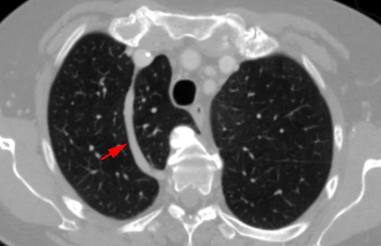
- Azigos fissürü sağ üst lobun mediastinal kenarında, hilusun üzerinde yer alır ve lobda ek bir ayrım oluşturur.
- Azigos veni azigos fissürü içinde seyreder.
- Azigos lobu, fissürün medialinde kalan sağ üst lob apikal parankimidir; bağımsız bronş ve damar ağacı olan gerçek bir lob değildir.
- Aksesuar fissürün tamamlanmış veya kısmi olması, akciğer parankimindeki çizginin kesintili seçilmesine yol açabilir.
- Fissürün çevresindeki parankim normal olabilir; eşlik eden konsolidasyon, kitle veya hacim kaybı ayrı değerlendirilmelidir.
Normalde
Standart anatomide sağ akciğerde oblik ve horizontal, sol akciğerde oblik fissür beklenir; azigos fissürü ve ek segmental çizgiler zorunlu normal sınırlara dahil değildir. Kıyaslarken ek çizginin ince-düzgün olup olmadığını, komşu parankimin havalı kalmasını ve azigos veninin fissür ucuyla ilişkisini kontrol et.
Normal BT ile kıyasla
Aynı bölgenin, kaynak veri setinde patoloji içermediği belirtilen bir BT incelemesini kesit kesit kaydırın; organ sınırlarını açarak anatomiyi hasta görüntüsüyle karşılaştırın. Küçük rastlantısal bulgular tümüyle dışlanmamıştır.
Kaynak: TotalSegmentator v2.01 veri seti (Wasserthal ve ark., Radiology: Artificial Intelligence 2023), CC BY 4.0, vaka s1278; organ sınırları veri setinin otomatik segmentasyonundan, birleştirilerek sadeleştirildi. Görüntüler 2,5 mm kesit aralığına yeniden örneklendi.
Ayırıcı tanı
- Apikal skar ayırıcı tanısında azigos fissürünün hizasını ve normal akciğer işaretlerinin korunmasını değerlendir; bu özellikler yanlış tanıyı önlemeye yardımcı olur.
- Azigos fissürü, fissür boyunca izlenen azigos veni ve karakteristik seyriyle tanınır. Azigos lobu pnömotoraksı taklit edebilir; azigos fissürünün hizası ve korunmuş akciğer işaretleri ayırımda yardımcıdır.
- Azigos lobu bül ile karıştırılabilir; varyantın karakteristik fissürünü ve korunmuş akciğer işaretlerini tanımak ayırıcı tanıya yardımcı olur.
- Mediastinal lenf nodu veya küçük kitle
- yumuşak doku odağının azigos venine ve fissürün kaudal ucuna uzanımı değerlendirilerek ayırt edilir.
- Normal oblik fissür, aksesuar fissürle karıştırılabilir; fissürün lobar sınırlarla ilişkisini aksiyel, koronal ve sagittal görüntülerde değerlendir.
Tuzaklar
- Azigos lobunu ayrı bir anatomik lob gibi adlandırmak bronkovasküler haritalamayı yanıltabilir; alanın sağ üst lob apikal segmentleriyle ilişkisini koru.
- Azigos veni kaudal ucunda nodül benzeri görülebilir; komşu kesitlerde venöz devamlılığını takip etmeden parankimal nodül olarak raporlama.
- Aksesuar fissürlerin BT'de tanınması güç olabilir; ince kolimasyon ve yüksek uzaysal çözünürlüklü rekonstrüksiyonlar fissürlerin seçilmesini kolaylaştırır.
- Bir çizgisel opasiteyi azigos fissürü olarak değerlendirirken fissürün hizasını ve normal akciğer işaretlerinin korunup korunmadığını incele; bu özellikler pnömotoraksla karışmayı önlemeye yardımcı olur.
- Aksesuar fissürlerin BT'de saptanması güç olabilir; kısmi fissürler parankimi tam ayırmadığından komşu loblarla devamlılık gösterebilir.
Kendini dene
Sağ üst lobun mediastinal kenarında, hilusun üzerinde azigos lobu ve azigos fissürü izleniyor. Bu görünüm nedir?
Cevabı göster
Azigos fissürü ve lobu. Azigos lobu sağ üst lobun mediastinal kenarında ve hilusun üzerinde yer alır; azigos veni azigos fissürü içinde seyreder. Azigos lobu bül ile karıştırılabilir; pnömotoraksı taklit eden apikal çizgisel görünümde karakteristik fissür ve korunmuş akciğer işaretleri tanıyı destekler.
Azigos lobunu oluşturan fissürün içinde hangi yapı seyreder?
Cevabı göster
Azigos veni. Azigos fissürü, fissür içinde seyreden azigos veninin yolunu gösterir.
Kaynaklar
Bu sayfadaki 36 cümle ve 2 quiz sorusu aşağıdaki kaynaklardan alıntılarla otomatik olarak karşılaştırıldı; 19 cümle bu karşılaştırmada düzeltildi (2026-10-09).
- Assessment of variability in pulmonary fissures using multidetector computed tomography: a short reviewpmc.ncbi.nlm.nih.gov
- Unveiling Hidden Variations in Lung Fissures: A Cadaveric Perspective on Surgical and Diagnostic Challengespmc.ncbi.nlm.nih.gov
- LobePrior segments lung lobes on computed tomography images in the presence of severe abnormalitiespmc.ncbi.nlm.nih.gov
- Azygos lobe: exploring the bronchial supply and clinical implicationspmc.ncbi.nlm.nih.gov
- Adult presentation of unilateral pulmonary venous atresia: CT findings in a rare congenital anomalypmc.ncbi.nlm.nih.gov
- Variations in human pulmonary fissures and lobes: a study conducted in nepalese cadavers.pubmed.ncbi.nlm.nih.gov
- [Saccular aneurysm of the azygos vein in a patient with azygos accessory fissure].pubmed.ncbi.nlm.nih.gov
- Non-azygos accessory fissure in right upper lobe associated with superior and inferior accessory fissures in right lower lobe.pubmed.ncbi.nlm.nih.gov
- Recognizing the azygos lobe: A case series detailing presentations and clinical implications.pubmed.ncbi.nlm.nih.gov
- Unveiling Hidden Variations in Lung Fissures: A Cadaveric Perspective on Surgical and Diagnostic Challenges.pubmed.ncbi.nlm.nih.gov
- Azygos lobe: exploring the bronchial supply and clinical implications.pubmed.ncbi.nlm.nih.gov
- Azygos lobe mimicking pneumothorax in trauma: a case report.pubmed.ncbi.nlm.nih.gov
- Surgical technique for minimally invasive segmentectomies of the right lung: anatomic rationale for a posterior approach.pubmed.ncbi.nlm.nih.gov
- LungSurg: A Generative AI System for Segmentation and Phase Classification in Thoracoscopic Lobectomy.pubmed.ncbi.nlm.nih.gov
- Thoracoscopic McKeown esophagectomy in a patient with an azygos lobe.pubmed.ncbi.nlm.nih.gov
- Personalized Management of Malignant and Non-Malignant Ectopic Mediastinal Thyroid: A Proposed 10-Item Algorithm Approach.pubmed.ncbi.nlm.nih.gov
- Imaging pitfalls, normal anatomy, and anatomical variants that can simulate disease on cardiac imaging as demonstrated on multidetector computed tomography.pubmed.ncbi.nlm.nih.gov
- Unveiling the synergetic benefits of the tunneling technique using stapler tractor in precise resection of lung segments: a retrospective cohort study.pubmed.ncbi.nlm.nih.gov
- Kerley A-lines represent thickened septal plates between lung segments in patients with lymphangitic carcinomatosis: confirmation using 3D-CT lung segmentation analysis.pubmed.ncbi.nlm.nih.gov
- First real-time imaging of bronchoscopic lung volume reduction by electrical impedance tomography.pubmed.ncbi.nlm.nih.gov
- Contrast-Enhanced CT Finding of Hot Spot Sign Secondary to Supra Hilar Mass Causing Superior Vena Cava Syndrome: A Case Report.pubmed.ncbi.nlm.nih.gov
- A practical approach to procedural planning for robot-assisted segmentectomy using a lobectomy-based surgical simulator.pubmed.ncbi.nlm.nih.gov
- Microsurgical clipping for coil-extruded recurrent aneurysms: Case series and technical pearls.pubmed.ncbi.nlm.nih.gov
- Foramen rotundum versus canal of the maxillary verve.pubmed.ncbi.nlm.nih.gov
- The expert consensus document on minimally invasive upper lobectomy for lung cancer in China.pubmed.ncbi.nlm.nih.gov
- Fleischner Society: Glossary of Terms for Thoracic Imaging.pubmed.ncbi.nlm.nih.gov
- ESR Essentials: imaging of common paediatric pulmonary diseases-practice recommendations by the European Society of Paediatric Radiology.pubmed.ncbi.nlm.nih.gov
- Intraoperative longitude-latitude-depth three-dimensional localization of pulmonary nodules.pubmed.ncbi.nlm.nih.gov
- Opposing Mitral Valve Clefts, or Perhaps an Isolated Quadricuspid Mitral Valve: A Case Report.pubmed.ncbi.nlm.nih.gov
- LobePrior segments lung lobes on computed tomography images in the presence of severe abnormalities.pubmed.ncbi.nlm.nih.gov
- Anatomical Variations and Morphometric Features of the Anterior Cerebral Artery: A Systematic Review and Meta-Analysis of 24,015 Cases.pubmed.ncbi.nlm.nih.gov
- Long-term safety and effectiveness of the "OptEase" vena cava filter.pubmed.ncbi.nlm.nih.gov
- Advancing human-centric AI for robust X-ray analysis through holistic self-supervised learning.pubmed.ncbi.nlm.nih.gov
- CXR-LT 2024: A MICCAI challenge on long-tailed, multi-label, and zero-shot disease classification from chest X-ray.pubmed.ncbi.nlm.nih.gov
- Current trends in image-guided chest interventions.pubmed.ncbi.nlm.nih.gov
- Advancements in Brain Aneurysm Management: Integrating Neuroanatomy, Physiopathology, and Neurosurgical Techniques.pubmed.ncbi.nlm.nih.gov
- Revisiting signs, strengths and weaknesses of Standard Chest Radiography in patients of Acute Dyspnea in the Emergency Department.pubmed.ncbi.nlm.nih.gov
- Preservation of the right bronchial artery by maintaining the azygos arch during minimally invasive esophagectomy: a two-center retrospective cohort study.pubmed.ncbi.nlm.nih.gov
- Real-time thoracic duct visualization in lung cancer surgery: feasibility of indocyanine green fluorescence imaging via inguinal lymph node injection.pubmed.ncbi.nlm.nih.gov
- State-of-the-Art Mediastinal Staging in Non-Small-Cell Lung Cancer: Integration of Combined Endosonographic Techniques with Updated IASLC TNM 9th Classification.pubmed.ncbi.nlm.nih.gov
- Hemiazygos vein aneurysm mimicking an enlarged lymph node.pubmed.ncbi.nlm.nih.gov
- Splenic pathologies: A pictorial review of common, wandering, twisting and rare presentations.pubmed.ncbi.nlm.nih.gov
- From asthma exacerbation to pulmonary mucoepidermoid carcinoma: A rare case report.pubmed.ncbi.nlm.nih.gov
- A clinically-recommended MR whole lung imaging protocol using free-breathing 3D isotropic zero echo time sequence.pubmed.ncbi.nlm.nih.gov
- Clinical TNM Lung Cancer Staging: A Diagnostic Algorithm with a Pictorial Review.pubmed.ncbi.nlm.nih.gov
- An old retained sharp intrapulmonary foreign body: a case report and literature review.pubmed.ncbi.nlm.nih.gov
- The Role of Innervation in Ocular Surface Homeostasis, Tissue Remodeling Following Nerve Injury, and the Therapeutic Potential of Hemocomponents in Neuronal and Cicatricial Pathologies.pubmed.ncbi.nlm.nih.gov
- Stepwise diagnostic algorithm for high-attenuation pulmonary abnormalities on CT.pubmed.ncbi.nlm.nih.gov
- Assessment of variability in pulmonary fissures using multidetector computed tomography: a short review.pubmed.ncbi.nlm.nih.gov
- Restoration of hepatopetal portal flow in a cat with left hepatic arteriovenous malformation following left hepatic division resection and staged attenuation of acquired portosystemic shunts.pubmed.ncbi.nlm.nih.gov
- Plastinated sectional anatomy applied in CT and MRI studies of the body of the crab-eating fox (Cerdocyon thous).pubmed.ncbi.nlm.nih.gov
- Asymptomatic central venous occlusion secondary to central venous catheter-use complicating pacemaker implantation: a leadless solution.pubmed.ncbi.nlm.nih.gov
- Impaired lymphatic drainage in a child with hydrops fetalis and a novel variant in the PROX1 gene: case report and review of literature.pubmed.ncbi.nlm.nih.gov
- Anomalous venoatrial connections - CT and MRI assessment.pubmed.ncbi.nlm.nih.gov
- Prenatal assessment of brain malformations on neuroimaging: an expert panel review.pubmed.ncbi.nlm.nih.gov
- Volumetric response after closure of a spinal CSF leak in patients with spontaneous intracranial hypotension: a multicompartmental longitudinal study.pubmed.ncbi.nlm.nih.gov
- Spontaneous Pneumothorax Due to Ruptured Bulla of the Azygos Lobe.pubmed.ncbi.nlm.nih.gov
- An In-Depth Review of the Azygos Vein and Its Clinical Significance.pubmed.ncbi.nlm.nih.gov
- Polysplenia syndrome in adulthood: A case report of incidental discovery.pubmed.ncbi.nlm.nih.gov
- Single Posthepatic Portosystemic Shunt Communicated With Internal Thoracic Vein and Azygos Continuation of the Caudal Vena Cava in a Dog.pubmed.ncbi.nlm.nih.gov
- Thoracic Vascular Variants and Anomalies: Imaging Findings, Review of the Embryology, and Clinical Features.pubmed.ncbi.nlm.nih.gov
- Chest Radiography Pearls in Select Adult Congenital Heart Disease.pubmed.ncbi.nlm.nih.gov
- Adult presentation of unilateral pulmonary venous atresia: CT findings in a rare congenital anomaly.pubmed.ncbi.nlm.nih.gov
- Chronic Subdural Hematoma: Pathophysiology, Diagnosis, and the Emerging Role of Middle Meningeal Artery Embolization.pubmed.ncbi.nlm.nih.gov
- International Society of Paediatric Surgical Oncology (IPSO) Surgical Practice Guidelines.pubmed.ncbi.nlm.nih.gov
- Geometric feature analysis for identifying and characterizing normal variations on chest radiographs.pubmed.ncbi.nlm.nih.gov
- Comprehensive Computed Tomography Evaluation of Gastric Cancer: 2026 Consensus Recommendation From the Korean Society of Abdominal Radiology.pubmed.ncbi.nlm.nih.gov
- Coexistence of Autoantibodies against the Golgi Complex and Ro52 Antigen in a Patient with Nonspecific Interstitial Pneumonia.pubmed.ncbi.nlm.nih.gov
- CT Findings of Azygos Venous System: Congenital Variants and Acquired Structural Changes.pubmed.ncbi.nlm.nih.gov
- Uniportal Video-Assisted Thoracoscopic Segmentectomy for Early-Stage Non-Small Cell Lung Cancer: Overview, Indications, and Techniques.pubmed.ncbi.nlm.nih.gov
- Irrigants and irrigation activation systems in Endodontics.pubmed.ncbi.nlm.nih.gov
- Rare Diseases of the Orbit.pubmed.ncbi.nlm.nih.gov
- Traumatic insemination and female counter-adaptation in Strepsiptera (Insecta).pubmed.ncbi.nlm.nih.gov
- A Cadaveric Study of Variations in Lung Fissures and Drainage Patterns of Pulmonary Veins at the Hilum With Its Clinical Implications.pubmed.ncbi.nlm.nih.gov
- Jugular Foramen Syndrome: Concurrent Neurological Deficits, Advanced Imaging Findings, Underlying Diagnoses, and Outcomes in 14 Dogs (2016-2024).pubmed.ncbi.nlm.nih.gov
- Ectopic olfactory neuroblastoma is associated with increased frequency of syndrome of inappropriate antidiuretic hormone secretion and reduced disease control: Case series with systematic review and pooled analysis.pubmed.ncbi.nlm.nih.gov
- Thoracic abnormal air collections in patients in the intensive care unit: radiograph findings correlated with CT.pubmed.ncbi.nlm.nih.gov
- Morphometric Analysis of Foramina in the Middle Cranial Fossa of Dogs: A Retrospective Cone-Beam CT Study.pubmed.ncbi.nlm.nih.gov
- Three-Dimensional Bronchovascular Modelling in Sublobar Pulmonary Resection: A Tool for Personalised Thoracic Surgery.pubmed.ncbi.nlm.nih.gov
- Study on Anatomical Variations in Fissures of Lung by CT Scan.pubmed.ncbi.nlm.nih.gov
- Sequential robotic-assisted thoracic surgery and transmanubrial approach for optimal resection of superior sulcus tumor: a case report.pubmed.ncbi.nlm.nih.gov
- Uniportal thoracoscopic mediastinal lymphadenectomy using appropriate surgical steps.pubmed.ncbi.nlm.nih.gov
- Congenital systemic venous return anomalies to the right atrium review.pubmed.ncbi.nlm.nih.gov
- Neglected right diaphragmatic hernia with transthoracic herniation of gallbladder and malrotated left liver lobe in an adult.pubmed.ncbi.nlm.nih.gov
- Pediatric Congenital Lung Malformation: Advanced Imaging Techniques in Pre- and Neonatal Evaluation.pubmed.ncbi.nlm.nih.gov
- Evaluation of the solitary pulmonary nodule: size matters, but do not ignore the power of morphology.pubmed.ncbi.nlm.nih.gov
- Clinical and radiological features of idiopathic interstitial pneumonias (IIPs): a pictorial review.pubmed.ncbi.nlm.nih.gov
- Imaging review of procedural and periprocedural complications of central venous lines, percutaneous intrathoracic drains, and nasogastric tubes.pubmed.ncbi.nlm.nih.gov
- Morphological variations of fissures, lobes, and hilar pattern of the lung in a select South African sample.pubmed.ncbi.nlm.nih.gov
- CT-guided percutaneous biopsy of intrathoracic lesions.pubmed.ncbi.nlm.nih.gov
Eğitim amaçlıdır; hasta başında tanı ve tedavi kararının yerine geçmez, tıbbi tavsiye değildir. Metinler yapay zekâ desteğiyle yazıldı ve otomatik bir adımda kaynak alıntılarıyla karşılaştırıldı; bu bir tıbbi doğrulama değildir ve içerik uzman radyolog incelemesinden geçmedi. Klinik kararlarda güncel kılavuzları, kurum protokollerini ve radyoloji raporunu esas alın.