Özet
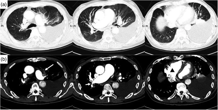
Kardiyojenik akciğer ödemi, sol kalp dolum basınçlarının yükselmesiyle interstisyel ve ardından alveoler sıvı birikiminin geliştiği akut ya da kronik konjesyon örüntüsüdür. Kontrastsız toraks BT'de düzgün septal kalınlaşma, bronkovasküler kılıflanma, damar dolgunluğu ve sıklıkla plevral efüzyon birlikte görülebilir; parankim ödeminin gösterilmesi için kontrast gerekmez. Yaygınlaşan alveoler ödem bilateral buzlu cam veya santral konsolidasyon oluşturup ARDS ve diğer alveoler hastalıklarla örtüşebilir. BT bulguları arasında ödem ve enfeksiyon örtüşebildiğinden, klinik bağlam ve ek görüntüleme bulguları ayırıcı tanıda önemlidir.
Faz ve pencere
- Kontrastsız tanısal
- Akciğer penceresinde düzgün interlobüler septal kalınlaşma, bronkovasküler kılıflarda kalınlaşma, santral/perihiler buzlu cam veya konsolidasyon; mediasten penceresinde kalp boyutu, plevral sıvı ve damar kalibresi incelenir. Ödem örüntüsünü göstermek için intravenöz kontrast fazı gerekli değildir.
Önerilen pencereler: Akciğer (G 1500 / M -600), Mediasten (G 350 / M 50).
BT bulguları
- Düzgün interlobüler septal kalınlaşma — interstisyum boyunca ince ve eşit kalınlıklı çizgiler olarak belirginleşir.
- Hidrostatik basınç artışı ve kapiller geçirgenlik değişiklikleri nedeniyle interstisyuma sızan sıvı, bronkovasküler demetler çevresinde birikerek kalınlaşma oluşturabilir.
- Sol kalp dolum basınçlarındaki artış, pulmoner venöz konjesyon ve hidrostatik ödeme yol açarak damar kalibre değişikliklerine neden olabilir.
- Perihiler buzlu cam — alveoler sıvı arttığında hilus çevresinde daha belirgin yamalı ya da yaygın opasite gelişebilir.
- Alveoler opasite — ödemde santral ağırlıklı yoğunluklar ve subpleural korunma örüntüsü gözlenebilir.
- Plevral efüzyon — ödemle birlikte tek veya çift taraflı plevral sıvı birikimi sık görülen bulgulardandır.
- Kardiyojenik akciğer ödemi, artmış sol kalp dolum basınçları ve tüm kalp hastalıklarının neden olabileceği hidrostasik sıvı birikimi ile karakterizedir.
- Hidrostatik yük artışı, plevral ve perikardiyal sıvı birikimine yol açabilir.
Normalde
Toraks radyolojisinde normal yapıların tanımı ve patolojik bulguların sınıflandırılması, Fleischner Derneği tarafından standartlaştırılan terminoloji sözlüğüne dayanır. Alt lob periferisinde şüpheli düzgün septaları, aynı düzeydeki karşı lobla ve normalde ince kalan bronş damar demetleriyle karşılaştırın; mediastinal pencerede kalp konturunu ve plevral sıvıyı ayrıca inceleyin.
Normal BT ile kıyasla
Aynı bölgenin, kaynak veri setinde patoloji içermediği belirtilen bir BT incelemesini kesit kesit kaydırın; organ sınırlarını açarak anatomiyi hasta görüntüsüyle karşılaştırın. Küçük rastlantısal bulgular tümüyle dışlanmamıştır.
Kaynak: TotalSegmentator v2.01 veri seti (Wasserthal ve ark., Radiology: Artificial Intelligence 2023), CC BY 4.0, vaka s1278; organ sınırları veri setinin otomatik segmentasyonundan, birleştirilerek sadeleştirildi. Görüntüler 2,5 mm kesit aralığına yeniden örneklendi.
Ayırıcı tanı
- ARDS
- alveol-kapiller geçirgenlik artışıyla ilişkili non-kardiyojenik ödem yapar; klinik tablo ve hidrostatik bulguların yokluğu ayrımı yönlendirir.
- Akut eozinofilik pnömoni
- düzgün septalar ve efüzyonla ödemi taklit edebilir; belirgin kalp-damar konjesyonu olmayabilir.
- Diffüz alveoler kanama
- santral buzlu cam ve konsolidasyon yapabilir; belirgin hidrostatik damar/septal bulgular tek başına beklenmez.
- Lenfanjitik yayılım, düzgün ya da nodüler interlobüler septal kalınlaşma yapabilir ve pulmoner interstisyumu tutabilir.
- Viral veya atipik enfeksiyon
- buzlu cam ve septal çizgiler görülebilir; bronşiolit nodülleri, fokal konsolidasyon ve klinik enfeksiyon öyküsü ayrımda yardımcıdır.
Tuzaklar
- Kalp boyutunun normal olması akut kardiyojenik ödemi dışlamaz; akciğer damarları, venöz konjesyon ve sıvı dağılımını ayrı değerlendirin.
- Tek taraflı pozisyon veya amfizem, ödemi asimetrik gösterebilir; her iki akciğerdeki damar ve septa bulgularını karşılaştırın.
- Düzgün septalar özgül değildir; AEP ve bazı diğer interstisyel süreçler de benzer çizgiler yapabilir.
- Plevral efüzyonun yanındaki bağımlı alt lob opasitesi pasif atelektazi olabilir; bronş açıklığı ve hacim kaybını kontrol edin.
Kendini dene
Nefes darlığında BT'de düzgün septalar, bronkovasküler kılıf kalınlaşması, damar dolgunluğu ve bilateral sıvı görülüyor. En uyumlu tanı hangisidir?
Cevabı göster
Kardiyojenik pulmoner ödem. Bu bulguların birlikte bulunması hidrostatik konjesyonu destekler. ARDS'de bağımlı heterojen opasite öne çıkabilir; kanama ve lenfanjitik yayılım tek başına damar dolgunluğu-plevral sıvı birlikteliğini açıklamaz.
HRCT'de düzgün interlobüler septal kalınlaşma, santral perihiler yumuşak doku kalınlaşması ve buzlu cam opasiteleri eşlik eden hastada eosinofili saptanıyor. En olası tanı hangisidir?
Cevabı göster
Akut eozinofilik pnömoni. Düzgün septal kalınlaşma, santral perihiler yumuşak doku kalınlaşması, buzlu cam opasiteleri ve eosinofili birlikteliği, balon lavaj ile doğrulanabilen akut eozinofilik pnömoni açısından düşündürmelidir.
İlgili konular
Kaynaklar
Bu sayfadaki 37 cümle ve 2 quiz sorusu aşağıdaki kaynaklardan alıntılarla tek tek karşılaştırıldı; 22 cümle bu karşılaştırmada düzeltildi (2026-10-08).
- Dengue Hemorrhagic Fever: A State-of-the-Art Review Focused in Pulmonary Involvement.pubmed.ncbi.nlm.nih.gov
- Pathophysiology, clinical features and radiological findings of differentiation syndrome/all-trans-retinoic acid syndrome.pubmed.ncbi.nlm.nih.gov
- Handheld ultrasound as an adjunct to physical examination in the diagnosis of cardiopulmonary disease.pubmed.ncbi.nlm.nih.gov
- Effectiveness of chest radiography, lung ultrasound and thoracic computed tomography in the diagnosis of congestive heart failure.pubmed.ncbi.nlm.nih.gov
- Ventilatory management of patients with acute respiratory distress syndrome and optimization of lung mechanics.pubmed.ncbi.nlm.nih.gov
- Value of Thoracic Echocardiography in the Diagnosis of Acute Heart Failure: Systematic Review and Meta-analysis.pubmed.ncbi.nlm.nih.gov
- Lung Imaging in Acute Hypoxemic Respiratory Failure: From Physics to Bedside Applications.pubmed.ncbi.nlm.nih.gov
- Lung B-line artefacts and their use.pubmed.ncbi.nlm.nih.gov
- Phenotypes and personalized medicine in the acute respiratory distress syndrome.pubmed.ncbi.nlm.nih.gov
- Lung ultrasonography findings in dogs with various underlying causes of cough.pubmed.ncbi.nlm.nih.gov
- What's Next After ARDS: Long-Term Outcomes.pubmed.ncbi.nlm.nih.gov
- 36th International Symposium on Intensive Care and Emergency Medicine : Brussels, Belgium. 15-18 March 2016.pubmed.ncbi.nlm.nih.gov
- The Significance of Subpleural Sparing in CT Chest: A State-of-the-Art Review.pubmed.ncbi.nlm.nih.gov
- Voriconazole-Induced acute interstitial pulmonary edema with angioedema: A case report.pubmed.ncbi.nlm.nih.gov
- Case report: cerebral venous sinus thrombosis and pulmonary embolism as the initial presentation in a child with asymptomatic primary nephrotic syndrome.pubmed.ncbi.nlm.nih.gov
- Primary Ciliary Dyskinesia.pubmed.ncbi.nlm.nih.gov
- Primary ciliary dyskinesia phenotypes and correlation with genotype.pubmed.ncbi.nlm.nih.gov
- Primary ciliary dyskinesia.pubmed.ncbi.nlm.nih.gov
- Diagnostic accuracy of ultra-low-dose chest computed tomography in an emergency department.pubmed.ncbi.nlm.nih.gov
- Idiopathic acute eosinophilic pneumonia.pubmed.ncbi.nlm.nih.gov
- Hypersensitivity Pneumonitis and Acute Respiratory Distress Syndrome From E-Cigarette Use.pubmed.ncbi.nlm.nih.gov
- [Expert recommendations for the diagnosis and treatment of interstitial lung disease caused by novel coronavirus pneumonia].pubmed.ncbi.nlm.nih.gov
- Hantavirus infection in emergency radiology: Imaging manifestations, pathophysiology, and infection control considerations.pubmed.ncbi.nlm.nih.gov
- Reversible Interlobular Septal 'Pearls' Associated with Takotsubo Cardiomyopathy and Heart Failure.pubmed.ncbi.nlm.nih.gov
- Artificial intelligence improves detection and classification of pulmonary venous hypertension related to left ventricular diastolic dysfunction by chest radiography.pubmed.ncbi.nlm.nih.gov
- Assessing volume status in heart failure: The role of renal duplex ultrasound in evaluating cardiorenal morbidity and heart failure mortality.pubmed.ncbi.nlm.nih.gov
- Cardiogenic Pulmonary Edema in Emergency Medicine.pubmed.ncbi.nlm.nih.gov
- Late Diagnosis of Noonan Syndrome Presenting With Infective Endocarditis: A Multidisciplinary Challenge in an Underserved Population.pubmed.ncbi.nlm.nih.gov
- Discriminating HFrEF vs HFpEF from chest radiographs: Mitigating demographic performance gaps via augmentation and multimodal fusion.pubmed.ncbi.nlm.nih.gov
- Diagnostic significance of lung ultrasound in evaluation of heart failure: A cross-sectional study in comparison with clinical assessment, proBNP, chest X-ray, and echocardiography.pubmed.ncbi.nlm.nih.gov
- Correlation of plasma N-terminal pro-brain natriuretic peptide (NT-proBNP) with radiographic features of congestion in chest CT scan of patients with COVID-19.pubmed.ncbi.nlm.nih.gov
- Reading chest radiographs in the critically ill (Part II): Radiography of lung pathologies common in the ICU patient.pubmed.ncbi.nlm.nih.gov
- The pleuroparenchymal fibroelastosis atlas reveals aberrant cell states and their zonation as an alternate roadmap to lung fibrosis.pubmed.ncbi.nlm.nih.gov
- Consensus statement on thoracic radiology terminology in Portuguese used in Brazil and in Portugal.pubmed.ncbi.nlm.nih.gov
- Imaging of pulmonary emphysema: a pictorial review.pubmed.ncbi.nlm.nih.gov
- Diffuse cystic lung diseases-a primer for radiologists.pubmed.ncbi.nlm.nih.gov
- Advances in the mechanisms, imaging characteristics and management strategies for immune checkpoint inhibitor-related pneumonitis.pubmed.ncbi.nlm.nih.gov
- Lung Ultrasound and High-Resolution Computed Tomography in Suspected COVID-19 Patients Admitted to the Emergency Department: A Comparison.pubmed.ncbi.nlm.nih.gov
- Acute exacerbation in fibrotic interstitial lung disease: an international working group report.pubmed.ncbi.nlm.nih.gov
- Medication-induced lung disease in children.pubmed.ncbi.nlm.nih.gov
- Management of pneumonia by the emergency radiologist.pubmed.ncbi.nlm.nih.gov
- Isolated pulmonary lymphangitic carcinomatosis as the initial manifestation of renal cell carcinoma.pubmed.ncbi.nlm.nih.gov
- Pulmonary lymphangitic carcinomatosis mimicking interstitial lung disease.pubmed.ncbi.nlm.nih.gov
- Multimodality Imaging for the Diagnosis and Evaluation of Pulmonary Sarcoidosis.pubmed.ncbi.nlm.nih.gov
- Host, Pathogen, and Pulmonary Anatomy in Pneumonia: An Integrated Imaging and Clinical Perspective.pubmed.ncbi.nlm.nih.gov
- Imaging in pulmonary infections of immunocompetent adult patients.pubmed.ncbi.nlm.nih.gov
- The Impact of Genetics on Pediatric Interstitial Lung Diseases: A Narrative Literature Review and Clinical Implications.pubmed.ncbi.nlm.nih.gov
- The Role of PoCUS in the Diagnosis and Early Monitoring of Cardiogenic Shock in the Emergency Department: A Narrative Review.pubmed.ncbi.nlm.nih.gov
- Point-of-care ultrasound to evaluate volume status in congestive heart failure.pubmed.ncbi.nlm.nih.gov
- Right Heart Catheterization: Best Practices and Specific Considerations.pubmed.ncbi.nlm.nih.gov
- Ultrasound use in the ICU for interventional pulmonology procedures.pubmed.ncbi.nlm.nih.gov
- Bioengineering and Clinical Translation of Human Lung and its Components.pubmed.ncbi.nlm.nih.gov
- Coexistence of Autoantibodies against the Golgi Complex and Ro52 Antigen in a Patient with Nonspecific Interstitial Pneumonia.pubmed.ncbi.nlm.nih.gov
- Tips and Tricks in Thoracic Radiology for Beginners: A Findings-Based Approach.pubmed.ncbi.nlm.nih.gov
- Fleischner Society: Glossary of Terms for Thoracic Imaging.pubmed.ncbi.nlm.nih.gov
- Lung Sonography in Critical Care Medicine.pubmed.ncbi.nlm.nih.gov
- The "pandemic" increase in lung ultrasound use in response to Covid-19: can we complement computed tomography findings? A narrative review.pubmed.ncbi.nlm.nih.gov
- Common and Rare Radiological Findings of Pulmonary and Thoracic Hydatid Cysts.pubmed.ncbi.nlm.nih.gov
- ESR Essentials: imaging of common paediatric pulmonary diseases-practice recommendations by the European Society of Paediatric Radiology.pubmed.ncbi.nlm.nih.gov
- Multimodality imaging of COVID-19 pneumonia: from diagnosis to follow-up. A comprehensive review.pubmed.ncbi.nlm.nih.gov
- Diagnostic Challenges in Takotsubo Syndrome: Bridging Mimics, Mechanisms, and Management.pubmed.ncbi.nlm.nih.gov
- Amphetamine-Associated Reversible Biventricular Cardiomyopathy Complicated by Acute Respiratory Failure: A Case Report.pubmed.ncbi.nlm.nih.gov
- Mesalazine-Induced Myocarditis: A Challenging Diagnostic Journey.pubmed.ncbi.nlm.nih.gov
- CT findings of lung injury during breast cancer treatment.pubmed.ncbi.nlm.nih.gov
- Interstitial Lung Disease and Cardiotoxicity Associated with Trastuzumab Deruxtecan, Sacituzumab Govitecan, and Trastuzumab Emtansine: A Narrative Review.pubmed.ncbi.nlm.nih.gov
- Immune Checkpoint Inhibitor-Related Pneumonitis in Renal Cell Carcinoma: Clinical Features, Mechanisms, and Lessons from Lung Cancer.pubmed.ncbi.nlm.nih.gov
- Mitral Valve Prolapse Presenting as Unilateral Pulmonary Edema Mimicking Pneumonia.pubmed.ncbi.nlm.nih.gov
- Carcinoid Heart Disease Presenting as Cardiogenic Shock: A Case Report.pubmed.ncbi.nlm.nih.gov
- Imaging in the COVID-19 era: Lessons learned during a pandemic.pubmed.ncbi.nlm.nih.gov
- Overview of treatment related complications in malignant pleural mesothelioma.pubmed.ncbi.nlm.nih.gov
- Right ventricular-protective ventilation in acute respiratory distress syndrome: phenotypes, monitoring and ventilatory management.pubmed.ncbi.nlm.nih.gov
- Emergency management of cerebral air gas embolism as a complication of CT-guided lung biopsy.pubmed.ncbi.nlm.nih.gov
- Fatal Cerebral Air Embolism Following Central Venous Catheter Mishandling in a Stroke Patient: A Case Report.pubmed.ncbi.nlm.nih.gov
- Lung ultrasound and the interstitial syndrome in tuberculosis endemic regions: illustrated insights from diverse clinical settings.pubmed.ncbi.nlm.nih.gov
- Remote dielectric sensing for pulmonary congestion assessment: current concepts and clinical perspectives.pubmed.ncbi.nlm.nih.gov
- Clinical correlates and short-term prognostic value of CT-detected subclinical pulmonary congestion in acute STEMI without overt heart failure.pubmed.ncbi.nlm.nih.gov
- Beyond resection: imaging findings of expected and complicated postoperative changes in lung cancer.pubmed.ncbi.nlm.nih.gov
- Myocardial Fibrosis Caused by Angiotensin II Implant in Rabbit Atherosclerosis Model Induced by High Cholesterol Diet.pubmed.ncbi.nlm.nih.gov
- Imaging in patients with acute dyspnea when cardiac or pulmonary origin is suspected.pubmed.ncbi.nlm.nih.gov
- Lung imaging methods: indications, strengths and limitations.pubmed.ncbi.nlm.nih.gov
- Imaging of Childhood Interstitial Lung Disease.pubmed.ncbi.nlm.nih.gov
- Pneumonitis Associated with Immune Checkpoint Inhibitors and Targeted Anticancer Therapies: A Retrospective Case Series of 12 Patients.pubmed.ncbi.nlm.nih.gov
- New CT-Detected Pulmonary Abnormalities During Breast Cancer Chemotherapy: Frequency, Imaging Features, and Cumulative-Exposure Associations.pubmed.ncbi.nlm.nih.gov
- Drug-Induced Interstitial Lung Disease in Lung Cancer Patients.pubmed.ncbi.nlm.nih.gov
- Expert consensus on cancer treatment-related lung injury.pubmed.ncbi.nlm.nih.gov
- COVID-19 Pneumonia: The Great Ultrasonography Mimicker.pubmed.ncbi.nlm.nih.gov
- Challenges in the Differential Diagnosis of COVID-19 Pneumonia: A Pictorial Review.pubmed.ncbi.nlm.nih.gov
- Pulmonary Complications in Children Following Hematopoietic Cell Transplantation: A Case Report and Review of the Diagnostic Approach.pubmed.ncbi.nlm.nih.gov
- From Silent to Life-Threatening: Giant Left Atrial Myxoma Presenting with Acute Pulmonary Edema-A Case Report.pubmed.ncbi.nlm.nih.gov
- Cardiogenic shock following acute MI in a young patient with familial hypercholesterolemia, and severe aortic stenosis: A case report.pubmed.ncbi.nlm.nih.gov
- Tricuspid Valve Academic Research Consortium Definitions for Tricuspid Regurgitation and Trial Endpoints.pubmed.ncbi.nlm.nih.gov
- Evaluation of Quantitative Computed Tomography Indices in Patients with Pneumonia and Acute Respiratory Failure in the Intensive Care Unit (ICU).pubmed.ncbi.nlm.nih.gov
- Thoracic trauma WSES-AAST guidelines.pubmed.ncbi.nlm.nih.gov
- Thoracic Computed Tomography to Assess ARDS and COVID-19 Lungs.pubmed.ncbi.nlm.nih.gov
- Reporting and management of incidental lung findings on computed tomography: beyond lung nodules.pubmed.ncbi.nlm.nih.gov
- Postoperative Pulmonary Complications: Clinical and Imaging Insightspubmed.ncbi.nlm.nih.gov
- Pericardial Diseases: International Position Statement on New Concepts and Advances in Multimodality Cardiac Imaging.pubmed.ncbi.nlm.nih.gov
- Management of the Diagnosis and Treatment of Pneumonia in an Aging Society.pubmed.ncbi.nlm.nih.gov
- Pulmonary Edema: A Pictorial Review of Imaging Manifestations and Current Understanding of Mechanisms of Disease.pubmed.ncbi.nlm.nih.gov
- Constrictive Pericarditis After Minimally Invasive Triple Valve Replacement: Insights From a Fatal Outcome.pubmed.ncbi.nlm.nih.gov
- Percutaneous Interventions for Heart Failure in Congenital Heart Disease.pubmed.ncbi.nlm.nih.gov
- Multimodal Imaging of Systemic Metastatic Myocardial and Vascular Calcification Associated with Renal Secondary Hyperparathyroidism in a Castrated Male Cat with End-Stage Chronic Kidney Disease: A Case Report.pubmed.ncbi.nlm.nih.gov
- Multimodality Imaging in Eosinophilic Myocarditis: A Rare Cause of Heart Failure.pubmed.ncbi.nlm.nih.gov
- Surgery for lung cancer: postoperative changes and complications-what the Radiologist needs to know.pubmed.ncbi.nlm.nih.gov
- Radiological contribution to the diagnosis of early postoperative complications after lung resection for primary tumor: a revisional study.pubmed.ncbi.nlm.nih.gov
- Lung Lymphatics in Edema, Inflammation, and Thrombosis.pubmed.ncbi.nlm.nih.gov
- Uncommon Causes of Interlobular Septal Thickening on CT Images and Their Distinguishing Features.pubmed.ncbi.nlm.nih.gov
- Chest radiography in adult critical care unit: A pictorial review.pubmed.ncbi.nlm.nih.gov
- Prone position enhances oxygenation and facilitates weaning from percutaneous extracorporeal membrane oxygenation in infants after the bidirectional Glenn procedure.pubmed.ncbi.nlm.nih.gov
- Ten Questions on Using Lung Ultrasonography to Diagnose and Manage Pneumonia in Hospital-at-Home Model: Part II-Confounders and Mimickers.pubmed.ncbi.nlm.nih.gov
Eğitim amaçlıdır; hasta başında tanı ve tedavi kararının yerine geçmez. İçerik yapay zekâ destekli bir süreçle güncel literatüre göre cümle cümle kaynaklarla karşılaştırıldı, ancak uzman radyolog incelemesinden geçmedi. Klinik kararlarda güncel kılavuzları, kurum protokollerini ve radyoloji raporunu esas alın.