Özet
Solunum hareketi toraks BT'de pulmoner damar sınırlarında bulanıklık veya çift kontur oluşturabilir. Toraks BT'de solunum hareketi pulmoner damar sınırlarında bulanıklık veya çift kontur oluşturabilir. Toraks BT'de artefaktların tanınması, yanlış pozitif yorumları önlemeye yardımcı olabilir. Artefaktı hastalık olarak yorumlamak yanlış pozitif değerlendirmeye yol açabilir; hareket artefaktları görüntü kalitesini düşürerek tanısal değerlendirmeyi güçleştirebilir.
Faz ve pencere
- Kontrastsız tanısal
- Solunum hareketi, toraks BT’de pulmoner vaskülatür sınırlarında bulanıklık veya çift kontur oluşturabilir. CTA’da solunum hareketi pulmoner vaskülatürde bulanıklık veya çift kontur oluşturarak damarların değerlendirilmesini güçleştirebilir.
Önerilen pencereler G genişlik, M merkez; değerler Hounsfield birimi (HU)
- AkciğerG 1500 / M −600
- MediastenG 350 / M +50
BT bulguları
- Vasküler kenar bulanıklığı — normalde keskin pulmoner damar sınırlarının solunum hareketi yönünde uzaması veya gölgelenmesi.
- Hareket artefaktı pulmoner damar kenarlarında bulanıklık veya çift kontur oluşturabilir; şüpheli damar bulgularını ince kesitlerde ve çok düzlemli reformasyonlarda değerlendirin.
- Hareket artefaktları, akciğer penceresinde parankimal veya vasküler anormalliklerin değerlendirilmesini ve saptanmasını güçleştirebilir.
- Solunum hareketi toraks BT'de pulmoner damar sınırlarında bulanıklık veya çift kontur oluşturabilir.
- Kesitler arası anatomik uyumsuzluk — damar veya bronş kenarındaki yer değiştirmiş görüntü bir kesitte ortaya çıkıp ardışık kesitlerde gerçek bir anatomik yapı olarak sürdürülemez.
- Kalp hareketine bağlı bulanıklık, lingula ve sol alt lobdaki damar ve bronş sınırlarını da etkileyebilir.
Normalde
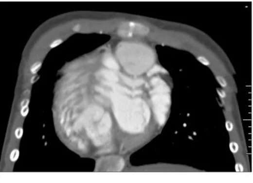
Solunum hareketi artefaktı, pulmoner damar sınırlarında bulanıklık veya çift kontur oluşturabilir. Solunum hareketi artefaktı, pulmoner vaskülatürün değerlendirilmesini güçleştirebilir. Hareket artefaktı pulmoner damar sınırlarında bulanıklık veya çift kontur oluşturabilir; kalp pulsasyonu da toraks BT’de hareket artefaktına yol açabilir.
Normal BT ile kıyasla
Aynı bölgenin, kaynak veri setinde patoloji içermediği belirtilen bir BT incelemesini kesit kesit kaydırın; organ sınırlarını açarak anatomiyi hasta görüntüsüyle karşılaştırın. Küçük rastlantısal bulgular tümüyle dışlanmamıştır.
Kaynak: TotalSegmentator v2.01 veri seti (Wasserthal ve ark., Radiology: Artificial Intelligence 2023), CC BY 4.0, vaka s1278; organ sınırları veri setinin otomatik segmentasyonundan, birleştirilerek sadeleştirildi. Görüntüler 2,5 mm kesit aralığına yeniden örneklendi.
Ayırıcı tanı
- Solunum hareketi toraks BT'de pulmoner damar sınırlarında bulanıklık veya çift kontur oluşturabilir.
- Hareket artefaktı, akciğer penceresinde parankimal ve vasküler anormalliklerin değerlendirilmesini güçleştirebilir; pulmoner damar sınırlarında bulanıklık veya çift kontur oluşturabilir.
- Bronşektazi, bronş ağacının anormal ve kalıcı genişlemesidir; hareket artefaktı ise pulmoner damar sınırlarında bulanıklık veya çift kontur oluşturabilir.
Tuzaklar
- Kalp hareketi, akciğer penceresinde parankim ve damar bulgularının değerlendirilmesini bozabilir; lingula veya sol alt lobda bulanıklık varsa kalp ve komşu damar sınırlarını da inceleyin.
- Bronşektazi, bronş ağacının anormal ve kalıcı genişlemesidir.
- Solunum hareketi artefaktı, pulmoner vaskülatürün değerlendirilmesini güçleştirebilir.
- CTPA’da solunum veya akım artefaktları yalancı pozitif emboli bulgularına yol açabilir; çok düzlemli değerlendirme bu taklitçileri ayırt etmeye yardımcı olabilir.
Kendini dene
Toraks BT anjiyografide solunum hareketi artefaktı hangi görünümü oluşturabilir?
Cevabı göster
Pulmoner damar kenarlarında bulanıklık ve çift kontur. Solunum hareketi artefaktı pulmoner vaskülatürde bulanıklık ve çift kontur oluşturabilir.
Toraks BT anjiyografide damar kenarlarında çift kontur ve bulanıklık görülmesi hangi artefaktla uyumludur?
Cevabı göster
Nefes tutma veya solunum hareketi artefaktı. Solunum hareketi artefaktı pulmoner vaskülatürde çift kontur ve bulanıklık oluşturabilir.
İlgili konular
Kaynaklar
Bu sayfadaki 32 cümle ve 2 quiz sorusu aşağıdaki kaynaklardan alıntılarla otomatik olarak karşılaştırıldı; 27 cümle bu karşılaştırmada düzeltildi (2026-10-09).
- Emphysema in an 11‐month‐old boy with alpha‐1 antitrypsin deficiencypmc.ncbi.nlm.nih.gov
- Acute Traumatic Aortic Injury: What the Radiologist Needs to Knowpmc.ncbi.nlm.nih.gov
- An integrated digital-to-surgical framework for patient-specific chest wall resection and reconstruction using 3D-printed titanium implants: proof-of-concept studypmc.ncbi.nlm.nih.gov
- When Neck Pain Is Bleeding: Spontaneous Cervical Epidural Hematoma Case Following Recent Binge Alcohol Intakepmc.ncbi.nlm.nih.gov
- Evaluation of artificial intelligence for pulmonary embolism detection on CTPApmc.ncbi.nlm.nih.gov
- Attenuation correction in 4D-PET using a single-phase attenuation map and rigidity-adaptive deformable registration.pubmed.ncbi.nlm.nih.gov
- PET/CT: Interpretative Pitfalls in the Thorax.pubmed.ncbi.nlm.nih.gov
- Simultaneous high-pitch multi-energy CT pulmonary angiography using a dual-source photon-counting-detector CT: A phantom experiment.pubmed.ncbi.nlm.nih.gov
- Estimation and correction of respiratory motion in pediatric renal SPECT through a data-driven center-of-mass method.pubmed.ncbi.nlm.nih.gov
- Increased Scan Speed and Pitch on Ultra-Low-Dose Chest CT: Effect on Nodule Volumetry and Image Quality.pubmed.ncbi.nlm.nih.gov
- Ultralow dose computed tomography as an alternative to conventional chest radiography for the evaluation of disease severity in paediatric cystic fibrosis.pubmed.ncbi.nlm.nih.gov
- CT-Guided Transthoracic Core-Needle Biopsy of Pulmonary Nodules: Current Practices, Efficacy, and Safety Considerations.pubmed.ncbi.nlm.nih.gov
- Impact of Contrast-Enhanced Mammography on Breast Imaging Reporting and Data System (BI-RADS) Reclassification: Correlation With Histopathology and Clinical Outcomes.pubmed.ncbi.nlm.nih.gov
- Duropathies as Unifying Concept-Part Two: A Narrative Overview of Clinical and Neuroradiological Features.pubmed.ncbi.nlm.nih.gov
- AI-driven multimodal retinal imaging for early detection and risk stratification of vascular and neurodegenerative diseases.pubmed.ncbi.nlm.nih.gov
- 2.5-mm articulated endoluminal forceps using a compliant mechanism.pubmed.ncbi.nlm.nih.gov
- Clinical correlates and short-term prognostic value of CT-detected subclinical pulmonary congestion in acute STEMI without overt heart failure.pubmed.ncbi.nlm.nih.gov
- Postoperative Respiratory Depression and Other Postoperative Pulmonary Complications in Veterinary Practice: A Narrative Review.pubmed.ncbi.nlm.nih.gov
- Small airway dysfunction: a shared, early, measurable, and potentially treatable trait across COPD, asthma, and fibrotic lung disease.pubmed.ncbi.nlm.nih.gov
- Simultaneous 3D structural-functional assessment of pediatric lung using 0.55-T MRI: a feasibility study.pubmed.ncbi.nlm.nih.gov
- Cinematic Rendering of Brain Magnetic Resonance Imaging: Photorealistic Visualization of Normal Neuroanatomy and Primary Brain Tumors.pubmed.ncbi.nlm.nih.gov
- Ultrashort echo time imaging of the lungs under high-frequency noninvasive ventilation: A new approach to lung imaging.pubmed.ncbi.nlm.nih.gov
- Current Insights into Clinical, Molecular, and Therapeutic Approaches to Acute Respiratory Distress Syndrome.pubmed.ncbi.nlm.nih.gov
- Imaging in the diagnosis and management of fibrosing interstitial lung diseases.pubmed.ncbi.nlm.nih.gov
- Thoracic Ultrasound for Pre-Procedural Dynamic Assessment of Non-Expandable Lung: A Non-Invasive, Real-Time and Multifaceted Diagnostic Tool.pubmed.ncbi.nlm.nih.gov
- Seeing the invisible: Three-dimensional liver modeling from simulation to the scalpel.pubmed.ncbi.nlm.nih.gov
- A survey of transformer-based architectures in medical image analysis: models, applications, and challenges.pubmed.ncbi.nlm.nih.gov
- A Lightweight, 3D-Printable, Low-Cost, "One-Click" Dorsal Skin Window for High-Quality Long-Term Multimodal Subcutaneous Tumor Imaging.pubmed.ncbi.nlm.nih.gov
- Development and validation of a dual-attention deep learning prediction model for noninvasive differentiation of pulmonary invasive mucinous adenocarcinoma from inflammatory pulmonary nodules.pubmed.ncbi.nlm.nih.gov
- Comparison of Postoperative Oxygenation and Dorsal Lung Ventilation in Patients Undergoing Craniotomy in Different Positions: Pulmonary Electrical Impedance Tomography Monitoring.pubmed.ncbi.nlm.nih.gov
- Bedside lung ultrasound versus chest CT in critically ill patients: a cross-sectional diagnostic accuracy study.pubmed.ncbi.nlm.nih.gov
- SPOT THE BOT: A comparative quality assessment of AI-generated written instructions for lung ultrasound training.pubmed.ncbi.nlm.nih.gov
- Diagnosis, Treatment, and Follow-Up of Tracheo/Bronchomalacia in Children: The Italian Multicenter Experience.pubmed.ncbi.nlm.nih.gov
- Childhood interstitial lung diseases: current understanding of the classification and imaging findings.pubmed.ncbi.nlm.nih.gov
- Imaging in non-cystic fibrosis bronchiectasis and current limitations.pubmed.ncbi.nlm.nih.gov
- ESR Essentials: imaging of common paediatric pulmonary diseases-practice recommendations by the European Society of Paediatric Radiology.pubmed.ncbi.nlm.nih.gov
- Lung imaging methods: indications, strengths and limitations.pubmed.ncbi.nlm.nih.gov
- High-affinity alphavbeta3 integrin targeted optical probe as a new imaging biomarker for early atherosclerosis: initial studies in Watanabe rabbits.pubmed.ncbi.nlm.nih.gov
- International Society of Paediatric Surgical Oncology (IPSO) Surgical Practice Guidelines.pubmed.ncbi.nlm.nih.gov
- Long-term safety and effectiveness of the "OptEase" vena cava filter.pubmed.ncbi.nlm.nih.gov
- Patterns of injury and violence in Yaoundé Cameroon: an analysis of hospital data.pubmed.ncbi.nlm.nih.gov
- State of the art of 18 F-FDG PET/CT application in inflammation and infection: a guide for image acquisition and interpretation.pubmed.ncbi.nlm.nih.gov
- Hypofractionated radiotherapy for localized prostate cancer.pubmed.ncbi.nlm.nih.gov
- Incidental Lung Lesions on the CT Component of Oncologic FDG PET/CT: A Pictorial Review of Interpretive Pitfalls and Diagnostic Clues.pubmed.ncbi.nlm.nih.gov
- Magnetic Resonance Imaging for Guidance and Follow-up of Thoracic Needle Biopsies and Thermal Ablations.pubmed.ncbi.nlm.nih.gov
- Imaging side effects and complications of chemotherapy and radiation therapy: a pictorial review from head to toe.pubmed.ncbi.nlm.nih.gov
- Postoperative Pulmonary Complications: Clinical and Imaging Insightspubmed.ncbi.nlm.nih.gov
- Uncommon Causes of Interlobular Septal Thickening on CT Images and Their Distinguishing Features.pubmed.ncbi.nlm.nih.gov
- Radiologic approach and progressive exploration of connective tissue disease-related interstitial lung disease: meeting the curiosity of rheumatologists.pubmed.ncbi.nlm.nih.gov
- Acute Traumatic Aortic Injury: What the Radiologist Needs to Know.pubmed.ncbi.nlm.nih.gov
- When Neck Pain Is Bleeding: Spontaneous Cervical Epidural Hematoma Case Following Recent Binge Alcohol Intake.pubmed.ncbi.nlm.nih.gov
- Case Report: Diabetic ketoacidosis following EGFR-TKIs therapy via aggravated insulin resistance.pubmed.ncbi.nlm.nih.gov
- Multifocal intracranial hemorrhage on emergency brain CT in a child with end-stage nephrotic syndrome.pubmed.ncbi.nlm.nih.gov
- Quantitative peri-lesional densitometry mapping via thin-slice volume rendering enhances differentiation of pneumonic-type lung cancer and inflammatory pneumonia.pubmed.ncbi.nlm.nih.gov
- Differentiating lung neuroendocrine neoplasms from tumor-like infection using CT in patients with ectopic ACTH syndrome.pubmed.ncbi.nlm.nih.gov
- The intersection of artificial intelligence and lung nodule research: current applications and future prospects.pubmed.ncbi.nlm.nih.gov
- Computer-based quantitative image texture analysis using multi-collinearity diagnosis in chest X-ray images.pubmed.ncbi.nlm.nih.gov
- Deep learning models for CT image classification: a comprehensive literature review.pubmed.ncbi.nlm.nih.gov
- Pneumothorax and Hemothorax: Practical Approaches for Radiologists.pubmed.ncbi.nlm.nih.gov
- Congenital Lung Malformations in Neonates: A 9-Year Tunisian Cohort Highlighting Diagnostic Challenges and Outcomes in a Resource-Limited Setting.pubmed.ncbi.nlm.nih.gov
- AI-based chest CT quantification of interstitial lung disease in idiopathic inflammatory myopathies: agreement with expert visual assessment in 107 patients.pubmed.ncbi.nlm.nih.gov
- Pulmonary Complications in Children Following Hematopoietic Cell Transplantation: A Case Report and Review of the Diagnostic Approach.pubmed.ncbi.nlm.nih.gov
- Prediction of Clinical Bronchiectasis from Asymptomatic Radiological Bronchiectasis.pubmed.ncbi.nlm.nih.gov
- CT Imaging Findings of Pulmonary Artery Stenosis: A Pictorial Review.pubmed.ncbi.nlm.nih.gov
- Dual-source computed tomography protocols for the pediatric chest - scan optimization techniques.pubmed.ncbi.nlm.nih.gov
- Practical Imaging Interpretation in Patients Suspected of Having Idiopathic Pulmonary Fibrosis: Official Recommendations from the Radiology Working Group of the Pulmonary Fibrosis Foundation.pubmed.ncbi.nlm.nih.gov
- Imaging of Childhood Interstitial Lung Disease.pubmed.ncbi.nlm.nih.gov
- A compound heterozygous combination of SLC34A2 variants in pulmonary alveolar microlithiasis: A case report and literature review.pubmed.ncbi.nlm.nih.gov
- Explosive multi-site metastasis temporally associated with acute exacerbation of idiopathic pulmonary fibrosis in a patient with surgically resected lung squamous cell carcinoma: a case report.pubmed.ncbi.nlm.nih.gov
- Case Report: Synchronous SCLC and minimally invasive adenocarcinoma in the same lobe: NGS-confirmed cross-histological molecular heterogeneity in an ultra-rare case.pubmed.ncbi.nlm.nih.gov
- Chest CT for suspected pulmonary complications of oncologic therapies: how I review and report.pubmed.ncbi.nlm.nih.gov
- Imaging in the COVID-19 era: Lessons learned during a pandemic.pubmed.ncbi.nlm.nih.gov
- Lung Imaging in Acute Hypoxemic Respiratory Failure: From Physics to Bedside Applications.pubmed.ncbi.nlm.nih.gov
- AWMF S2k Guideline: Diagnosis and Assessment of Lung Disease Caused by Occupational Silica Dust Exposure (Silicosis) - Disease No. 4101 as per Occupational Diseases Ordinance (BKV) Annex 1.pubmed.ncbi.nlm.nih.gov
- Deep learning-based motion correction: cardiac motion artifact and image quality improvements on chest CT.pubmed.ncbi.nlm.nih.gov
- Nonstop gated cone-beam computed tomography for respiratory gating pancreatic cancer radiotherapy using dual-domain deep learning reconstruction.pubmed.ncbi.nlm.nih.gov
- A multisystem, cardio-renal investigation of post-COVID-19 illness.pubmed.ncbi.nlm.nih.gov
- Characterizing heterogeneity and subphenotyping acute respiratory distress syndrome with computed tomography.pubmed.ncbi.nlm.nih.gov
- A rare case of H syndrome with severe multisystem involvement: Clinical challenges in low-resource healthcare settings.pubmed.ncbi.nlm.nih.gov
- Multimodality imaging in the detection and management of coronary and peripheral arterial disease in patients with cancer receiving cardiotoxic antineoplastic treatments: A clinical consensus statement of the ESC Council of Cardio-Oncology and the European Association of Cardiovascular Imaging (EACVI) of the ESC.pubmed.ncbi.nlm.nih.gov
- Evaluation of patients diagnosed with primary Sjogren's syndrome with and without pulmonary involvement.pubmed.ncbi.nlm.nih.gov
- Contributing factors to pediatric COVID-19 and MIS-C during the initial waves: A systematic review of 92 case reports.pubmed.ncbi.nlm.nih.gov
- Lung Imaging in COPD Part 1: Clinical Usefulness.pubmed.ncbi.nlm.nih.gov
- Lung ultrasound in cardiac rehabilitation: expert consensus on protocols, clinical use, and integration into patient management from the working group on cardiac rehabilitation and cardiovascular prevention of the Italian society of cardiology.pubmed.ncbi.nlm.nih.gov
- Precise discrimination of mycobacterial pulmonary diseases via multimodal machine learning integrating chest CT and clinical markers.pubmed.ncbi.nlm.nih.gov
- Common and Rare Radiological Findings of Pulmonary and Thoracic Hydatid Cysts.pubmed.ncbi.nlm.nih.gov
- Direct visualization of postoperative aortobronchial fistula on computed tomography.pubmed.ncbi.nlm.nih.gov
- Correlation analysis of chest HRCT quantitative parameters, exhaled nitric oxide, and pulmonary function in patients with chronic obstructive pulmonary disease.pubmed.ncbi.nlm.nih.gov
- Clinical usability of 3D gradient-echo-based ultrashort echo time imaging: Is it enough to facilitate diagnostic decision in real-world practice?pubmed.ncbi.nlm.nih.gov
- Lipoid Pneumonia: HRCT and MRI Spectrum, Diagnostic Pitfalls, and Imaging-Based Diagnostic Workflow.pubmed.ncbi.nlm.nih.gov
- Correlation of Thoracic HRCT Scores with Right Heart Mechanics and TAPSE/sPAP Ratio in Pulmonary Alveolar Proteinosis.pubmed.ncbi.nlm.nih.gov
- Diffuse cystic lung diseases-a primer for radiologists.pubmed.ncbi.nlm.nih.gov
- Objective assessment of diagnostic image quality in CT scans: what radiologists and researchers need to know.pubmed.ncbi.nlm.nih.gov
- Improvement in Image Quality and Efficiency of Non-contrast Thoracic MR Angiography: Comparison of a Highly Accelerated Dixon-based Technique with the Conventional Fat-suppressed Technique.pubmed.ncbi.nlm.nih.gov
- Future Applications of Cardiothoracic CT.pubmed.ncbi.nlm.nih.gov
- Society for Cardiovascular Magnetic Resonance/European Society of Cardiovascular Imaging/American Society of Echocardiography/Society for Pediatric Radiology/North American Society for Cardiovascular Imaging Guidelines for the use of cardiovascular magnetic resonance in pediatric congenital and acquired heart disease : Endorsed by The American Heart Association.pubmed.ncbi.nlm.nih.gov
- Society for Cardiovascular Magnetic Resonance/European Society of Cardiovascular Imaging/American Society of Echocardiography/Society for Pediatric Radiology/North American Society for Cardiovascular Imaging Guidelines for the Use of Cardiac Magnetic Resonance in Pediatric Congenital and Acquired Heart Disease: Endorsed by The American Heart Association.pubmed.ncbi.nlm.nih.gov
- 2022 ACC/AHA guideline for the diagnosis and management of aortic disease: A report of the American Heart Association/American College of Cardiology Joint Committee on Clinical Practice Guidelines.pubmed.ncbi.nlm.nih.gov
- Society for Cardiovascular Magnetic Resonance 2024 Cases of SCMR case series.pubmed.ncbi.nlm.nih.gov
- Standardized image interpretation and post processing in cardiovascular magnetic resonance: Society for Cardiovascular Magnetic Resonance (SCMR) board of trustees task force on standardized post processing.pubmed.ncbi.nlm.nih.gov
- Neurologic complications of coronavirus and other respiratory viral infections.pubmed.ncbi.nlm.nih.gov
- Lung ultrasound versus chest radiography for the detection of early postoperative pulmonary complications in children undergoing cardiac surgery.pubmed.ncbi.nlm.nih.gov
- Granulomatosis with polyangiitis: clinical characteristics and updates in diagnosis.pubmed.ncbi.nlm.nih.gov
- Imaging of coronavirus disease (COVID-19): a pictorial review.pubmed.ncbi.nlm.nih.gov
- Guidelines on the Use of Therapeutic Apheresis in Clinical Practice-Evidence-Based Approach From the Writing Committee of the American Society for Apheresis: The Tenth Special Issue.pubmed.ncbi.nlm.nih.gov
- Surgery for lung cancer: postoperative changes and complications-what the Radiologist needs to know.pubmed.ncbi.nlm.nih.gov
- EACR 2025 Congress: Innovative Cancer Science, 16-19 June 2025.pubmed.ncbi.nlm.nih.gov
- The role of radiologic assessment in evaluating and monitoring respiratory function in amyotrophic lateral sclerosis (ALS) patients: a narrative review.pubmed.ncbi.nlm.nih.gov
- The impact of technical factors on the chest radiographic findings in COVID-19 pneumonia.pubmed.ncbi.nlm.nih.gov
- Lung ultrasound in a nutshell. Lines, signs, some applications, and misconceptions from a radiologist's point of view.pubmed.ncbi.nlm.nih.gov
- The abstracts of VIII National Congress AdET (Accademia di Ecografia Toracica).pubmed.ncbi.nlm.nih.gov
- Research progress in computer-aided diagnosis systems for lung cancer.pubmed.ncbi.nlm.nih.gov
- Application of an improved YOLOv5s-based deep learning model for automated detection of pulmonary adenocarcinoma <i>in situ</i> and minimally invasive adenocarcinoma.pubmed.ncbi.nlm.nih.gov
- Consensus statement on thoracic radiology terminology in Portuguese used in Brazil and in Portugal.pubmed.ncbi.nlm.nih.gov
- Radiological imaging and interventional procedures of the thorax in children.pubmed.ncbi.nlm.nih.gov
Eğitim amaçlıdır; hasta başında tanı ve tedavi kararının yerine geçmez, tıbbi tavsiye değildir. Metinler yapay zekâ desteğiyle yazıldı ve otomatik bir adımda kaynak alıntılarıyla karşılaştırıldı; bu bir tıbbi doğrulama değildir ve içerik uzman radyolog incelemesinden geçmedi. Klinik kararlarda güncel kılavuzları, kurum protokollerini ve radyoloji raporunu esas alın.