Özet
İntramural özofagus hematomu, kanın özofagus duvarı içinde birikerek duvarı genişlettiği, akut göğüs ağrısı ve disfajiyle ortaya çıkabilen nadir bir durumdur. BT, özofagus duvarındaki hematomla uyumlu kitleyi ve lümen basısını gösterebilir; aort diseksiyonu ayırıcı tanıda olduğunda, kaynaklarda hematomu ve diseksiyonu değerlendirmek için kontrastlı toraks BT anjiyografi kullanılmıştır. Kanama antikoagülasyon, travma, kusma veya girişimle ilişkili olabilir; BT'de varis ya da tümör taklidi oluşturabilir. Hematomu atlayıp yanlış biçimde aortik acil veya tümör olarak yorumlamak uygun klinik yaklaşımı geciktirebilir; perforasyon ve aortoözofageal fistül bulguları ayrıca aranmalıdır.
Faz ve pencere
- Kontrastsız tanısal
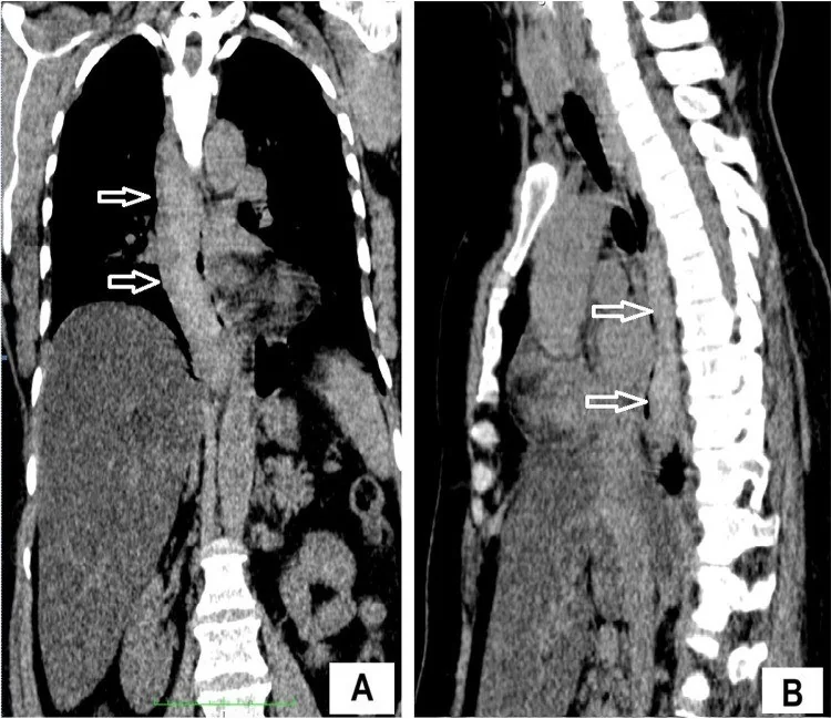
- Kontrastsız seride özofagus duvarında hiperdens (yüksek atenüasyonlu) düzgün sınırlı mural kitle veya kalınlaşma izlenir; bu kitle lümeni basılayabilir.
- Arteriyel tanısal
- Kontrastlı BT'de hematom, özofagus boyunca uzanan kontrastlanmayan yumuşak doku kitlesi ve lümende dıştan bası şeklinde görülebilir.
Önerilen pencereler: Mediasten (G 350 / M 50), Yumuşak doku (G 400 / M 40), Anjiyo (G 600 / M 150).
BT bulguları
- Özofagus duvarını eksantrik veya çevresel biçimde genişleten düzgün sınırlı mural kitle
- Kontrastsız BT’de hematom, özofagus duvarında uzunlamasına uzanan homojen hiperdensite şeklinde görülebilir.
- Uzunlamasına uzanabilen duvar kalınlaşması ve özofagus lümeninde bası ya da ileri daralma
- IV kontrast sonrası hematom bileşeninde belirgin iç kontrastlanma olmaması
- Kontrastlı BT’de intramural hematom, bazı olgularda kontrast kaçağıyla birlikte görülebilir.
- Aortoözofageal fistül olasılığında BT'de aort duvarı çıkıntısı, ektopik gaz ve periaortik yağ dokuda kirlenme gibi bulgular değerlendirilebilir.
- Perforasyon eşlik ediyorsa özofagus duvar kesintisi, ekstraluminal gaz ya da mediastinal sıvı-koleksiyon
- Lezyon özofagus duvarında eksantrik veya çevresel kalınlaşma şeklinde izlenebilir ve lümende dıştan basıya yol açabilir.
Normalde
Aynı torasik seviyedeki normal özofagus, kontrastsız seride ince duvarlı ve lümeni basısız tüp şeklindedir; duvar içinde kan yoğunluğunda ayrı bir kitle bulunmaz. Kıyaslarken mural içeriğin atenüasyonunu komşu kan havuzuyla karşılaştır, lümen açıklığını izle ve IV kontrast sonrasında hematomun kontrastlanıp kontrastlanmadığını değerlendir.
Normal BT ile kıyasla
Aynı bölgenin, kaynak veri setinde patoloji içermediği belirtilen bir BT incelemesini kesit kesit kaydırın; organ sınırlarını açarak anatomiyi hasta görüntüsüyle karşılaştırın. Küçük rastlantısal bulgular tümüyle dışlanmamıştır.
Kaynak: TotalSegmentator v2.01 veri seti (Wasserthal ve ark., Radiology: Artificial Intelligence 2023), CC BY 4.0, vaka s1278; organ sınırları veri setinin otomatik segmentasyonundan, birleştirilerek sadeleştirildi. Görüntüler 2,5 mm kesit aralığına yeniden örneklendi.
Ayırıcı tanı
- Özofagus varisi
- portal venöz fazda kontrastlanan serpijinöz kanallar oluşturur; kontrastsızda yüksek atenüasyonlu tek mural kitle görünümü beklenmez.
- Özofagus karsinomu
- BT'de kitle oluşturan ya da duvar kalınlaşması gösteren özofageal bir malignitesidir; lenf düğümü metastazı görülebilir ve intramural hematomun ayırıcı tanısında malignite dışlanmalıdır.
- Özofagus duplikasyon kisti
- aberran foregut gelişiminden kaynaklanan konjenital bir kist olup sıkça posterior mediastende yerleşir; BT'de diğer mediastinal kitlelerden kesin ayrımı her zaman kolay değildir.
- Özofagus perforasyonu
- ekstraluminal oral kontrast, mediastinal gaz ve çevre sıvısı ön plandadır; tek başına mural hematomla açıklanmaz.
Tuzaklar
- Portal venöz kanla benzer yoğunluktaki kontrastlanan varisler mural hematom sanılabilir; kontrastsız görüntüde yüksek atenüasyon ve kontrast sonrası boyanmama hematomu destekler.
- Tümör de duvarı kalınlaştırıp lümeni daraltabilir; hematomda düzgün sınır ve kan yoğunluğu, tümörde ise düzensiz infiltratif kontur ve solid kontrastlanma ayrımda yardımcıdır.
- Aortoözofageal fistül şüphesinde BT'de aort duvarı çıkıntısı, ektopik gaz ve periaortik yağ dokuda kirlenme gibi bulgulara dikkat edin.
- Perforasyon şüphesinde oral kontrastlı görüntüleme yapılabilir; ekstraluminal hava veya kontrast ekstravazasyonu perforasyon değerlendirmesinde aranabilir.
- Ani göğüs ağrısında aort diseksiyonu ayırıcı tanıda yer alır; kaynaklarda intramural özofagus hematomunu değerlendirirken kontrastlı toraks BT anjiyografi kullanılmıştır.
- Kontrastlı BT’de özofagus boyunca uzanan intramural hiperdensite veya kontrastlanmayan yumuşak doku kitlesi ve lümen basısı hematomla uyumlu olabilir.
Kendini dene
Ani göğüs ağrısı ve disfajisi olan hastada BT, özofagus boyunca uzanan homojen hiperdens mural oluşum ve lümende dıştan bası gösteriyor. En olası tanı nedir?
Cevabı göster
İntramural hematom. BT’de özofagus duvarındaki homojen hiperdens görünüm hematomu düşündürür; hematom lümene dıştan bası yapabilir. Göğüs ağrısı ve disfaji intramural özofagus hematomunda görülebilir; BT’de duvar içindeki homojen hiperdens görünüm tanıyı destekler.
Kontrastlı BT’de özofagus boyunca uzanan, kontrastlanmayan yumuşak doku kitlesi ve lümende dıştan bası görülüyor. Aort diseksiyonu ve özofagus rüptürü saptanmıyor. En olası tanı nedir?
Cevabı göster
İntramural hematom. Özofagus boyunca uzanan kontrastlanmayan kitle ve lümende dıştan bası intramural özofagus hematomunu düşündürür. Kontrastlı BT’de bu bulgular hematomla uyumlu olabilir.
Kaynaklar
Bu sayfadaki 41 cümle ve 2 quiz sorusu aşağıdaki kaynaklardan alıntılarla otomatik olarak karşılaştırıldı; 18 cümle bu karşılaştırmada düzeltildi (2026-10-09).
- Intramural Esophageal Hematoma as a Complication of Percutaneous Coronary Intervention: A Case Reportpmc.ncbi.nlm.nih.gov
- “Spontaneous Intramural Esophageal Hematoma Associated with Uncontrolled Hypertension: Case Report and Review”pmc.ncbi.nlm.nih.gov
- When pain persists after peroral endoscopic myotomy: endoscopic management of a large postprocedural intramural esophageal hematomapmc.ncbi.nlm.nih.gov
- Intramural Hematoma of the Esophagus: A Case Report and Literature Reviewpmc.ncbi.nlm.nih.gov
- Intramural Esophageal Hematoma With Associated Hemothoraxpmc.ncbi.nlm.nih.gov
- Spontaneous Paraesophageal Hematoma.pubmed.ncbi.nlm.nih.gov
- Spontaneous esophageal submucosal hematoma.pubmed.ncbi.nlm.nih.gov
- Intramural Esophageal Hematoma With Associated Hemothorax.pubmed.ncbi.nlm.nih.gov
- Spontaneous Intramural Esophageal Hematoma Secondary to Thrombolysis in the Setting of Pulmonary Embolism.pubmed.ncbi.nlm.nih.gov
- Probable intramural esophageal hematoma mimicking esophageal abscess.pubmed.ncbi.nlm.nih.gov
- Spontaneous intramural esophageal hematoma (IEH) secondary to anticoagulation and/or thrombolysis therapy in the setting of a pulmonary embolism: a case report.pubmed.ncbi.nlm.nih.gov
- Intramural Hematoma of the Esophagus: A Case Report and Literature Review.pubmed.ncbi.nlm.nih.gov
- Intramural Esophageal Hematoma as a Complication of Percutaneous Coronary Intervention: A Case Report.pubmed.ncbi.nlm.nih.gov
- Intramural Esophageal Hematoma Mimicking Acute Coronary Syndrome: Complication of Warfarin Use in a Mechanical Heart Valve Patient.pubmed.ncbi.nlm.nih.gov
- Successful Endoscopic Fenestration for Giant Esophageal Hematoma After Failed Conservative Therapy.pubmed.ncbi.nlm.nih.gov
- Idiopathic esophageal intramural hematoma associated with hemothorax leading to esophageal perforation.pubmed.ncbi.nlm.nih.gov
- Esophageal Hematomapubmed.ncbi.nlm.nih.gov
- Acute aortic syndromes after esophageal endoscopic submucosal dissection.pubmed.ncbi.nlm.nih.gov
- An Overview of the Diagnostic and Treatment Strategies for Aortic Disease.pubmed.ncbi.nlm.nih.gov
- Visceral Arterial Pseudoaneurysms-A Clinical Review.pubmed.ncbi.nlm.nih.gov
- 2024 European Heart Rhythm Association/Heart Rhythm Society/Asia Pacific Heart Rhythm Society/Latin American Heart Rhythm Society expert consensus statement on catheter and surgical ablation of atrial fibrillation.pubmed.ncbi.nlm.nih.gov
- Esophageal emergencies: another important cause of acute chest pain.pubmed.ncbi.nlm.nih.gov
- C-Reactive Protein: Pathophysiology, Diagnosis, False Test Results and a Novel Diagnostic Algorithm for Clinicians.pubmed.ncbi.nlm.nih.gov
- Computed Tomography Imaging of Thoracic Aortic Surgery: Distinguishing Life-Saving Repairs from Life-Threatening Complications.pubmed.ncbi.nlm.nih.gov
- MDCT Imaging of Non-Traumatic Thoracic Aortic Emergencies and Its Impact on Diagnosis and Management-A Reappraisal.pubmed.ncbi.nlm.nih.gov
- 2022 ACC/AHA guideline for the diagnosis and management of aortic disease: A report of the American Heart Association/American College of Cardiology Joint Committee on Clinical Practice Guidelines.pubmed.ncbi.nlm.nih.gov
- Primary Cardiac Angiosarcoma in a Young Male: Rare Presentation With Right Atrial Endocardial Dissection and Right Coronary Artery Tumor Fistula.pubmed.ncbi.nlm.nih.gov
- Malignancy-mimicking Kayexalate-associated extraintestinal mass and literature review.pubmed.ncbi.nlm.nih.gov
- Thoracic aortic pseudoaneurysm with esophageal compression due to brucellosis: a rare case report.pubmed.ncbi.nlm.nih.gov
- A Complicated Multiloculated Pyogenic Liver Abscess With Spontaneous Rupture and Life-Threatening Hemorrhage Successfully Managed With Transarterial Embolization: A Case Report.pubmed.ncbi.nlm.nih.gov
- Radiologic Interventions for Gastrointestinal Bleeding.pubmed.ncbi.nlm.nih.gov
- Iatrogenic portobiliary fistula treated with covered stent following percutaneous transhepatic biliary drainage.pubmed.ncbi.nlm.nih.gov
- Aortoesophageal fistula as a late-onset complication of infected aortic arch aneurysm caused by Salmonella enterica serotype Choleraesuis: a case report.pubmed.ncbi.nlm.nih.gov
- Primary aortoesophageal fistula: a rare cause of acute upper gastrointestinal bleeding.pubmed.ncbi.nlm.nih.gov
- Endo-hepatology: Bridging the gap between lumen and liver.pubmed.ncbi.nlm.nih.gov
- Characteristics of patients with epithelioid hemangioendothelioma (EHE): a retrospective analysis of the Charité- Universitätsmedizin Berlin.pubmed.ncbi.nlm.nih.gov
- Safety and preliminary efficacy of allogeneic umbilical cord-derived mesenchymal stem cells administered via the hepatic artery in patients with liver cirrhosis: A phase I open-label trial.pubmed.ncbi.nlm.nih.gov
- Life-threatening bleeding from dissecting Intramural Hematoma of Esophagus (IHE) treated by trans arterial embolization.pubmed.ncbi.nlm.nih.gov
- Surgical treatment of an esophageal bronchogenic cyst with massive upper digestive tract hematoma without esophagectomy: a case report and the review of the literature.pubmed.ncbi.nlm.nih.gov
- Imaging the posterior mediastinum: a multimodality approach.pubmed.ncbi.nlm.nih.gov
- The injured esophagus: imaging spectrum of esophageal emergencies.pubmed.ncbi.nlm.nih.gov
- Lung squamous cell carcinoma complicated with immune checkpoint inhibitor-associated active tuberculosis: a case report and literature review.pubmed.ncbi.nlm.nih.gov
- Immune-Related Cystitis Following Immune Checkpoint Inhibitor Therapy: A Report of Two Cases.pubmed.ncbi.nlm.nih.gov
- Computed tomography features and clinicopathological characteristics of resectable esophageal sarcomatoid carcinoma: a retrospective study.pubmed.ncbi.nlm.nih.gov
- Foregut mediastinal cysts: a narrative review.pubmed.ncbi.nlm.nih.gov
- Case Report: Giant esophageal duplication cyst mimicking cardiac compression in an adult.pubmed.ncbi.nlm.nih.gov
- Successful resection of a large duplication cyst via retropleural thoracotomy approach in a 3-month-old infant from Kabul, Afghanistan: a case report.pubmed.ncbi.nlm.nih.gov
- Clinical Features and Factors Contributing to Preoperative Misdiagnosis of Gastric Gastrointestinal Stromal Tumors: A Retrospective Analysis of 27 Cases.pubmed.ncbi.nlm.nih.gov
- Clinical, imaging and surgical outcomes of secondary intussusceptions in children: a retrospective analysis based on the benign and malignant pathologic lead points.pubmed.ncbi.nlm.nih.gov
- Natural Course of Gastric Subepithelial Tumors.pubmed.ncbi.nlm.nih.gov
- Extrinsic Left Atrial Compression: An Echocardiography-Guided Diagnosis Illustrated by Two Clinical Cases and a Structured Review of Published Cases.pubmed.ncbi.nlm.nih.gov
- Small Intestine Tumors: Diagnostic Role of Multiparametric Ultrasound.pubmed.ncbi.nlm.nih.gov
- Comparison of Endoscopic Ultrasound and CT Scan in the Diagnosis of Esophageal Duplication Cysts.pubmed.ncbi.nlm.nih.gov
- Imaging modalities (MRI, CT, PET/CT), indications, differential diagnosis and imaging characteristics of cystic mediastinal masses: a review.pubmed.ncbi.nlm.nih.gov
- Pericardial Diseases: International Position Statement on New Concepts and Advances in Multimodality Cardiac Imaging.pubmed.ncbi.nlm.nih.gov
- Patterns of injury and violence in Yaoundé Cameroon: an analysis of hospital data.pubmed.ncbi.nlm.nih.gov
- Long-term safety and effectiveness of the "OptEase" vena cava filter.pubmed.ncbi.nlm.nih.gov
- Diagnostic Value of EUS-FNA in the Differential Diagnosis of Esophageal Strictures Lacking Typical Malignant Features.pubmed.ncbi.nlm.nih.gov
- Gastric bronchogenic cyst pooled case analysis: a case report and systematic review.pubmed.ncbi.nlm.nih.gov
- Presumed Infected Bronchogenic Cyst Presenting as Acute Pericarditis: A Case Report.pubmed.ncbi.nlm.nih.gov
- Endoscopic Ultrasound and Gastric Sub-Epithelial Lesions: Ultrasonographic Features, Tissue Acquisition Strategies, and Therapeutic Management.pubmed.ncbi.nlm.nih.gov
- Cardiac Masses and Pseudomasses: An Overview about Diagnostic Imaging and Clinical Background.pubmed.ncbi.nlm.nih.gov
- Imaging of the aortic root on high-pitch non-gated and ECG-gated CT: awareness is the key!pubmed.ncbi.nlm.nih.gov
- Acute Traumatic Aortic Injury: What the Radiologist Needs to Know.pubmed.ncbi.nlm.nih.gov
- Imaging insights in veno-venous and veno-arterial extracorporeal membrane oxygenation (ECMO): CT protocols, underlying pathophysiology, and main complications.pubmed.ncbi.nlm.nih.gov
- An atypical case of fatal 'esophageal apoplexy': post-mortem findings and differential diagnosis.pubmed.ncbi.nlm.nih.gov
- Aortoesophageal fistula complicated by mycotic aneurysm secondary to endoscopic procedures: A case report.pubmed.ncbi.nlm.nih.gov
- Radiological features of uncommon aneurysms of the cardiovascular system.pubmed.ncbi.nlm.nih.gov
- Surgical management and clinical reflections on acute type A intramural hematoma with a focal intimal tear in an adult with double aortic arch: a case report.pubmed.ncbi.nlm.nih.gov
- Aortic Dissection Mimicry Under Extracorporeal Membrane Oxygenation (ECMO) After Cardiac Arrest: A Case Report of Emergency Imaging Dilemmas.pubmed.ncbi.nlm.nih.gov
- Spectrum of Multidetector Computed Tomography Imaging Findings in Iatrogenic Abdominopelvic Injuries: A Comprehensive Pictorial Review.pubmed.ncbi.nlm.nih.gov
- Endoscopic treatment of upper gastrointestinal postsurgical leaks: a narrative review.pubmed.ncbi.nlm.nih.gov
- Common, Less Common, and Unexpected Complications after Bariatric Surgery: A Pictorial Essay.pubmed.ncbi.nlm.nih.gov
- Endovascular stent-graft treatment for aortoesophageal fistula induced by an esophageal fishbone: Two cases report.pubmed.ncbi.nlm.nih.gov
- A rare case of posttraumatic aortic rupture, treated with an endovascular stent graft implantation and complicated with esophageal rupture.pubmed.ncbi.nlm.nih.gov
- Undiagnosed aortoesophageal fistula causing intramural hematoma of the esophagus.pubmed.ncbi.nlm.nih.gov
- Secondary Aortopulmonary Fistula After Thoracic Endovascular Aortic Repair: Case Report.pubmed.ncbi.nlm.nih.gov
- Predictors of outcome and different management of aortobronchial and aortoesophageal fistulas.pubmed.ncbi.nlm.nih.gov
- "Spontaneous Intramural Esophageal Hematoma Associated with Uncontrolled Hypertension: Case Report and Review".pubmed.ncbi.nlm.nih.gov
- Characterizing a CT esophagram protocol after flexible endoscopic diverticulotomy for Zenker's diverticulum: a retrospective series.pubmed.ncbi.nlm.nih.gov
- Multimodality imaging of adult gastric emergencies: A pictorial review.pubmed.ncbi.nlm.nih.gov
- Case Report: a hypermetabolic mediastinal mass (SUVmax 16.7) mimicking malignancy with spontaneous resolution: an inflammatory myofibroblastic proliferation and diagnostic pitfall.pubmed.ncbi.nlm.nih.gov
- Resection of C3 osteochondroma in a 20-year-old male with the combination of virtual reality and 3D printing for preoperative planning: illustrative case.pubmed.ncbi.nlm.nih.gov
- Uni-portal non-coaxial spinal endoscopic surgery on percutaneous hollow screw fixation for atypical hangman's fracture: a novel minimally invasive technique case report.pubmed.ncbi.nlm.nih.gov
- Radiologic Imaging of Traumatic Bowel and Mesenteric Injuries: A Comprehensive Up-to-Date Review.pubmed.ncbi.nlm.nih.gov
- The utility of a dual-phase, dual-energy CT protocol in patients presenting with overt gastrointestinal bleeding.pubmed.ncbi.nlm.nih.gov
- Conservative Management for Left Atrial Intramural Hematoma After Cryoballoon Ablation.pubmed.ncbi.nlm.nih.gov
- Idiopathic esophageal submucosal hematoma after flow diverter stent placement for an unruptured intracranial aneurysm: illustrative case.pubmed.ncbi.nlm.nih.gov
- Inflammatory Aortopathies in Rheumatic Diseases: A State-of-the-Art Review.pubmed.ncbi.nlm.nih.gov
- Role of Computed Tomography in Cardiac Electrophysiology.pubmed.ncbi.nlm.nih.gov
- Thoracic Diseases: Technique and Applications of Dual-Energy CT.pubmed.ncbi.nlm.nih.gov
- Role of Spiral and Multislice Computed Tomography in the evaluation of traumatic and spontaneous oesophageal perforation. Our experience.pubmed.ncbi.nlm.nih.gov
- Radiological Evaluation of Bowel Ischemia.pubmed.ncbi.nlm.nih.gov
Eğitim amaçlıdır; hasta başında tanı ve tedavi kararının yerine geçmez, tıbbi tavsiye değildir. Metinler yapay zekâ desteğiyle yazıldı ve otomatik bir adımda kaynak alıntılarıyla karşılaştırıldı; bu bir tıbbi doğrulama değildir ve içerik uzman radyolog incelemesinden geçmedi. Klinik kararlarda güncel kılavuzları, kurum protokollerini ve radyoloji raporunu esas alın.