Özet
Mediastinal metastaz, torasik veya toraks dışı primer tümörün mediastinal lenf düğümlerine ya da plevra ve perikarda yayılımını ifade eder; BT görünümü primer tümöre ve yayılım yoluna göre değişir. İntravenöz kontrastlı toraks BT, düğümlerin kısa aksını ve morfolojisini, plevral/perikardiyal nodülleri, efüzyonları ve komşu mediastinal yapılara uzanımı aynı incelemede haritalar. Düğüm büyüklüğü tek başına metastazı kanıtlamaz; küçük düğümlerde hastalık olabilir, büyük düğümler benign süreçlere bağlı olabilir ve kesin nodal evreleme gerektiğinde doku örneklemesi kullanılır. Plevral veya perikardiyal yayılımın atlanması evreyi, prognoz değerlendirmesini ve tedavi planını değiştirebilir.
Faz ve pencere
- Kontrastlı tanısal
- Venöz faz kontrastlı toraks görüntülerinde mediastinal düğümlerin kısa aksı, kontrastlanma heterojenliği/nekroz, damarlarla sınırları ve plevra-perikard nodüllerinin kontrastlanması değerlendirilir; eşlik eden sıvı ve kitle etkisi aynı seride görünür.
Önerilen pencereler G genişlik, M merkez; değerler Hounsfield birimi (HU)
- MediastenG 350 / M +50
- Yumuşak dokuG 400 / M +40
- AkciğerG 1500 / M −600
BT bulguları
- Büyümüş mediastinal lenf düğümü — kısa aks artışı şüphe doğurur; tek başına malignite ölçütü değildir.
- Yuvarlak şekil ve heterojenite lenf düğümünde malignite şüphesini artırabilir; BT bulguları tek başına metastazı doğrulamaz.
- Nodal konglomera — komşu lenf düğümleri arasındaki yağ planlarının kaybolduğu, birleşik yumuşak doku kitlesi.
- Plevral metastaz — kontrastlanan nodüler veya lentiküler plevral kalınlaşma; eşlik eden tek taraflı efüzyon görülebilir.
- Perikardiyal nodül veya kalınlaşma — perikard yüzeyinde fokal yumuşak doku odakları ve bazen perikardiyal efüzyon.
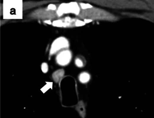
- Mediastinal komşu yapılara uzanım — damar lümeninde daralma/çevrelenme, özofagus duvarında düzensizlik veya trakeobronşiyal bası.
- Eşlik eden torasik metastatik odaklar — primer kitle, karşı akciğer nodülleri, kemik lezyonu ya da adrenal/hepatik odaklar evreleme bağlamını güçlendirir.
- Mediastinal düğümleri IASLC lenf düğümü istasyon haritasına göre adlandırın; istasyon bilgisi evreleme için, kısa aks ölçümü ise BT değerlendirmesi için kullanılabilir.
Ölçütler ve sınıflamalar
- Mediastinal lenf nodunda tarihsel kısa eksen anormallik eşiği (metastazı tek başına göstermez)
- ≥10 mm (tek başına metastaz tanısı koydurmaz)
- Akciğer kanseri TNM-9, M kategorisi
- M1a: ayrı kontralateral akciğer tümör nodülü veya plevral/perikardiyal metastaz (nodül ya da malign efüzyon); M1b: tek ekstratorasik metastaz veya nonregional metastatik lenf düğümü; M1c1: tek organ sisteminde çoklu ekstratorasik metastaz; M1c2: birden çok organ sisteminde çoklu ekstratorasik metastaz. Bu sınıflama akciğer kanserine özgüdür.
Normalde
Normal mediastende görülebilen lenf düğümlerinin kısa aksı istasyona göre değişebilir; benign internal mammary düğümler oval veya lobüler, yağlı hiluslu ve sınırlı konturlu olabilir; plevra ince ve düzgün bir yumuşak doku yapısıdır. Şüpheli düğümün kısa aksını ölçüp aynı zincirdeki karşı taraf düğümleriyle kıyaslayın; plevral/perikardiyal yüzeylerde nodüler kalınlaşma ve yeni sıvı olup olmadığını ayrıca gözden geçirin.
Normal BT ile kıyasla
Aynı bölgenin, kaynak veri setinde patoloji içermediği belirtilen bir BT incelemesini kesit kesit kaydırın; organ sınırlarını açarak anatomiyi hasta görüntüsüyle karşılaştırın. Küçük rastlantısal bulgular tümüyle dışlanmamıştır.
Kaynak: TotalSegmentator v2.01 veri seti (Wasserthal ve ark., Radiology: Artificial Intelligence 2023), CC BY 4.0, vaka s1278; organ sınırları veri setinin otomatik segmentasyonundan, birleştirilerek sadeleştirildi. Görüntüler 2,5 mm kesit aralığına yeniden örneklendi.
Ayırıcı tanı
- Reaktif mediastinal lenfadenopati
- genellikle eşlik eden enfeksiyon/ödem bağlamında, daha homojen düğümler ve zaman içinde gerileme gösterebilir.
- Lenfoma
- çok sayıda istasyonda hacimli, birleşen düğümler yapabilir; damarları çevrelese de lümeni koruyabilmesi metastatik invazyondan ayrımda yardımcıdır.
- Sarkoidoz
- simetrik bilateral hiler ve sağ paratrakeal düğümler, bazen yumurta kabuğu tarzı kalsifikasyon ile birlikte olabilir.
- Fibrozan mediastinit mediasteni diffüz veya lokal olarak infiltre edebilir ve mediastinal yapılarda daralmaya yol açabilir; kalsifikasyon bazı olgularda görülür.
- Plevral mezotelyoma
- yaygın çevresel, fissüral ve mediastinal plevra kalınlaşması yapabilir; BT tek başına primer plevral tümör ile metastatik plevrayı her zaman ayıramaz.
Tuzaklar
- 10 mm kısa aks eşiğini metastaz tanısı gibi kullanmayın; morfoloji, bilinen primer, önceki incelemeler ve gerektiğinde biyopsiyle birlikte yorumlayın.
- Kontrastlı BT'de nodal nekroz enfeksiyon ve bazı lenfomalarda da görülebilir; primer tümör öyküsü ve dağılım olmadan histolojiyi kesinleştirmez.
- Kanser hastasındaki her plevral veya perikardiyal sıvı malign değildir; BT'de nodül yoksa klinik ve sıvı incelemesi evreleme yorumunu değiştirebilir.
- Primer kitleye bitişik tümör ile büyümüş düğüm tek yumuşak doku kitlesi gibi görünebilir; lenf nodu istasyonlarını ve damar/bronş sınırlarını ayrı ayrı izleyin.
- BT’de düğüm boyutu ve morfolojisi nodal metastazı kesinleştirmez; evreleme gerektiğinde PET/BT ve EBUS/EUS eşliğinde örnekleme ile doku doğrulaması değerlendirilir.
Kendini dene
Akciğer karsinomu olan hastada santral nekrozlu subkarinal düğüm ve kontrastlanan plevral nodüller görülüyor. Plevral yayılımın TNM-9 M kategorisi nedir?
Cevabı göster
M1a. Plevral metastatik nodüller akciğer kanserinde M1a grubundadır. M1b tek ekstratorasik metastazı, M1c1 tek organ sisteminde çoklu ekstratorasik metastazı, M1c2 birden çok organ sistemindeki çoklu ekstratorasik metastazı tanımlar.
BT'de simetrik hiler ve mediastinal lenfadenopatiyle birlikte perilenfatik dağılımlı akciğer mikronodülleri hangi hastalık için karakteristik bir örüntüdür?
Cevabı göster
Sarkoidoz. Simetrik hiler ve mediastinal lenfadenopatiyle birlikte perilenfatik dağılımlı akciğer mikronodülleri sarkoidoz için karakteristiktir; bu örüntü tek başına kesin tanı koydurmaz.
Kaynaklar
Bu sayfadaki 40 cümle ve 2 quiz sorusu aşağıdaki kaynaklardan alıntılarla otomatik olarak karşılaştırıldı; 5 cümle bu karşılaştırmada düzeltildi (2026-10-09).
- Tumors of the pulmonary artery and veins.pubmed.ncbi.nlm.nih.gov
- Staging lymph node metastases from lung cancer in the mediastinum.pubmed.ncbi.nlm.nih.gov
- Methods for staging non-small cell lung cancer: Diagnosis and management of lung cancer, 3rd ed: American College of Chest Physicians evidence-based clinical practice guidelines.pubmed.ncbi.nlm.nih.gov
- Clinical utility of F-18 FDG PET-CT in the initial evaluation of lung cancer.pubmed.ncbi.nlm.nih.gov
- Hepatocellular carcinoma with thoracic metastases presenting as hemothorax: A case report and literature review.pubmed.ncbi.nlm.nih.gov
- Mediastinal recurrence after curative resection of colon cancer: A case report and review of literature.pubmed.ncbi.nlm.nih.gov
- A vocal cord palsy caused by the uterine cancer metastatic tumor in the mediastinum revealed in a patient with a thyroid lesion: a case report and review of the literature.pubmed.ncbi.nlm.nih.gov
- CT-guided trans-sternal iodine-125 seeds implantation for masses in the anterior or middle mediastinum.pubmed.ncbi.nlm.nih.gov
- The Value of a Systematic Protocol Using Endobronchial Ultrasound and Endoscopic Ultrasound in Staging of Lung Cancer for Patients with Imaging iN0-N1 Disease.pubmed.ncbi.nlm.nih.gov
- Detection of Mediastinal Lymph Node Metastases Using Indocyanine Green (ICG) Fluorescence Imaging in an Orthotopic Implantation Model.pubmed.ncbi.nlm.nih.gov
- Stereotactic body radiotherapy with helical TomoTherapy for medically inoperable early stage primary and second-primary non-small-cell lung neoplasm: 1-year outcome and toxicity analysis.pubmed.ncbi.nlm.nih.gov
- Hepatoid adenocarcinoma of the lung: Presenting mediastinal metastasis without transfer to the liver.pubmed.ncbi.nlm.nih.gov
- Sorafenib Improves Survival in Metastatic Hepatocellular Carcinoma: A Case Report.pubmed.ncbi.nlm.nih.gov
- Rare Case of Sellar and Suprasellar Metastasis from Ewing's Sarcoma of Tibia.pubmed.ncbi.nlm.nih.gov
- [Mediastinal Staging].pubmed.ncbi.nlm.nih.gov
- Pancreatic liposarcoma: a case report.pubmed.ncbi.nlm.nih.gov
- [Not Available].pubmed.ncbi.nlm.nih.gov
- [Contrast-enhanced multislice CT features and predominant anatomic distribution of mediastinal malignant lymphoma].pubmed.ncbi.nlm.nih.gov
- Whole-body magnetic resonance imaging in paediatric Hodgkin lymphoma - evaluation of quantitative magnetic resonance metrics for nodal staging.pubmed.ncbi.nlm.nih.gov
- Value of Assessing Peripheral Vascularization with Micro-Flow Imaging, Resistive Index and Absent Hilum Sign as Predictor for Malignancy in Lymph Nodes in Head and Neck Squamous Cell Carcinoma.pubmed.ncbi.nlm.nih.gov
- Lymph Node Assessment in Prostate Cancer: Evaluation of Iodine Quantification With Spectral Detector CT in Correlation to PSMA PET/CT.pubmed.ncbi.nlm.nih.gov
- Classification and staging of thymoma.pubmed.ncbi.nlm.nih.gov
- Present indications of surgical exploration of the mediastinum.pubmed.ncbi.nlm.nih.gov
- Using the Endoscope for Endobronchial Ultrasound in the Esophagus.pubmed.ncbi.nlm.nih.gov
- Simultaneous occurrence of Hodgkin's disease and tubercular lymphadenitis in the same cervical lymph node: a rare presentation.pubmed.ncbi.nlm.nih.gov
- Correlations between computed tomography and positron emission tomography/computed tomography findings and pathology in 6 cases of pulmonary epithelioid angiosarcoma.pubmed.ncbi.nlm.nih.gov
- Computed tomography characterization of neuroendocrine tumors of the thymus can aid identification and treatment.pubmed.ncbi.nlm.nih.gov
- Prediction of lymph node metastasis using the combined criteria of helical CT and mRNA expression profiling for non-small cell lung cancer.pubmed.ncbi.nlm.nih.gov
- Predicting malignancy in mediastinal lymph nodes by endobronchial ultrasound: a new ultrasound scoring system.pubmed.ncbi.nlm.nih.gov
- Ultrasound of superficial lymph nodes.pubmed.ncbi.nlm.nih.gov
- Cervical lymph nodes.pubmed.ncbi.nlm.nih.gov
- Imaging and Management of Internal Mammary Lymph Nodes.pubmed.ncbi.nlm.nih.gov
- Imaging of breast cancer-related changes after nonsurgical therapy.pubmed.ncbi.nlm.nih.gov
- Choroidal Metastasis as the Initial Presentation of Lung Cancer: A Case Report.pubmed.ncbi.nlm.nih.gov
- CT differentiation of diffuse malignant peritoneal mesothelioma and peritoneal carcinomatosis.pubmed.ncbi.nlm.nih.gov
- Pericardial Fat and Primary Tumor Radiomics for Predicting Occult N2 Disease and Survival in Clinical Stage I Non-Small Cell Lung Cancer: Multicenter Study and Biologic Correlation.pubmed.ncbi.nlm.nih.gov
- A PET-CT score for discriminating malignant from benign pleural effusions.pubmed.ncbi.nlm.nih.gov
- Good response of malignant pleural effusion from carcinoma of unknown primary site to the anti-tuberculosis therapy: a case report.pubmed.ncbi.nlm.nih.gov
- Minilaparoscopy-Assisted Modified Neck Dissection Through Bilateral Breast Approach.pubmed.ncbi.nlm.nih.gov
- Consensus definition of stage III non-small-cell lung cancer technical resectability to standardise inclusion criteria for clinical trials: a multisocietal EORTC-Lung Cancer Group collaboration.pubmed.ncbi.nlm.nih.gov
- Preoperative intrathoracic lymph node staging in patients with non-small-cell lung cancer: accuracy of integrated positron emission tomography and computed tomography.pubmed.ncbi.nlm.nih.gov
- Peritoneal carcinomatosis secondary to metastatic lung cancer complicated with acute suppurative appendicitis: A case report and literature review.pubmed.ncbi.nlm.nih.gov
- Onsite cytopathology evaluation and ancillary studies beneficial in EUS-FNA of pancreatic, mediastinal, intra-abdominal, and submucosal lesions.pubmed.ncbi.nlm.nih.gov
- Primary orbital ganglioneuroblastoma: A case report.pubmed.ncbi.nlm.nih.gov
- A 21-Year-Old Immune-Competent Man With Recurrent Cough.pubmed.ncbi.nlm.nih.gov
- Sarcoidosis or cancer? That is the question.pubmed.ncbi.nlm.nih.gov
- Pleural Mesothelioma: Ninth Edition of the TNM Staging Classification.pubmed.ncbi.nlm.nih.gov
- The International Association for the Study of Lung Cancer Staging Project for Lung Cancer: Proposals for the Revision of the M Descriptors in the Forthcoming Ninth Edition of the TNM Classification for Lung Cancer.pubmed.ncbi.nlm.nih.gov
- The International Association for the Study of Lung Cancer Thymic Epithelial Tumors Staging Project: Proposal for a Stage Classification for the Forthcoming (Ninth) Edition of the TNM Classification of Malignant Tumors.pubmed.ncbi.nlm.nih.gov
- The International Association for the Study of Lung Cancer Lung Cancer Staging Project: Proposals for Revision of the TNM Stage Groups in the Forthcoming (Ninth) Edition of the TNM Classification for Lung Cancer.pubmed.ncbi.nlm.nih.gov
- The Proposed Ninth Edition TNM Classification of Lung Cancer.pubmed.ncbi.nlm.nih.gov
- The International Association for the Study of Lung Cancer (IASLC) Staging Project for Lung Cancer: Recommendation to Introduce Spread Through Air Spaces as a Histologic Descriptor in the Ninth Edition of the TNM Classification of Lung Cancer. Analysis of 4061 Pathologic Stage I NSCLC.pubmed.ncbi.nlm.nih.gov
- ¹¹C-acetate PET/CT imaging: physiologic uptake, variants, and pitfalls.pubmed.ncbi.nlm.nih.gov
- Imaging Evaluation of Mediastinal and Hilar Lymphadenopathy: Approach, Classification, and Differential Diagnosis.pubmed.ncbi.nlm.nih.gov
- Occupational hazard in an immunosuppressed patient.pubmed.ncbi.nlm.nih.gov
- Treatment of Primary Mediastinal B-Cell Lymphoma With R-CEOP (Rituximab, Cyclophosphamide, Etoposide, Vincristine, and Prednisone).pubmed.ncbi.nlm.nih.gov
- A case of a periaortic lymphoma presenting with the features of descending thoracic aorta dissection.pubmed.ncbi.nlm.nih.gov
- Case Report: Pediatric mediastinal actinomycosis mimicking lymphoma diagnosed by tissue metagenomic next-generation sequencing.pubmed.ncbi.nlm.nih.gov
- Computed tomography findings in fibrosing mediastinitis.pubmed.ncbi.nlm.nih.gov
- Learning from the pulmonary veins.pubmed.ncbi.nlm.nih.gov
- Ptosis and Miosis Associated with Fibrosing Mediastinitis.pubmed.ncbi.nlm.nih.gov
- Malignant mesothelioma.pubmed.ncbi.nlm.nih.gov
- Neoplastic and nonneoplastic conditions of serosal membrane origin: CT findings.pubmed.ncbi.nlm.nih.gov
- Diagnosis of malignant pericarditis: a single centre experience.pubmed.ncbi.nlm.nih.gov
- Pictorial Review of Pleural Disease: Multimodality Imaging and Differential Diagnosis.pubmed.ncbi.nlm.nih.gov
- PET/CT imaging of lung cancer.pubmed.ncbi.nlm.nih.gov
- Additional value of volumetric and texture analysis on FDG PET assessment in paediatric Hodgkin lymphoma: an Italian multicentric study protocol.pubmed.ncbi.nlm.nih.gov
- Diffuse aggressive histologies of non-hodgkin lymphoma: treatment and biology of limited disease.pubmed.ncbi.nlm.nih.gov
- Primary thyroid lymphoma: a case series.pubmed.ncbi.nlm.nih.gov
- F-18 FDG PET/CT findings of intrathoracic lymphadenopathy in EBUS-TBNA-proven anthracosis.pubmed.ncbi.nlm.nih.gov
- Normal radiological lymph node appearance in the thorax.pubmed.ncbi.nlm.nih.gov
- [A case of neurosarcoidosis with recurrent brainstem infarction, obstructive hydrocephalus and brainstem atrophy].pubmed.ncbi.nlm.nih.gov
- Gemstone Spectral CT Virtual Noncontrast Images and Iodine Maps for the Characterization of Thyroid Lesions and Distinguishing Thyroid Papillary Carcinoma from Nodular Goiter.pubmed.ncbi.nlm.nih.gov
- Early diagnosis of miliary tuberculosis in a hemodialysis patient by combining two interferon-γ-release assays: a case report.pubmed.ncbi.nlm.nih.gov
- CT Chest findings in IgG4-related disease.pubmed.ncbi.nlm.nih.gov
- Respiratory manifestations in amyloidosis.pubmed.ncbi.nlm.nih.gov
- Amyloidosis: Modern Cross-sectional Imaging.pubmed.ncbi.nlm.nih.gov
- The IASLC Lung Cancer Staging Project: data elements for the prospective project.pubmed.ncbi.nlm.nih.gov
- The new tumor, node, and metastasis staging system.pubmed.ncbi.nlm.nih.gov
- The IASLC Lung Cancer Staging Project: Proposals for the Revision of the M Descriptors in the Forthcoming Eighth Edition of the TNM Classification of Lung Cancer.pubmed.ncbi.nlm.nih.gov
- Staging Lung Cancer: Metastasis.pubmed.ncbi.nlm.nih.gov
- M1b Disease in the 8th Edition of TNM Staging of Lung Cancer: Pattern of Single Extrathoracic Metastasis and Clinical Outcome.pubmed.ncbi.nlm.nih.gov
- Clinical relevance of the M1b and M1c descriptors from the proposed TNM 8 classification of lung cancer.pubmed.ncbi.nlm.nih.gov
- Sarcoidosis of the lacrimal gland: the prominence of the differential diagnosis.pubmed.ncbi.nlm.nih.gov
- Pulmonary Sarcoidosis: Imaging and Diagnostic Limitations.pubmed.ncbi.nlm.nih.gov
- Radiologic Features of World Trade Center-related Sarcoidosis in Exposed NYC Fire Department Rescue Workers.pubmed.ncbi.nlm.nih.gov
- Sarcoidosis: A Diagnosis of Exclusion.pubmed.ncbi.nlm.nih.gov
- Sarcoidosis from Head to Toe: What the Radiologist Needs to Know.pubmed.ncbi.nlm.nih.gov
- Clinical presentation of sarcoidosis and diagnostic work-up.pubmed.ncbi.nlm.nih.gov
- Necrotic mediastinal lymphadenopathy: Tuberculosis or Sarcoidosis, a diagnostic conundrum - A case report and review of the literature.pubmed.ncbi.nlm.nih.gov
- Evaluation of the diagnostic utility of endobronchial ultrasound-guided transbronchial needle aspiration for metastatic mediastinal tumors.pubmed.ncbi.nlm.nih.gov
- Pleuroparenchymal sarcoidosis: A rare manifestation mimicking metastatic lung cancer.pubmed.ncbi.nlm.nih.gov
- Giant Intrapulmonary Solitary Fibrous Tumour With a Possible Liver Metastasis: A Rare Case.pubmed.ncbi.nlm.nih.gov
- Advanced cystic lung cancer complicated with pulmonary lymphangitic carcinomatosis: a case report.pubmed.ncbi.nlm.nih.gov
- Nodal dissection in thymic surgery: a narrative review of the evolution of practice and evidence in the era of the 9th edition TNM classification (2000-2025).pubmed.ncbi.nlm.nih.gov
- Impact of structured reporting on interpretability of PSMA PET/CT in prostate cancer staging-towards standardised clinical implementation.pubmed.ncbi.nlm.nih.gov
- The first CT-based classification system for pulmonary actinomycosis: correlating imaging patterns with therapeutic strategies and prognosis in a highly misdiagnosed disease.pubmed.ncbi.nlm.nih.gov
- Neoadjuvant chemotherapy enabling resection of stage IVA thymoma with myasthenia gravis and systemic lupus erythematosus: a case report.pubmed.ncbi.nlm.nih.gov
- The extent of lymph node dissection in early-stage non-small cell lung cancer: current controversies, evidence gaps, and future directions in the immunotherapy era-a narrative review.pubmed.ncbi.nlm.nih.gov
- Predictive modeling of peripheral blood EGFR positivity in patients with advanced primary non-small cell lung cancer.pubmed.ncbi.nlm.nih.gov
- Analysis of MRI features correlated with TNM staging in thymic epithelial tumors.pubmed.ncbi.nlm.nih.gov
- Novel strategies in radiation therapy: high-dose radiotherapy for oligometastatic/oligo-progressive thymic tumors.pubmed.ncbi.nlm.nih.gov
- Congenital pulmonary airway malformation mimicking lung cancer in adults: a pitfall in oncologic diagnosis.pubmed.ncbi.nlm.nih.gov
- Preliminary investigation of the clinical value of thoracic ultrasound in pediatric pleuropulmonary blastoma: a comparative analysis with computed tomography and pathology.pubmed.ncbi.nlm.nih.gov
- Analysis of the clinicopathological characteristics of pulmonary sclerosing pneumocytoma (PSP).pubmed.ncbi.nlm.nih.gov
- Diagnostic accuracy of the Canada Lymph Node Score for predicting malignant mediastinal lymphadenopathy in patients undergoing endobronchial ultrasound-guided transbronchial needle aspiration in a tuberculosis-endemic setting: A cross-sectional study.pubmed.ncbi.nlm.nih.gov
- Development and validation of a nomogram integrating multi-dimensional clinical factors for predicting lung cancer-related mediastinal/hilar lymph node metastasis before endobronchial ultrasound-guided transbronchial needle aspiration.pubmed.ncbi.nlm.nih.gov
- Differentiating pulmonary sarcoidosis from mediastinal lymph node metastasis via conventional computed tomography features and quantitative imaging parameters: a retrospective study.pubmed.ncbi.nlm.nih.gov
- Beyond the lung: recognizing incidental breast lesions on chest computed tomography.pubmed.ncbi.nlm.nih.gov
- CT imaging features of pulmonary SMARCA4-deficient undifferentiated carcinoma: a retrospective case series.pubmed.ncbi.nlm.nih.gov
Eğitim amaçlıdır; hasta başında tanı ve tedavi kararının yerine geçmez, tıbbi tavsiye değildir. Metinler yapay zekâ desteğiyle yazıldı ve otomatik bir adımda kaynak alıntılarıyla karşılaştırıldı; bu bir tıbbi doğrulama değildir ve içerik uzman radyolog incelemesinden geçmedi. Klinik kararlarda güncel kılavuzları, kurum protokollerini ve radyoloji raporunu esas alın.