Özet
Primer özofagus lenfoması nadirdir; bildirilen bir olguda BT'de çok segmentli duvar kalınlaşması ve trakeal bası izlenmiştir. BT, özofagus duvarındaki çok segmentli kalınlaşmayı ve trakeal basıyı gösterebilir; lenfoma tanısı histopatolojik ve immünohistokimyasal incelemeyle konur. Özofagus lenfomasının görüntüleme bulguları özgül değildir; karsinom, özofajit ve özofagus tüberkülozu ile ayırıcı tanı gerekir. Özofagus lenfoması karsinomla karışabileceğinden, patolojik doğrulama yanlış tanı riskini azaltmak için önemlidir.
Okuma sırası
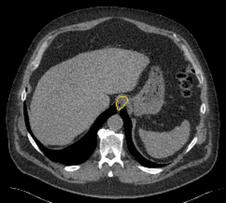
- orta torasik özofagus duvarı. Karina altından başlayıp özofagus duvarının çevresini izle; yumuşak dokunun tek odak mı yoksa uzun segmentli çevresel bir tabaka mı olduğunu belirle.
- özofagus-subkarinal arayüzü. Duvar kitlesinin komşu subkarinal nodal dokuya kesintisiz bağlanıp bağlanmadığını ardışık kesitlerde kontrol et.
- özofagus lümeni ve subkarinal istasyon. Özofagus çevresindeki lümen çapını ve ayrı kalan lenf düğümlerini göster; açık lümen infiltratif hastalığı dışlamaz.
- çok istasyonlu lenfatik zincirler ve parankimal organlar. Son olarak boyun altı, hiler ve üst abdominal nodal istasyonları ve dalak-karaciğer parankimini aynı dağılım haritasına ekle.
Faz ve pencere
- Portal tanısal
- Toraks BT'de özofagus yumuşak doku kitlesi, kontrast sonrası heterojen kontrastlanması, komşu yapılarda bası veya yer değiştirme ve mediastinal ya da perigastrik lenf düğümleri değerlendirilebilir.
Önerilen pencereler G genişlik, M merkez; değerler Hounsfield birimi (HU)
- MediastenG 350 / M +50
- BatınG 400 / M +50
- Yumuşak dokuG 400 / M +40
BT bulguları
- Bir veya birden fazla segmentte çevresel, uzunlamasına ilerleyen duvar kalınlaşması; lümen kısmen daralabilir veya duvar kitlesine rağmen açık kalabilir.
- Duvar içinde ya da lümene doğru kabaran düşük yoğunluklu/heterojen yumuşak doku kitlesi.
- Özofagus duvarındaki doku ile bitişik mediastinal lenf düğümleri arasında sınırın silinmesi veya düğümden duvara doğrudan uzanım.
- Paratrakeal, subkarinal, hiler, üst abdominal veya retroperitoneal zincirlerde birden çok bölgede lenfadenopati.
- Özofagus çevresinde ekstralüminal yumuşak doku; hava yolu basısı, fistül veya nadiren lümene uzanım.
- Dalak, karaciğer ve diğer lenfatik istasyonlarda eşzamanlı hastalık şüphesi araştırılır.
Normalde
BT, özofagus duvarındaki çok segmentli kalınlaşmayı gösterebilir; lenfoma tanısı görüntüyle değil, histopatolojik ve immünohistokimyasal incelemeyle konur.
Normal BT ile kıyasla
Aynı bölgenin, kaynak veri setinde patoloji içermediği belirtilen bir BT incelemesini kesit kesit kaydırın; organ sınırlarını açarak anatomiyi hasta görüntüsüyle karşılaştırın. Küçük rastlantısal bulgular tümüyle dışlanmamıştır.
Kaynak: TotalSegmentator v2.01 veri seti (Wasserthal ve ark., Radiology: Artificial Intelligence 2023), CC BY 4.0, vaka s1278; organ sınırları veri setinin otomatik segmentasyonundan, birleştirilerek sadeleştirildi. Görüntüler 2,5 mm kesit aralığına yeniden örneklendi.
Ayırıcı tanı
- Yassı hücreli karsinom
- çoğu kez fokal/asimetrik kitle ve belirgin segmental darlık.
- Adenokarsinom
- distal özofagus ya da bileşke merkezli kitle ve üst abdomen bölgesel yayılımı.
- GIST veya leiomyom
- sınırlı submukozal kitle, genellikle yaygın nodal zincir tutulumu olmaksızın.
- Mediastinal adenopati
- özofagusa dıştan bası yapabilir veya özofagus duvarına uzanabilir; bu bulgular tek başına metastatik kökeni göstermez.
- Mediastinal lenfadenopati özofagus duvarına uzanabilir; BT'de subkarinal yumuşak doku kitlesi özofagustan ayrılamayabilir.
Tuzaklar
- Çevresel duvar kalınlaşması lenfomaya özgü değildir; aynı görüntü karsinom veya özofajitte görülebilir, histolojiyi BT görünümünden çıkarsama.
- Duvarı tutan birleşik lenf düğümü primer özofagus kitlesi izlenimi verir; mediastinal istasyonları kraniokaudal sırayla inceleyip olası başlangıç noktasını bul.
- Lümen açık kaldığı için geniş infiltratif hastalık hafif darlıklı görünebilir; yalnız obstrüksiyon derecesine bakarak yaygınlığı küçümseme.
- Özofagus lenfomasının görüntüleme bulguları özgül değildir; karsinom, özofajit ve özofagus tüberkülozu ile örtüşebilir, kesin tanı için histopatolojik ve immünohistokimyasal inceleme gerekir.
Kendini dene
BT'de özofagus duvarında çok segmentli kalınlaşma saptanıyor. Kesin lenfoma tanısı için hangi inceleme gerekir?
Cevabı göster
Histopatolojik ve immünohistokimyasal inceleme. BT'de çok segmentli duvar kalınlaşması görülebilir; lenfoma tanısı histopatolojik ve immünohistokimyasal incelemeyle konur.
Duvar kitlesiyle bitişik subkarinal yumuşak doku arasındaki sınır kaybolmuş. Hangi yorum en yerindedir?
Cevabı göster
Nodal hastalığın duvara uzanımı olabilir. Lenf düğümü ve duvar lezyonu arasındaki devamlılık sekonder yayılım olasılığını doğurur. BT bu görüntüyle histolojik kökeni kanıtlayamaz; reaktif ve neoplastik süreçler görüntü bulgularında örtüşebilir, kesin tanı histopatolojik doğrulama gerektirir.
Kaynaklar
Bu sayfadaki 33 cümle ve 2 quiz sorusu aşağıdaki kaynaklardan alıntılarla otomatik olarak karşılaştırıldı; 11 cümle bu karşılaştırmada düzeltildi (2026-10-09).
- Computed tomography and barium swallow findings of esophageal squamous cell carcinoma with suspected bronchoesophageal fistula in a patient with chronic graft-versus-host disease: A case reportpmc.ncbi.nlm.nih.gov
- Simultaneous idiopathic mediastinal and retroperitoneal fibrosis presenting with acute respiratory failure: A case report and literature reviewpmc.ncbi.nlm.nih.gov
- Hepatosplenic sarcoidosispmc.ncbi.nlm.nih.gov
- Anterior cervical abscess caused by migration of an ingested fish bone in a child: A case reportpmc.ncbi.nlm.nih.gov
- Case Report: Spectral CT characterization of giant esophageal schwannoma with reactive lymphoid hyperplasia mimicking lymphoproliferative diseasepmc.ncbi.nlm.nih.gov
- Imaging of oligodendroglioma.pubmed.ncbi.nlm.nih.gov
- Imaging of the spleen: what the clinician needs to know.pubmed.ncbi.nlm.nih.gov
- Axillary lymph node metastasis in second oropharyngeal cancer.pubmed.ncbi.nlm.nih.gov
- Surgery for adrenocortical carcinoma: When and how?pubmed.ncbi.nlm.nih.gov
- Pathways of lymphatic spread in gynecologic malignancies.pubmed.ncbi.nlm.nih.gov
- MRI for response assessment in metastatic bone disease.pubmed.ncbi.nlm.nih.gov
- [ 124 I]CLR1404 PET/CT in High-Grade Primary and Metastatic Brain Tumors.pubmed.ncbi.nlm.nih.gov
- [Sentinel node in melanoma and breast cancer. Current considerations].pubmed.ncbi.nlm.nih.gov
- Sentinel node approach in prostate cancer.pubmed.ncbi.nlm.nih.gov
- A-kinase anchoring protein 2 is required for calcitonin-mediated invasion of cancer cells.pubmed.ncbi.nlm.nih.gov
- PET imaging of recurrent and metastatic prostate cancer with novel tracers.pubmed.ncbi.nlm.nih.gov
- A spontaneously metastatic model of bladder cancer: imaging characterization.pubmed.ncbi.nlm.nih.gov
- Understanding the effect of mechanical forces on ovarian cancer progression.pubmed.ncbi.nlm.nih.gov
- Impact of high glucose on metastasis of colon cancer cells.pubmed.ncbi.nlm.nih.gov
- Primary gastrointestinal malignancies in childhood and adolescence--an Asian perspective.pubmed.ncbi.nlm.nih.gov
- A BAFfling liver aspirate: Metastatic high grade SMARCA4 deficient undifferentiated gastroesophageal junction carcinoma masquerading as a hematolymphoid malignancy.pubmed.ncbi.nlm.nih.gov
- Robotic-Assisted Thoracoscopic Circumferential Resection of Congenital Esophageal Stenosis in Under 10 kg Patient.pubmed.ncbi.nlm.nih.gov
- Surgical treatment of a circumferential oesophagogastric mass associated with a peritoneopericardial diaphragmatic hernia in a Maine Coon cat.pubmed.ncbi.nlm.nih.gov
- Esophageal leiomyoma within an epinephric diverticulum.pubmed.ncbi.nlm.nih.gov
- Rare Giant Pedunculated Liposarcoma of the Hypopharynx: Case Report and Review of Literature.pubmed.ncbi.nlm.nih.gov
- Esophageal cancer in an adult with congenital esophageal stenosis: a case report.pubmed.ncbi.nlm.nih.gov
- Esophageal Carcinoma Cuniculatum Associated with Non-Necrotizing Granulomatous Inflammation and Lymphadenopathy: Clinicopathologic Features and Diagnostic Challenges.pubmed.ncbi.nlm.nih.gov
- Esophageal cancer: evaluation with triple-phase dynamic CT--initial experience.pubmed.ncbi.nlm.nih.gov
- Diagnosis and management of Barrett esophagus: European Society of Gastrointestinal Endoscopy (ESGE) Guideline.pubmed.ncbi.nlm.nih.gov
- Surgical techniques.pubmed.ncbi.nlm.nih.gov
- Western strategy for EGJ carcinoma.pubmed.ncbi.nlm.nih.gov
- Immune Checkpoint Inhibitor-Related Aortitis Treated with Nivolumab for Esophageal Squamous Cell Carcinoma.pubmed.ncbi.nlm.nih.gov
- Imaging and clinicopathologic features of esophageal gastrointestinal stromal tumors.pubmed.ncbi.nlm.nih.gov
- [Oesophageal GIST at the left tracheobronchial angle: resection with right-sided VATS].pubmed.ncbi.nlm.nih.gov
- Sarcoidosis with esophageal involvement: an uncommon clinical enigma.pubmed.ncbi.nlm.nih.gov
- Corrosive-Induced Carcinoma of Esophagus: Esophagographic and CT Findings.pubmed.ncbi.nlm.nih.gov
- Esophageal tuberculosis induced dysphagia: a case report.pubmed.ncbi.nlm.nih.gov
- Paranasal sinus cancer.pubmed.ncbi.nlm.nih.gov
- CT imaging features of skeletal muscle metastasis.pubmed.ncbi.nlm.nih.gov
- High-dose hyperfractionated simultaneous integrated boost radiotherapy versus standard-dose radiotherapy for limited-stage small-cell lung cancer in China: a multicentre, open-label, randomised, phase 3 trial.pubmed.ncbi.nlm.nih.gov
- Remediastinoscopy.pubmed.ncbi.nlm.nih.gov
- Primary lymphomas in the gastrointestinal tract.pubmed.ncbi.nlm.nih.gov
- Suspected oesophageal cancer in a 33 year old lady.pubmed.ncbi.nlm.nih.gov
- Case Report: Spectral CT characterization of giant esophageal schwannoma with reactive lymphoid hyperplasia mimicking lymphoproliferative disease.pubmed.ncbi.nlm.nih.gov
- Localised malignant mesothelioma presenting as an esophageal mass: An unusual manifestation.pubmed.ncbi.nlm.nih.gov
- Case Report: a hypermetabolic mediastinal mass (SUVmax 16.7) mimicking malignancy with spontaneous resolution: an inflammatory myofibroblastic proliferation and diagnostic pitfall.pubmed.ncbi.nlm.nih.gov
- Incidentally Detected Mediastinal Mass by Transesophageal Echocardiography in a Patient With Severe Mitral Stenosis.pubmed.ncbi.nlm.nih.gov
- Medullary Renal Cell Carcinoma: A Case Study.pubmed.ncbi.nlm.nih.gov
- Case Report: Locally invasive thyroid metastases from renal cell carcinoma: surgery after neoadjuvant therapy.pubmed.ncbi.nlm.nih.gov
- Probable cavernous sinus-adjacent meningeal metastasis from esophageal squamous cell carcinoma: A case report.pubmed.ncbi.nlm.nih.gov
- Unprecedented non-luminal esophageal adenocarcinoma invading the spine.pubmed.ncbi.nlm.nih.gov
- Solitary esophageal metastasis ten years after curative resection of stage I rectal adenocarcinoma: A case report.pubmed.ncbi.nlm.nih.gov
- Esophageal adenosquamous carcinoma with mucinous carcinoma-predominant lymph node metastases: a case report.pubmed.ncbi.nlm.nih.gov
- Multidisciplinary Treatment for Advanced Pancreatic Adenocarcinoma Arising from Esophageal Heterotopic Pancreas in a Young Adult: A Case Report.pubmed.ncbi.nlm.nih.gov
- Thoracoscopic Reconstruction with the Non-Flap Tunnel Technique for Esophagogastric Junction Cancer Requiring High Intramediastinal Anastomosis: A Case Report.pubmed.ncbi.nlm.nih.gov
- Lymphoma of the cervix invading the uterine body with distant lymphadenopathy: MRI and FDG-PET/CT findings.pubmed.ncbi.nlm.nih.gov
- Superior Vena Cava Syndrome: Etiologies, Manifestations, and Treatments.pubmed.ncbi.nlm.nih.gov
- Rigid bronchoscopy and silicone stents in the management of central airway obstruction.pubmed.ncbi.nlm.nih.gov
- C-Reactive Protein: Pathophysiology, Diagnosis, False Test Results and a Novel Diagnostic Algorithm for Clinicians.pubmed.ncbi.nlm.nih.gov
- Third ventricle non-Hodgkin lymphoma: An unusual presentation of primary central nervous system lymphoma.pubmed.ncbi.nlm.nih.gov
- Synchronous medullary carcinoma of the pancreas and non-ampullary duodenal adenocarcinoma.pubmed.ncbi.nlm.nih.gov
- Pulmonary plasma cell disorders: histopathology, diagnosis, and clinical perspectives.pubmed.ncbi.nlm.nih.gov
- Unusual manifestations of extra-pulmonary tuberculosis: a pictorial essay.pubmed.ncbi.nlm.nih.gov
- Beyond squamous cell carcinoma: MRI appearance of uncommon anal neoplasms and mimickers.pubmed.ncbi.nlm.nih.gov
- Performance of Large Language Models (ChatGPT and Gemini Advanced) in Gastrointestinal Pathology and Clinical Review of Applications in Gastroenterology.pubmed.ncbi.nlm.nih.gov
- Gastrointestinal Kaposi Sarcoma: Histopathological Features and Diagnostic Challenges - Insights from a Single Center.pubmed.ncbi.nlm.nih.gov
- The Predilection of Specific Diseases to Affect Different Sections of the Esophagus.pubmed.ncbi.nlm.nih.gov
- Dysphagia Secondary to Extrinsic Esophageal Compression From an Anterior Mediastinal Squamous Cell Carcinoma.pubmed.ncbi.nlm.nih.gov
- Retrospective single-center cytohistologic correlation study of fine-needle aspiration cytology for excised superficial enlarged lymph node lesions in different disease entities.pubmed.ncbi.nlm.nih.gov
- Chronic intestinal pseudo-obstruction secondary to intestinal leiomyositis in a ragdoll cat.pubmed.ncbi.nlm.nih.gov
- Stricturing Diseases of the Gastrointestinal Tract-Current Clinical Practice.pubmed.ncbi.nlm.nih.gov
- Robotic-assisted total gastrectomy for refractory hypoproteinemia in Menetrier's disease: a case report with operative video and literature review.pubmed.ncbi.nlm.nih.gov
- Rethinking Common Diagnoses: Idiopathic Multicentric Castleman Disease Presenting as TAFRO Syndrome: A Case Report.pubmed.ncbi.nlm.nih.gov
- When Unilateral Leg Swelling Is Not Deep Vein Thrombosis: A Case of Disseminated Kaposi Sarcoma Revealing Advanced Human Immunodeficiency Virus.pubmed.ncbi.nlm.nih.gov
- Diffuse Large B-cell Lymphoma of the Breast Presenting 40 Years After Breast-Conserving Therapy: A Case Report.pubmed.ncbi.nlm.nih.gov
- State-of-the-Art Mediastinal Staging in Non-Small-Cell Lung Cancer: Integration of Combined Endosonographic Techniques with Updated IASLC TNM 9th Classification.pubmed.ncbi.nlm.nih.gov
- Beyond the Nodes: Atypical Presentations of Non-Hodgkin's Lymphoma Across Organs.pubmed.ncbi.nlm.nih.gov
- Approach to mediastinal cytopathology: a diagnostic overview.pubmed.ncbi.nlm.nih.gov
- Beyond the Typical Atypical Imaging Features of Leptomeningeal Enhancement: A Case Series.pubmed.ncbi.nlm.nih.gov
- Optimizing Care for Veterans at Risk of Cancer From Camp Lejeune Water Exposure.pubmed.ncbi.nlm.nih.gov
- Primary esophageal marginal zone lymphoma with respiratory symptoms: a case report and review of the literature.pubmed.ncbi.nlm.nih.gov
- Case Report: Sinonasal plasmablastic lymphoma in a human immunodeficiency virus-negative elderly woman diagnosed by immunohistochemistry.pubmed.ncbi.nlm.nih.gov
- The hidden path of mycobacterium: a case series of rare and elusive manifestations of gastrointestinal tuberculosis.pubmed.ncbi.nlm.nih.gov
- A Case Report of an Unusual Cause of Abnormal Uterine Bleeding: Primary Cervical Lymphoma Misdiagnosed As Hormonal Dysfunction.pubmed.ncbi.nlm.nih.gov
- Diagnostic Imaging of Extrapulmonary Tuberculosis Across Organ Systems.pubmed.ncbi.nlm.nih.gov
- Hemoptysis in Cancer Patients.pubmed.ncbi.nlm.nih.gov
- Neoplastic pathology at the crossroads between neck imaging and cardiothoracic imaging.pubmed.ncbi.nlm.nih.gov
- A case report of SMARCA4-deficient undifferentiated gastric carcinoma diagnosed by endoscopic ultrasound-guided fine needle aspiration.pubmed.ncbi.nlm.nih.gov
- Invasive mucinous adenocarcinoma of the lung: integrating molecular landscape, imaging phenotypes, and translational therapeutic strategies.pubmed.ncbi.nlm.nih.gov
- Beyond Description: A Functional GIUS-Based Algorithm for Enteral Feeding Decisions in the ICU.pubmed.ncbi.nlm.nih.gov
- Gastrointestinal Ultrasound: Measurements and Normal Findings - What Do You Need to Know?pubmed.ncbi.nlm.nih.gov
- Computed Tomographic Findings of Liver Cirrhosis in Dogs: Comparison between Compensated and Decompensated Cirrhosis.pubmed.ncbi.nlm.nih.gov
- Boerhaave Syndrome Caused by Pancreatitis-Induced Gastric Outlet Obstruction: A Case Report.pubmed.ncbi.nlm.nih.gov
- Computed Tomographic Findings and Clinical Manifestations of Acute Severe Hypersensitivity to a Non-Ionic Iodinated Contrast Agent in a Castrated Male Dog: A Case Report.pubmed.ncbi.nlm.nih.gov
- Exploring the Role of Post-Viral Phenomenon in the Pathogenesis of Achalasia: Insights From a Clinical Case.pubmed.ncbi.nlm.nih.gov
Eğitim amaçlıdır; hasta başında tanı ve tedavi kararının yerine geçmez, tıbbi tavsiye değildir. Metinler yapay zekâ desteğiyle yazıldı ve otomatik bir adımda kaynak alıntılarıyla karşılaştırıldı; bu bir tıbbi doğrulama değildir ve içerik uzman radyolog incelemesinden geçmedi. Klinik kararlarda güncel kılavuzları, kurum protokollerini ve radyoloji raporunu esas alın.