
Özet
Özofagus varisleri portal hipertansiyon nedeniyle gelişen, özofagus duvarı içindeki genişlemiş venöz kollaterallerdir ve sirozlu hastada üst gastrointestinal kanamanın önemli nedenlerindendir. Portal venöz fazlı BT, lümen çevresinde ya da lümene kabaran ve portal venöz kanla benzer biçimde kontrastlanan kıvrımlı damarları gösterebilir; aynı incelemede gastrik ve diğer portosistemik kollateraller de haritalanabilir. BT varislerin varlığını ortaya koyabilir, ancak kanama riskinin ve endoskopik görünümün değerlendirilmesinde endoskopinin yerini almaz. Varislerin fark edilmemesi portal hipertansiyonun ve olası kanama kaynağının raporda atlanmasına neden olabilir.
Faz ve pencere
- Portal tanısal
- Portal venöz fazda özofagus duvarı içindeki ve paraözofageal yağdaki yuvarlak, tübüler veya serpijinöz kanallar portal ve mezenterik venlerle benzer düzeyde kontrastlanır; inceleme damar devamlılığını izlemeye elverişlidir.
Önerilen pencereler G genişlik, M merkez; değerler Hounsfield birimi (HU)
- MediastenG 350 / M +50
- BatınG 400 / M +50
- AnjiyoG 600 / M +150
BT bulguları
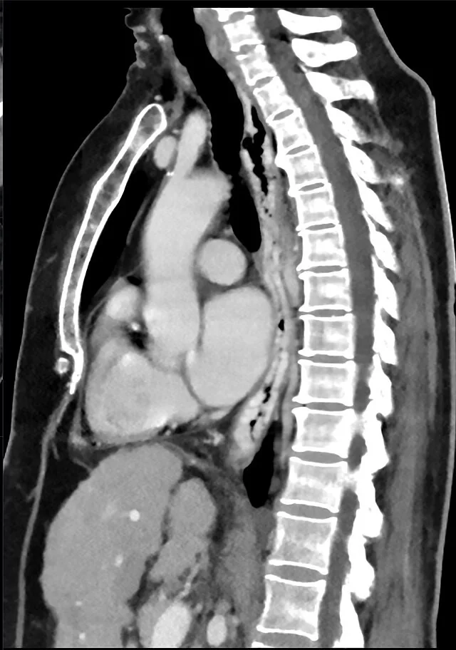
- Özofagus lümeni boyunca duvar içinde lümene kabaran yuvarlak veya kıvrımlı kontrastlanan venöz kanallar
- Özofagus dış çevresindeki paraözofageal kollaterallerin azygos, hemiazygos veya vertebral venöz pleksusa uzanan serpijinöz seyri
- Distal özofagus ve gastroözofageal bileşkede belirginleşen submukozal venöz yapılar
- Sol gastrik venin genişlemesi ve gastroözofageal bileşke çevresinde gastrik kollateraller
- Portal hipertansiyon eşlikçileri olarak splenomegali, asit, karaciğer kontur nodülaritesi veya portosistemik şantlar
Normalde
Aynı torasik düzeyde normal özofagus çevresinde portal venöz fazda lümen boyunca uzanan serpijinöz, kanla dolu kontrastlanan kanallar görülmez; özofagus duvarı düzgün yumuşak doku konturundadır. Kıyaslarken lümene kabaran damarları özofagus lümeninden ayır ve kesitler boyunca sol gastrik ven ya da azygos sistemiyle devamlılığını izle.
Normal BT ile kıyasla
Aynı bölgenin, kaynak veri setinde patoloji içermediği belirtilen bir BT incelemesini kesit kesit kaydırın; organ sınırlarını açarak anatomiyi hasta görüntüsüyle karşılaştırın. Küçük rastlantısal bulgular tümüyle dışlanmamıştır.
Kaynak: TotalSegmentator v2.01 veri seti (Wasserthal ve ark., Radiology: Artificial Intelligence 2023), CC BY 4.0, vaka s0975; organ sınırları veri setinin otomatik segmentasyonundan, birleştirilerek sadeleştirildi. Görüntüler 2,5 mm kesit aralığına yeniden örneklendi.
Ayırıcı tanı
- Özofagus intramural hematomu
- kontrastsızda yüksek atenüasyonlu duvar içi kitle ve lümen basısı yapar; varis gibi portal venöz kanla kontrastlanmaz.
- Özofagus leiomyomu
- düzgün sınırlı solid duvar kitlesi olarak lümeni iter; kesitler boyunca venöz ağa devam etmez.
Tuzaklar
- Paraözofageal varisler özofagus duvarındaki varislerden ayrıdır; her iki kompartımanı da incele ve damarları çok düzlemli görüntülerde izle.
- Tek bir kesitte damar kesiti lenf nodu ya da kitle gibi görünebilir; seri kesitlerde tübüler-serpijinöz devamlılık ve venöz faz kontrastlanması aranmalıdır.
- BT'de varis görülmesi endoskopik kanama riskini tek başına derecelendirmez; aktif kanama odağı yokluğu varis yokluğu anlamına gelmez.
- BT’de görülen varisler portal hipertansiyonun ve kollaterallerin anatomik değerlendirmesine katkı sağlar; varislerin endoskopik varlığını ve derecesini belirlemede üst endoskopi referans yöntemdir.
Kendini dene
Sirozlu hastanın portal venöz fazlı BT'sinde distal özofagus çevresinde portal venle aynı düzeyde kontrastlanan serpijinöz kanallar görülüyor. Tanı nedir?
Cevabı göster
Özofagus varisleri. Portal venöz fazda venöz kanla benzer kontrastlanan, lümen çevresinde kıvrımlı kanallar varisleri destekler. Özofagus varisleri BT'de düzgün sınırlı, yuvarlak, tübüler veya serpijinöz yapılar olarak görülür ve komşu portal ve mezenterik venlerle benzer kontrastlanır.
Özofagusun yanında görülen yuvarlak yumuşak doku odağı kesitler boyunca azygos venine bağlanıyor ve portal fazda kontrastlanıyor. En olası yapı nedir?
Cevabı göster
Paraözofageal varis. Tübüler devamlılık, azygos bağlantısı ve venöz kontrastlanma paraözofageal kollateral lehinedir. Paraözofageal varisler özofagusu çevreleyen çoklu venöz kollaterallerdir ve koroner veni azygos, hemiazygos venler ile vertebral pleksusa bağlar.
Kaynaklar
Bu sayfadaki 32 cümle ve 2 quiz sorusu aşağıdaki kaynaklardan alıntılarla otomatik olarak karşılaştırıldı; 2 cümle bu karşılaştırmada düzeltildi (2026-10-09).
- Portal hypertension: Imaging of portosystemic collateral pathways and associated image-guided therapypmc.ncbi.nlm.nih.gov
- Capsule Endoscopy for Portal Hypertensive Enteropathy.pubmed.ncbi.nlm.nih.gov
- Computed Tomography Images of Spontaneous Portosystemic Shunt in Liver Cirrhosis.pubmed.ncbi.nlm.nih.gov
- Elastography-based screening for esophageal varices in patients with advanced chronic liver disease.pubmed.ncbi.nlm.nih.gov
- Νon-invasive screening for esophageal varices in patients with liver cirrhosis.pubmed.ncbi.nlm.nih.gov
- Congenital hepatic fibrosis: case report and review of literature.pubmed.ncbi.nlm.nih.gov
- Invasive and noninvasive methods to diagnose portal hypertension and esophageal varices.pubmed.ncbi.nlm.nih.gov
- [Bacillus cereus septicemia and necrotizing fasciitis in a patient with liver cirrhosis: a case report].pubmed.ncbi.nlm.nih.gov
- Computed tomography in evaluating gastroesophageal varices in patients with portal hypertension: A meta-analysis.pubmed.ncbi.nlm.nih.gov
- Noninvasive assessment of portal hypertension in patients with cirrhosis.pubmed.ncbi.nlm.nih.gov
- Multidetector CT anatomy of drainage routes of gastric varices: a pictorial review.pubmed.ncbi.nlm.nih.gov
- Hepatic myelopathy with spastic paraparesis: report of two cases and review of the literature.pubmed.ncbi.nlm.nih.gov
- Case report and systematic literature review of a novel etiology of sinistral portal hypertension presenting with UGI bleeding: Left gastric artery pseudoaneurysm compressing the splenic vein treated by embolization of the pseudoaneurysm.pubmed.ncbi.nlm.nih.gov
- [A case of extrahepatic portal vein aneurysm with massive thrombosis].pubmed.ncbi.nlm.nih.gov
- Rectal arterio-portal fistula: an unusual cause of persistent bleeding per rectum following a proximal spleno-renal shunt.pubmed.ncbi.nlm.nih.gov
- Massive Esophagogastric Variceal Bleeding from Chronic Portal Vein Thrombosis in a Non-Cirrhotic Young Man: A Resource-Limited Endoscopic Management Dilemma.pubmed.ncbi.nlm.nih.gov
- Ectopic Duodenal Varices Secondary to Portal Hypertension Caused by Primary Hepatic Neuroendocrine Tumor.pubmed.ncbi.nlm.nih.gov
- Bleeding Stomal Varices and Vascular Interventional Radiology Management: A Case Report.pubmed.ncbi.nlm.nih.gov
- Narrative review of the current management of ectopic varices: an update.pubmed.ncbi.nlm.nih.gov
- Use of a recanalized umbilical vein to embolize bleeding varices: A case report.pubmed.ncbi.nlm.nih.gov
- Successful Endoscopic Sealing of Gastric Perforation With Tissue Adhesive Following Endoscopic Treatment for Gastric Fundal Variceal Bleeding: A Case Report.pubmed.ncbi.nlm.nih.gov
- The characteristics of liver nodules in biliary atresia native liver survivors.pubmed.ncbi.nlm.nih.gov
- A non-invasive nomogram for high-risk esophageal varices in cirrhosis without transient elastography: development and internal validation.pubmed.ncbi.nlm.nih.gov
- An unusual superior mesenteric arteriovenous malformation causing obscure gastrointestinal bleeding: a case report.pubmed.ncbi.nlm.nih.gov
- Umbilical vein recanalization with a diameter greater than 6.48mm may promote deterioration of liver reserve function: A retrospective cohort study.pubmed.ncbi.nlm.nih.gov
- Imaging features, diagnosis, and clinical management of hepatic involvement in hereditary hemorrhagic telangiectasia: a case series report and literature review.pubmed.ncbi.nlm.nih.gov
- Hiatal Hernia-Related Esophageal Varices Misdiagnosed as Cancer: Salvaged With Clipping.pubmed.ncbi.nlm.nih.gov
- Case Report: CT manifestations of acute portal vein thrombosis: cases report and literature review.pubmed.ncbi.nlm.nih.gov
- Cystic lesions and their mimics involving the intrahepatic bile ducts and peribiliary space: diagnosis, complications, and management.pubmed.ncbi.nlm.nih.gov
- Balloon-occluded Retrograde Transvenous Obliteration for Gastric Varices Draining into the Gastrocaval Shunt.pubmed.ncbi.nlm.nih.gov
- Understanding Portosystemic Collateral Anatomy.pubmed.ncbi.nlm.nih.gov
- Peroral Endoscopic Cardial Constriction with Band Ligation for Gastroesophageal Reflux Disease: A Prospective Pilot Study.pubmed.ncbi.nlm.nih.gov
- Upper gastrointestinal endoscopy: evolving classification tools.pubmed.ncbi.nlm.nih.gov
- Pathophysiological management of gastric varices: From hemodynamics to targeted therapies.pubmed.ncbi.nlm.nih.gov
- Masked polycythemia vera presenting as non-cirrhotic portal hypertension with isolated gastric varices: a case report.pubmed.ncbi.nlm.nih.gov
- Management of parastomal variceal bleeding in an adult with metastatic liver cancer: A case-based review.pubmed.ncbi.nlm.nih.gov
- Portal vein recanalization and transjugular intrahepatic portosystemic shunt in chronic portal vein thrombosis: a narrative review.pubmed.ncbi.nlm.nih.gov
- The Gastric Vein Variants: An Evidence-Based Systematic Review of Prevalence and Clinical Considerations.pubmed.ncbi.nlm.nih.gov
- CT Findings of Azygos Venous System: Congenital Variants and Acquired Structural Changes.pubmed.ncbi.nlm.nih.gov
- Endoscopic Ultrasonography In Diagnostic Endohepatology: The Hepatologist's Swiss Army Knife.pubmed.ncbi.nlm.nih.gov
- Stricturing Diseases of the Gastrointestinal Tract-Current Clinical Practice.pubmed.ncbi.nlm.nih.gov
- Preclinical modeling of Loeys-Dietz syndrome: insights into mechanisms and therapy.pubmed.ncbi.nlm.nih.gov
- Severe Rectal Syphilis in the Setting of Profound HIV Immunosuppression: A Case Report Highlighting ERG/CD38 Immunophenotyping and a Review of the Literature.pubmed.ncbi.nlm.nih.gov
- Inflammatory Aortopathies in Rheumatic Diseases: A State-of-the-Art Review.pubmed.ncbi.nlm.nih.gov
- Computed Tomography Imaging of Thoracic Aortic Surgery: Distinguishing Life-Saving Repairs from Life-Threatening Complications.pubmed.ncbi.nlm.nih.gov
- Radiologic Imaging of Traumatic Bowel and Mesenteric Injuries: A Comprehensive Up-to-Date Review.pubmed.ncbi.nlm.nih.gov
- Acute gastrointestinal hemorrhage as the sole initial manifestation of gastric leiomyosarcoma: a case report highlighting the discrepancy between preoperative assessment and final pathology.pubmed.ncbi.nlm.nih.gov
- Primary malignant melanoma of the esophagus: differentiation from esophageal squamous cell carcinoma and leiomyoma using dynamic contrast-enhanced CT findings.pubmed.ncbi.nlm.nih.gov
- Gastrointestinal Ultrasound: Measurements and Normal Findings - What Do You Need to Know?pubmed.ncbi.nlm.nih.gov
- The diagnostic value of endoscopic ultrasound for esophageal subepithelial lesions: A review.pubmed.ncbi.nlm.nih.gov
- Downhill Varices and Apical Lung Cancer Without Superior Vena Cava Syndrome.pubmed.ncbi.nlm.nih.gov
- Role of Endoscopic Ultrasound in Liver Disease: Where Do We Stand?pubmed.ncbi.nlm.nih.gov
- Value of two-phase dynamic multidetector computed tomography in differential diagnosis of post-inflammatory strictures from esophageal cancer.pubmed.ncbi.nlm.nih.gov
- Comparison of Outcomes From Sequential Endoscopic Therapy in 60 Patients With Cirrhosis and Esophagogastric Varices Treated at a Provincial and a Tertiary District Hospital.pubmed.ncbi.nlm.nih.gov
- Machine Learning in Predicting the Risk of Esophagogastric Variceal Bleeding Among Patients With Liver Cirrhosis: Systematic Review and Meta-Analysis.pubmed.ncbi.nlm.nih.gov
- Combined endoscopic ultrasound and conventional endoscopy improves outcomes in bleeding GOV2 and IGV1 gastric varices: A multicenter retrospective propensity-matched cohort study.pubmed.ncbi.nlm.nih.gov
- Fontan-associated liver disease in children following surgical correction of congenital heart defects.pubmed.ncbi.nlm.nih.gov
- Portal Vein Thrombosis in Patients Without Cirrhosis: Current Practical Approaches and Treatment Strategies.pubmed.ncbi.nlm.nih.gov
- Prevalence and Risk Factors for Portal Cavernoma in Adult Patients with Portal Vein Thrombosis.pubmed.ncbi.nlm.nih.gov
- Intestinal Obstruction as Initial Presentation of Idiopathic Portal and Mesenteric Venous Thrombosis: Diagnosis, Management, and Literature Review.pubmed.ncbi.nlm.nih.gov
- Computed tomography virtual oesophagography for the grading of oesophageal varices in cirrhotic liver disease patients with upper gastrointestinal endoscopic examination as the gold standard: a diagnostic validation study.pubmed.ncbi.nlm.nih.gov
- Comparison of sequential CT arterioportography-arteriosplenography with standard cross-sectional imaging and endoscopy in children with portal hypertension.pubmed.ncbi.nlm.nih.gov
- Exceptionally prolonged retention of a silicone-covered esophageal stent before TIPS for refractory variceal bleeding: a case report.pubmed.ncbi.nlm.nih.gov
- Cervical Esophageal Characteristics in Smokers Versus Non-Smokers: An Ultrasonographic Comparative Analysis.pubmed.ncbi.nlm.nih.gov
- Involved-Field Irradiation Versus Elective Nodal Irradiation in Patients With Locally Advanced Esophageal Squamous Cell Carcinoma Treated With Neoadjuvant Chemoradiotherapy.pubmed.ncbi.nlm.nih.gov
- Intramural Esophageal Hematoma as a Complication of Percutaneous Coronary Intervention: A Case Report.pubmed.ncbi.nlm.nih.gov
- Huge esophagogastric submucosal hematoma associated with arterial malformations: A case report.pubmed.ncbi.nlm.nih.gov
- Thoracotomy for Esophageal Pseudodiverticulum Following Cardiac Surgery.pubmed.ncbi.nlm.nih.gov
- Case Report: Complete endoscopic submucosal dissection for occult superficial esophageal squamous cell carcinoma concealed by a large esophageal leiomyoma.pubmed.ncbi.nlm.nih.gov
- Robotic-assisted versus video-assisted thoracoscopic enucleation for esophageal leiomyoma: A propensity-weighted retrospective cohort study.pubmed.ncbi.nlm.nih.gov
- Robot-Assisted Partial Cystectomy in a 10-Year-Old: Managing a Rare Bladder Leiomyoma With Minimally Invasive Surgery.pubmed.ncbi.nlm.nih.gov
- Clinical Applications of Endobronchial Ultrasound (EBUS) Scope: Challenges and Opportunities.pubmed.ncbi.nlm.nih.gov
- Multidisciplinary Management of Acute Esophageal Necrosis Secondary to Alcoholic Lactic Acidosis: A Case Report.pubmed.ncbi.nlm.nih.gov
- Cystic Fibrosis in an Adolescent: A "Miranda Warning" Against Blaming TB-A Case-Based Scholarly Update.pubmed.ncbi.nlm.nih.gov
- Role of Transcatheter Arterial Embolization in Acute Refractory Non-variceal Upper Gastrointestinal Bleeding Not Controlled by Endoscopy: A Single-Center Experience and a Literature Review.pubmed.ncbi.nlm.nih.gov
- Acute Traumatic Aortic Injury: What the Radiologist Needs to Know.pubmed.ncbi.nlm.nih.gov
- Imaging insights in veno-venous and veno-arterial extracorporeal membrane oxygenation (ECMO): CT protocols, underlying pathophysiology, and main complications.pubmed.ncbi.nlm.nih.gov
- Small Intestine Tumors: Diagnostic Role of Multiparametric Ultrasound.pubmed.ncbi.nlm.nih.gov
- Endoscopic combined esophageal semi-tunnel and gastric exposed resection for a large transesophagogastric leiomyoma: a case report.pubmed.ncbi.nlm.nih.gov
- Case Report: Spectral CT characterization of giant esophageal schwannoma with reactive lymphoid hyperplasia mimicking lymphoproliferative disease.pubmed.ncbi.nlm.nih.gov
- Giant Mid-Esophageal Leiomyoma in a Young Adult Managed by Video-Assisted Thoracoscopic Enucleation: A Case Report.pubmed.ncbi.nlm.nih.gov
- Controversies in EUS: Do we need miniprobes?pubmed.ncbi.nlm.nih.gov
- Patterns of injury and violence in Yaoundé Cameroon: an analysis of hospital data.pubmed.ncbi.nlm.nih.gov
- Findings of Endoscopic US and CT of Esophageal Disease.pubmed.ncbi.nlm.nih.gov
- Incremental Value of Magnetic Resonance Imaging in Further Characterizing Hypodense Mediastinal and Paracardiac Lesions Identified on Computed Tomography.pubmed.ncbi.nlm.nih.gov
- Severe Infectious Complications after Endoscopic Ultrasound-Guided Fine Needle Aspiration of Suspected Mediastinal Duplication Cysts: A Case Series.pubmed.ncbi.nlm.nih.gov
- Comparison of Endoscopic Ultrasound and CT Scan in the Diagnosis of Esophageal Duplication Cysts.pubmed.ncbi.nlm.nih.gov
- Surgical treatment of an esophageal bronchogenic cyst with massive upper digestive tract hematoma without esophagectomy: a case report and the review of the literature.pubmed.ncbi.nlm.nih.gov
- Tips and Tricks in Thoracic Radiology for Beginners: A Findings-Based Approach.pubmed.ncbi.nlm.nih.gov
- Special Issue: EACR 2026 Congress: Innovative Cancer Science, 8-11 June 2026.pubmed.ncbi.nlm.nih.gov
- Imaging the posterior mediastinum: a multimodality approach.pubmed.ncbi.nlm.nih.gov
- Clinical applications of 4D flow magnetic resonance imaging in hepatic vascular disorders: practical biomarkers, protocol design, and translational implementation.pubmed.ncbi.nlm.nih.gov
- Can "Attenuation Subtraction", a Computed Tomography Scan-Based Factor, be Used as a Predictor of High-Risk Esophageal Varices in Cirrhotic Patients? A Retrospective Cohort Study.pubmed.ncbi.nlm.nih.gov
Eğitim amaçlıdır; hasta başında tanı ve tedavi kararının yerine geçmez, tıbbi tavsiye değildir. Metinler yapay zekâ desteğiyle yazıldı ve otomatik bir adımda kaynak alıntılarıyla karşılaştırıldı; bu bir tıbbi doğrulama değildir ve içerik uzman radyolog incelemesinden geçmedi. Klinik kararlarda güncel kılavuzları, kurum protokollerini ve radyoloji raporunu esas alın.