
Özet
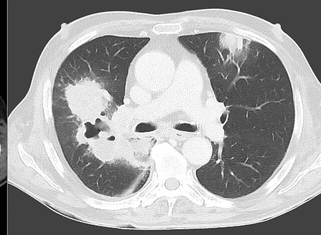
Kavite; akciğer konsolidasyonu, nodülü veya kitlesi içinde görülen, havayla dolu alçak atenüanslı bir boşluktur. BT’de kavite duvarının kalınlığı ile çevresindeki sentrilobüler nodüller ve konsolidasyon gibi bulgular değerlendirilir; bunlar klinik bilgilerle birlikte enfeksiyöz, sistemik ve malign nedenlerin ayırıcı tanısına katkı sağlar. Hava-sıvı seviyesi tek başına apseyi kanıtlamaz; ince duvar da maligniteyi dışlamaz. Apse ve septik emboli gibi süreçlerde lokal ampiyem, bronkoplevral fistül ve plevral effüzyon gibi komplikasyonlar gelişebilir.
Faz ve pencere
- Kontrastsız tanısal
- BT’de kavite duvar kalınlığı, hava-sıvı seviyesi ve çevresindeki sentrilobüler nodüller ile konsolidasyon değerlendirilebilir.
Önerilen pencereler G genişlik, M merkez; değerler Hounsfield birimi (HU)
- AkciğerG 1500 / M −600
- MediastenG 350 / M +50
BT bulguları
- Kavite — konsolidasyon, nodül veya kitle içindeki gaz ya da düşük atenüasyonlu boşluğun çevresinde tanımlanabilir duvar bulunması.
- Duvarın en kalın kesimde ölçülen kalınlığı ile iç yüzün düzgün, lobüle veya düzensiz olması; tek başına kesin benign-malign ayrımı sağlamaz.
- Tek veya çok sayıda kavite; tek lezyonun lobu ve çoklu lezyonların dağılımı ayırıcı tanıyı yönlendirir.
- Kavitede yatay hava-sıvı seviyesi; hava yolu bağlantısı veya sıvı içeriğiyle oluşabilir ve özgül değildir.
- Çevresel sentrilobüler nodül ve tree-in-bud örüntüsü; bronşioler yayılım gösteren enfeksiyonu destekler.
- Kavite çevresinde konsolidasyon, buzlu cam veya nekrotik parankim; enfeksiyon ve kanama eşlik edebilir.
- Komşu bronşun kaviteye açılması, plevral kalınlaşma, plevral sıvı ya da hava-sıvı içeren plevral koleksiyon.
Ölçütler ve sınıflamalar
- İnce duvarlı akciğer kisti için tipik duvar
- <2 mm
- Tek kaviter lezyonlarda benign hastalık için yüksek özgüllüklü duvar kalınlığı
- <7 mm
- Tek kaviter lezyonlarda malignite için yüksek özgüllüklü duvar kalınlığı
- >24 mm
Normalde
Kavite, konsolidasyon, kitle veya nodül içinde görülen gaz dolu ya da düşük atenüasyonlu bir alandır. Kaviteyi değerlendirirken boşluğun konsolidasyon, kitle veya nodül içinde yer alıp almadığını belirle.
Normal BT ile kıyasla
Aynı bölgenin, kaynak veri setinde patoloji içermediği belirtilen bir BT incelemesini kesit kesit kaydırın; organ sınırlarını açarak anatomiyi hasta görüntüsüyle karşılaştırın. Küçük rastlantısal bulgular tümüyle dışlanmamıştır.
Kaynak: TotalSegmentator v2.01 veri seti (Wasserthal ve ark., Radiology: Artificial Intelligence 2023), CC BY 4.0, vaka s1278; organ sınırları veri setinin otomatik segmentasyonundan, birleştirilerek sadeleştirildi. Görüntüler 2,5 mm kesit aralığına yeniden örneklendi.
Ayırıcı tanı
- Akciğer apsesi
- nekrotik merkez çevresinde kalın, çoğu kez çevresel kontrastlanan duvar ve komşu konsolidasyon görülür; kontrastlı inceleme istenmişse duvarı değerlendir.
- Kavitasyon yapan bronşiyal karsinom
- düzensiz, değişken kalınlıkta duvarlı tek kitle olarak ortaya çıkabilir; bronşiyal karsinomların %10-15'i kaviter seyir gösterir.
- Tüberküloz
- üst loblarda kavite, çevresinde sentrilobüler nodüller ve bronkojenik yayılım bulguları görülebilir.
- Septik pulmoner emboli
- iki taraflı periferik nodüllerin bir kısmında kavitasyon ve ayrı plevra tabanlı kama opasiteler olabilir.
- Granülomatozis ile polianjiit
- birden fazla kaviteli nodül, eşlik eden vasküler ve üst hava yolu hastalığı bağlamında değerlendirilir.
- Nekrotizan pnömoni ve kronik pulmoner aspergilloz
- kaviter görünüm oluşturabilir; dağılım, eşlik eden parankim bulguları ve klinik bağlam ayırıcı tanıya katkı sağlar.
Tuzaklar
- Duvar kalınlığı olasılığı değiştirir fakat eşiklerin dışındaki ince duvarlı karsinomlar ve kalın duvarlı enfeksiyonlar tanımlanmıştır; tek ölçümle benign ya da malign hükmü verme.
- Hava-sıvı seviyesi hem apse hem kaviteli tümör veya bronşla ilişkili diğer boşluklarda görülebilir; çevre konsolidasyon ve duvarın kontrastlanması ayırıcı bağlam sağlar.
- Kaviteyi bül veya kist sanmak, özellikle çok ince duvar ve çevre parankim hastalığı yokluğunda mümkündür; bronş ilişkisini ve eşlik eden fibrotik mimariyi kontrol et.
- Periferal lokalize apselere komşu plevrada kalınlaşma veya ampiyem gelişebilir; bu durum akciğer içi kavite ile karışabilir.
Kendini dene
Öksürüğü ve ateşi olan bir hastada, duvarı kalın ve çevresel olarak kontrastlanan, içinde hava-sıvı seviyesi bulunan bir kavite ile komşu konsolidasyon görülüyor. En olası tanı hangisidir?
Cevabı göster
Akciğer apsesi. Nekrotik merkez çevresinde çevresel duvar kontrastlanması ve komşu konsolidasyon apseyi destekler. Kavite içeriği benign ve malign lezyonları ayırmada genellikle az yardımcıdır; hava-sıvı seviyesi bronkojenik kistlerde ve bazı akciğer adenokarsinomlarında da görülebilir.
Bronşiyal karsinomların yaklaşık ne kadarı kaviter özellik gösterir?
Cevabı göster
%10–15. Kaynağa göre bronşiyal karsinomların %10–15'i kaviter özellik gösterir.
İlgili konular
- Skuamöz hücreli akciğer kanseri
- Kavitasyonlu pulmoner metastazlar
- Akciğer apsesi / nekrotik enfeksiyon
Kaynaklar
Bu sayfadaki 38 cümle ve 2 quiz sorusu aşağıdaki kaynaklardan alıntılarla otomatik olarak karşılaştırıldı; 11 cümle bu karşılaştırmada düzeltildi (2026-10-09).
- Differential Diagnosis of Cavitary Lung Lesionspmc.ncbi.nlm.nih.gov
- Traumatic pulmonary pseudocyst: An underreported entity.pubmed.ncbi.nlm.nih.gov
- Radiological approach to cavitary lung lesions.pubmed.ncbi.nlm.nih.gov
- Unusual presentation of disseminated Nocardia abscessus infection in a patient with AIDS.pubmed.ncbi.nlm.nih.gov
- Pulmonary sequestration: a retrospective analysis of 2625 cases in China.pubmed.ncbi.nlm.nih.gov
- [Hydatid cyst disease mimicking metastatic lung disease: a case report].pubmed.ncbi.nlm.nih.gov
- Primary multidrug-resistant tuberculosis versus drug-sensitive tuberculosis in non-HIV-infected patients: Comparisons of CT findings.pubmed.ncbi.nlm.nih.gov
- Propionibacterium acnes : an uncommon cause of lung abscess in chronic obstructive pulmonary disease complicated with bullous emphysema.pubmed.ncbi.nlm.nih.gov
- Diagnostic yield of additional conventional transbronchial lung biopsy following radial endobronchial ultrasound lung biopsy for peripheral pulmonary lesions.pubmed.ncbi.nlm.nih.gov
- A Case of Lung Cancer Originating from Cavitary Mycobacterium xenopi Infection.pubmed.ncbi.nlm.nih.gov
- Unusual case of cavitary lung metastasis from squamous cell carcinoma of the uterine cervix.pubmed.ncbi.nlm.nih.gov
- Cystic, nodular and cavitary metastases to the lungs in a patient with endometrial stromal sarcoma of the uterus.pubmed.ncbi.nlm.nih.gov
- Solitary thin-walled cystic lung cancer with extensive extrapulmonary metastasis: A case report and review of the literature.pubmed.ncbi.nlm.nih.gov
- Computed tomography for the diagnosis of solitary thin-walled cavity lung cancer.pubmed.ncbi.nlm.nih.gov
- Case of a 33-Year-Old Woman With Hemoptysis and Migrant Nodular Cavitary Lesions.pubmed.ncbi.nlm.nih.gov
- Diagnostic Value of Non-contrast Lung CT in Evaluating Pulmonary Cavitary Lesions in Children and Adolescents: With a Specific Focus in Perforated Hydatid Cyst, Necrotizing Pneumonia, and Lung Abscess.pubmed.ncbi.nlm.nih.gov
- Pulmonary artery floating sign: a novel predictor of fatal haemoptysis in pulmonary mucormycosis and its early intervention strategy.pubmed.ncbi.nlm.nih.gov
- Common and Rare Radiological Findings of Pulmonary and Thoracic Hydatid Cysts.pubmed.ncbi.nlm.nih.gov
- Pathogen-associated radiological and serum inflammatory findings in pediatric necrotizing pneumonia: a retrospective analysis.pubmed.ncbi.nlm.nih.gov
- First endogenous case of <i>Blastomyces dermatitidis</i> ARDS in an immunocompetent man in Newfoundland and Labrador: Case report and mini-review.pubmed.ncbi.nlm.nih.gov
- Precise discrimination of mycobacterial pulmonary diseases via multimodal machine learning integrating chest CT and clinical markers.pubmed.ncbi.nlm.nih.gov
- Anatomic Pulmonary Lobectomy via Video-Assisted Thoracoscopic Surgery in an Adult Patient With Congenital Lobar Emphysema: A Case Report.pubmed.ncbi.nlm.nih.gov
- Evaluation of Cavitary Lung Lesions with <sup>18</sup>F-FDG PET/CT: Differentiating Benign from Malignant Lesions.pubmed.ncbi.nlm.nih.gov
- Congenital pulmonary airway malformation mimicking lung cancer in adults: a pitfall in oncologic diagnosis.pubmed.ncbi.nlm.nih.gov
- Pulmonary Abscess With Contiguous Extension to the Chest Wall in an Immunocompetent Young Woman: A Case Report.pubmed.ncbi.nlm.nih.gov
- Trans-thoracic Extension of a Lung Abscess With a Pleurocutaneous Fistula: A Report of a Rare Case.pubmed.ncbi.nlm.nih.gov
- Transforming pulmonary care with applications of endobronchial valves beyond emphysema: a narrative review.pubmed.ncbi.nlm.nih.gov
- Acute and chronic complications of cesarean delivery: An update for the general radiologist.pubmed.ncbi.nlm.nih.gov
- International practices and variability in right heart echocardiography: results from the RVNet(Work) international survey.pubmed.ncbi.nlm.nih.gov
- Stereotactic body radiotherapy for recurrent hemoptysis due to chronic pulmonary aspergillosis: a case report and systematic review of the literature.pubmed.ncbi.nlm.nih.gov
- Analysis of risk factors for early detection of cystic lung cancer based on CT genomics and its relationship with pathological invasion.pubmed.ncbi.nlm.nih.gov
- Role of Multidetector Computed Tomography in Differentiation of Benign and Malignant Cavitary Lung Lesions With a Histopathological Correlation: A Retrospective Cross-Sectional Study.pubmed.ncbi.nlm.nih.gov
- Computed tomographic characteristics of cavitary pulmonary adenocarcinoma in 3 dogs and 2 cats.pubmed.ncbi.nlm.nih.gov
- A pathological analysis of pediatric pulmonary cystic/cavitary lesions.pubmed.ncbi.nlm.nih.gov
- Management of pneumonia by the emergency radiologist.pubmed.ncbi.nlm.nih.gov
- Carcinoma of Unknown Primary Origin Presenting as Rapidly Progressive Multifocal Skeletal Lesions and Fatal Systemic Thrombosis in a Dog: A Case Report.pubmed.ncbi.nlm.nih.gov
- Delayed Hemopneumothorax and Multiple Rib Fractures Following Blunt Chest Trauma.pubmed.ncbi.nlm.nih.gov
- <i>Streptococcus intermedius</i> lung abscess in children: a case report and literature review.pubmed.ncbi.nlm.nih.gov
- Ultrasonographic Assessment of the Lungs in Asiatic Elephants (Elephas maximus).pubmed.ncbi.nlm.nih.gov
- Successful treatment of <i>Mycobacterium abscessus</i> pulmonary disease with limited resection and perioperative chemotherapy containing clofazimine: a case report.pubmed.ncbi.nlm.nih.gov
- Branches of Deception: A Tree-in-Bud Pattern Revealing Advanced Non-small Cell Lung Cancer (NSCLC).pubmed.ncbi.nlm.nih.gov
- Host, Pathogen, and Pulmonary Anatomy in Pneumonia: An Integrated Imaging and Clinical Perspective.pubmed.ncbi.nlm.nih.gov
- Case Report: Delayed appearance of the ablation margin after CT-guided microwave ablation of a solid pulmonary nodule.pubmed.ncbi.nlm.nih.gov
- Contrast-enhanced ultrasound-guided precision fibrinolysis for refractory loculated chylothorax after lung transplantation: a case report.pubmed.ncbi.nlm.nih.gov
- Retrospective Review Is the Best View: A Primer for Residents and Radiologists on Missed Findings in Chest Radiography.pubmed.ncbi.nlm.nih.gov
- Incidental Lung Lesions on the CT Component of Oncologic FDG PET/CT: A Pictorial Review of Interpretive Pitfalls and Diagnostic Clues.pubmed.ncbi.nlm.nih.gov
- Density homogeneity as a crucial CT indicator for differentiating malignant and benign subcentimeter solid pulmonary nodules: A retrospective multi-center study.pubmed.ncbi.nlm.nih.gov
- Case Report: Pulmonary benign metastasizing leiomyoma with mild <sup>18</sup>F-FDG uptake occurring 30 years after hysterectomy.pubmed.ncbi.nlm.nih.gov
- Beyond smoking status: risk factors for pulmonary nodules ≥5 mm in a Chinese community screening program.pubmed.ncbi.nlm.nih.gov
- Predictive Factors and Nomogram for Malignant Pulmonary Nodules (≤ 1 cm).pubmed.ncbi.nlm.nih.gov
- Non-invasive detection of hTERT mRNA in deep-cough swabs for early diagnosis of lung cancer.pubmed.ncbi.nlm.nih.gov
- AI driven hybrid convolutional and transformer based deep learning architecture for precise lung nodule classification.pubmed.ncbi.nlm.nih.gov
- Lung Cancers Associated with Cystic Airspaces.pubmed.ncbi.nlm.nih.gov
- Splenic hamartomas in children.pubmed.ncbi.nlm.nih.gov
- Thoracotomy for Urgent Repair of Lung Torsion Identified as an Incidental Finding.pubmed.ncbi.nlm.nih.gov
- Uniportal robotic-assisted left upper lobectomy with pulmonary artery reconstruction for a central lung cancer: a step-by-step surgical technique.pubmed.ncbi.nlm.nih.gov
- Microscopic anatomy of the lungs of domestic animals, mice, and rats.pubmed.ncbi.nlm.nih.gov
- Pathologic and Radiologic Correlation of Adult Cystic Lung Disease: A Comprehensive Review.pubmed.ncbi.nlm.nih.gov
- International Harmonization of Nomenclature and Diagnostic Criteria (INHAND): Non-proliferative and Proliferative Lesions of the Non-human Primate ( M. fascicularis ).pubmed.ncbi.nlm.nih.gov
- Intestinal Perforation Caused by Intestinal Tuberculosis of a Nepalese Man Living in a Low Tuberculosis-Burden Country: A Case Report.pubmed.ncbi.nlm.nih.gov
- Post-tuberculosis morbidities and their associated mortality: moving from challenges to solutions.pubmed.ncbi.nlm.nih.gov
- Missed Lung Cancers on Chest Radiograph: An Illustrative Review of Common Blind Spots on Chest Radiograph with Emphasis on Various Radiologic Presentations of Lung Cancers.pubmed.ncbi.nlm.nih.gov
- Management of life-threatening hemoptysis in the ICU.pubmed.ncbi.nlm.nih.gov
- The first CT-based classification system for pulmonary actinomycosis: correlating imaging patterns with therapeutic strategies and prognosis in a highly misdiagnosed disease.pubmed.ncbi.nlm.nih.gov
- Primary pulmonary mucosa-associated lymphoid tissue lymphoma presenting as a right hilar mass: A case report.pubmed.ncbi.nlm.nih.gov
- Pulmonary cryptococcosis with or without immunocompromise: a retrospective comparative study of clinical and CT features and influential factors for lesion absorption.pubmed.ncbi.nlm.nih.gov
- The Role of CEUS in the Diagnosis and Follow-Up of Pleuropulmonary Diseases and Interventional Procedures.pubmed.ncbi.nlm.nih.gov
- Two in one: Overlapping CT findings of COVID-19 and underlying lung diseases.pubmed.ncbi.nlm.nih.gov
- COVID19 pneumonia with cavitation and cystic lung changes: multi-detector computed tomography spectrum of a gamut of etiologies.pubmed.ncbi.nlm.nih.gov
- From presumed infection to systemic vasculitis: de novo granulomatosis with polyangiitis in pregnancy - a case report.pubmed.ncbi.nlm.nih.gov
- Diagnostic Journey to Granulomatosis with Polyangiitis with Rare Hepatic and Suspected Splenic Involvement: A Case Report.pubmed.ncbi.nlm.nih.gov
- Immunoglobulin G4 (IgG4)-Related Lung Disease Mimicking Pulmonary Malignancy: A Case Report With Histopathological Characterisation and Review of Diagnostic Criteria.pubmed.ncbi.nlm.nih.gov
- MRI insights into endometrial stromal tumors.pubmed.ncbi.nlm.nih.gov
- Gossypiboma Following Cesarean Section Presenting as Bilateral Abdominal Abscesses: A Case Report.pubmed.ncbi.nlm.nih.gov
- Cavitary pulmonary disease.pubmed.ncbi.nlm.nih.gov
- Empyema Versus Lung Abscess: A Case Report.pubmed.ncbi.nlm.nih.gov
- Advances and challenges in the management of haemoptysis in the emergency department: A narrative review.pubmed.ncbi.nlm.nih.gov
- Atlas of gross, histologic, and immunohistologic anatomy of the lymphoid system in Egyptian rousettes (Rousettus aegyptiacus).pubmed.ncbi.nlm.nih.gov
- Robot-Assisted Laparoscopic Hiatal Hernia Repair in a Patient With Right Aortic Arch: Implications for Surgical Planning and Symptom Resolution.pubmed.ncbi.nlm.nih.gov
- AWMF S2k Guideline: Diagnosis and Assessment of Lung Disease Caused by Occupational Silica Dust Exposure (Silicosis) - Disease No. 4101 as per Occupational Diseases Ordinance (BKV) Annex 1.pubmed.ncbi.nlm.nih.gov
- Aggressiveness-guided nodule management for lung cancer screening in Europe-justification for follow-up intervals and definition of growth.pubmed.ncbi.nlm.nih.gov
- Chinese expert consensus on the diagnosis and management of bronchopleural fistula after pulmonary resection (2026 edition).pubmed.ncbi.nlm.nih.gov
- Fatal multi-organ failure following delayed diagnosis of ruptured pulmonary hydatid cyst in an 11-year-Old boy: The airway management dilemma ('intubate or not intubate').pubmed.ncbi.nlm.nih.gov
- Clinical classification of chronic obstructive pulmonary disease with invasive pulmonary aspergillosis: a retrospective study.pubmed.ncbi.nlm.nih.gov
- Lung cancer associated with cystic airspaces: Current concept, pathogenesis, imaging features, pathological basis, and management.pubmed.ncbi.nlm.nih.gov
- Esophago-tracheobronchial fistula induced by anlotinib: A case report.pubmed.ncbi.nlm.nih.gov
- Fleischner Society: Glossary of Terms for Thoracic Imaging.pubmed.ncbi.nlm.nih.gov
- Diagnostic approach for incidental pulmonary nodules.pubmed.ncbi.nlm.nih.gov
- Cavitary Pulmonary Adenocarcinoma in a Young Adult With a History of Treated Hodgkin Lymphoma: A Case Report.pubmed.ncbi.nlm.nih.gov
- A Rare Case of Salmonella Lung Abscess in an Immunocompromised Patient.pubmed.ncbi.nlm.nih.gov
- Pulmonary <i>Nocardia paucivorans</i> Infection Mimicking Lung Cancer in an Immunocompetent Patient: A Case Report.pubmed.ncbi.nlm.nih.gov
- Diffuse cystic lung diseases-a primer for radiologists.pubmed.ncbi.nlm.nih.gov
- Case Report: Necrotizing pulmonary destruction and fatal gangrene: the imperative of early recognition.pubmed.ncbi.nlm.nih.gov
- Imaging features of lung cancer with cystic airspaces: clinical utility, challenges, and perspectives.pubmed.ncbi.nlm.nih.gov
- Atelectasis secondary to two separate video-assisted thoracoscopic wedge resection of the lung lobe and pleurodesis under general anesthesia in the same patient: a case report.pubmed.ncbi.nlm.nih.gov
- Recurrent hepatic hydatid disease presenting with biliptysis and suspected bronchobiliary fistula: successful management via thoracotomy, lung wedge resection, and diaphragmatic/subdiaphragmatic cyst evacuation.pubmed.ncbi.nlm.nih.gov
- Superior sulcus tumor with vertebral invasion following initial diagnosis of cavitary lung disease: a case report.pubmed.ncbi.nlm.nih.gov
- Towards a more realistic anthropomorphic chest phantom using 3D-printed and cork-integrated components.pubmed.ncbi.nlm.nih.gov
- Lipoid Pneumonia: HRCT and MRI Spectrum, Diagnostic Pitfalls, and Imaging-Based Diagnostic Workflow.pubmed.ncbi.nlm.nih.gov
- Non-Contrast Radiation-Free NIR Lung Imaging.pubmed.ncbi.nlm.nih.gov
- Machine learning-based model assists in differentiating Mycobacterium avium Complex Pulmonary Disease from Pulmonary Tuberculosis: A Multicenter Study.pubmed.ncbi.nlm.nih.gov
- Image Findings from Dual-phase Computed Tomography Pulmonary Angiography for Diagnosing Tuberculosis-associated Fibrosing Mediastinitis.pubmed.ncbi.nlm.nih.gov
- Bronchial Dieulafoy disease: A case report and literature review.pubmed.ncbi.nlm.nih.gov
- Beyond the Usual Suspects: Rare Causes of Hemoptysis.pubmed.ncbi.nlm.nih.gov
- Structural and functional characteristics of healthy and injured porcine lungs during deflation: a quantitative CT imaging analysis.pubmed.ncbi.nlm.nih.gov
- AI-Based Quantitative HRCT for In-Hospital Adverse Outcomes and Exploratory Assessment of Reinfection in COVID-19.pubmed.ncbi.nlm.nih.gov
- Ultrasound imaging of lung disease and its relationship to histopathology: An experimentally validated simulation approach.pubmed.ncbi.nlm.nih.gov
- Practical Imaging Interpretation in Patients Suspected of Having Idiopathic Pulmonary Fibrosis: Official Recommendations from the Radiology Working Group of the Pulmonary Fibrosis Foundation.pubmed.ncbi.nlm.nih.gov
- Quantitative CT imaging and advanced visualization methods: potential application in novel coronavirus disease 2019 (COVID-19) pneumonia.pubmed.ncbi.nlm.nih.gov
- Pulmonary cavitary lesions: a captivating visual review.pubmed.ncbi.nlm.nih.gov
- What Findings on Chest CTs Can Delay Diagnosis of Pleuropulmonary Paragonimiasis?pubmed.ncbi.nlm.nih.gov
Eğitim amaçlıdır; hasta başında tanı ve tedavi kararının yerine geçmez, tıbbi tavsiye değildir. Metinler yapay zekâ desteğiyle yazıldı ve otomatik bir adımda kaynak alıntılarıyla karşılaştırıldı; bu bir tıbbi doğrulama değildir ve içerik uzman radyolog incelemesinden geçmedi. Klinik kararlarda güncel kılavuzları, kurum protokollerini ve radyoloji raporunu esas alın.