Özet
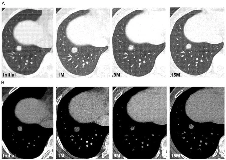
Part-solid nodülde buzlu cam zemin içinde parankim izlerini örten gerçek solid bileşen bulunur. İnce kesit kontrastsız BT’de toplam lezyon çapı ile solid kısmın ayrı değerlendirilmesi, adenokarsinom spektrumundaki invaziv bileşenle ilişkili değişimi izletir. Solid bileşenin yeni ortaya çıkması veya büyümesi, toplam çap sabit kalırken gözden kaçabilir.
Faz ve pencere
- Kontrastsız tanısal
- İnce kesit BT’de buzlu cam zemin ile parankimi örten solid odak ayrı değerlendirilir; toplam nodül ve solid bileşen çapları ölçülür.
Önerilen pencereler: Akciğer (G 1500 / M -600), Mediasten (G 350 / M 50).
BT bulguları
- Damar/bronş izleri buzlu cam kısımda seçilir, solid odakta örtülür.
- Toplam nodül çapı ile solid bileşenin çapı ayrı kaydedilir.
- Toplam nodül ve solid bileşen çapları ince kesit BT’de ölçülür; solid bileşenin sınırlarının net belirlenmesi için mediastinal pencere kullanımı önerilir. Mediastinal pencere, lezyon içindeki yağ veya kalsifikasyon varlığını doğrulamaya yardımcı olabilir.
- Lobülasyon, spikülasyon, plevral çekinti ve hava bronkogramı eşlik edebilir.
- Seri incelemede yeni solid alan veya solid komponent artışı, toplam çap değişmese de kaydedilir.
Ölçütler ve sınıflamalar
- Subsolid nodülde ince kesit ve ölçüm
- Kesit kalınlığı <3 mm, ideal olarak 1–1,5 mm; toplam nodül ve solid bileşen ayrı ölçülür.
- Fleischner Society 2017 subsolid nodüller
- Part-solid nodül, hem buzlu cam hem de solid bileşen içerdiğinden ayrı değerlendirilmelidir; yönetimi hastanın risk faktörlerine ve nodül özelliklerine göre kişiselleştirilir.
- ACR Lung-RADS
- Tarama nodülü kategorisi toplam boyutun yanı sıra solid bileşenin boyut ve değişimine bağlıdır.
- TNM 9. baskı solid-subsolid ölçümü
- Klinik T evrelemesinde part-solid primer tümörün boyutu, solid bileşenin en uzun çapına dayanılarak belirlenir; bu yaklaşım güncel TNM evreleme sistemlerinde korunmaktadır.
Normalde
Normal akciğerde damar ve bronş konturları kesintisiz seçilir. Part-solid nodülde damarlar buzlu cam kısmında korunur, solid kısımda silinir; sınırı akciğer ve mediasten pencerelerinde karşılaştır.
Normal BT ile kıyasla
Aynı bölgenin, kaynak veri setinde patoloji içermediği belirtilen bir BT incelemesini kesit kesit kaydırın; organ sınırlarını açarak anatomiyi hasta görüntüsüyle karşılaştırın. Küçük rastlantısal bulgular tümüyle dışlanmamıştır.
Kaynak: TotalSegmentator v2.01 veri seti (Wasserthal ve ark., Radiology: Artificial Intelligence 2023), CC BY 4.0, vaka s1278; organ sınırları veri setinin otomatik segmentasyonundan, birleştirilerek sadeleştirildi. Görüntüler 2,5 mm kesit aralığına yeniden örneklendi.
Ayırıcı tanı
- Saf GGN
- tüm odakta damar izleri korunur, gerçek solid odak yoktur.
- Fokal enfeksiyon
- yamalı buzlu cam, sentrilobüler nodüller veya zaman içinde gerileme gösterebilir.
- Kısmi atelektazi, özellikle tümöre bağlı kollapsla solid görünüm taklit edebilir.
- İnvaziv solid nodül
- buzlu cam bileşen olmadan parankimi örter.
Tuzaklar
- Solid bileşeni toplam nodül çapıyla karıştırmayın; solid bileşen, akciğer penceresindeki damar konturlarıyla örtüşebileceğinden mediastinal pencerede ayrı ölçülür. Mediastinal pencerede solid bileşen, akciğer penceresine göre daha belirgin görünebilir ve daha geniş ölçülebilir.
- Kalın kesitler küçük nodülleri gizleyebilir; ince kesit görüntüleme (≤1,5 mm) tespit ve karakterizasyon için gereklidir.
- BT solid komponenti histolojik invaziv komponentle bire bir eşleştirmez.
Kendini dene
Buzlu cam odağın içinde damar kesitlerinin silindiği bölüm var. Lezyon nasıl adlandırılır?
Cevabı göster
Part-solid nodül. Buzlu cam ve parankim izlerini örten solid bileşenin birlikteliği part-solid tanımıdır.
Seri BT’de toplam çap benzer, fakat yeni solid odak oluşmuş. Hangi değişiklik kaydedilmeli?
Cevabı göster
Solid komponentin yeni oluşumu ve çapı. Solid komponentin ortaya çıkması veya artması, toplam çap sabit olsa da morfolojik değişimdir.
Bu konunun yer aldığı turlar
İlgili konular
Kaynaklar
Bu sayfadaki 38 cümle ve 2 quiz sorusu aşağıdaki kaynaklardan alıntılarla tek tek karşılaştırıldı; 10 cümle bu karşılaştırmada düzeltildi (2026-10-08).
- CT prediction of malignancy in part-solid pulmonary nodules based on vascular interruption and distortion.pubmed.ncbi.nlm.nih.gov
- Evaluating the Patient With a Pulmonary Nodule: A Review.pubmed.ncbi.nlm.nih.gov
- Pulmonary Nodules.pubmed.ncbi.nlm.nih.gov
- Probability of cancer in pulmonary nodules detected on first screening CT.pubmed.ncbi.nlm.nih.gov
- Initial Pulmonary Nodule Management for the Generalist: A Brief Review.pubmed.ncbi.nlm.nih.gov
- Low-dose CT screening among never-smokers with or without a family history of lung cancer in Taiwan: a prospective cohort study.pubmed.ncbi.nlm.nih.gov
- Accurate diagnosis of pulmonary nodules using a noninvasive DNA methylation test.pubmed.ncbi.nlm.nih.gov
- [Not Available].pubmed.ncbi.nlm.nih.gov
- Ground-glass nodules of the lung in never-smokers and smokers: clinical and genetic insights.pubmed.ncbi.nlm.nih.gov
- Management of Ground-Glass Nodules: When and How to Operate?pubmed.ncbi.nlm.nih.gov
- Cancer Risk in Subsolid Nodules in the National Lung Screening Trial.pubmed.ncbi.nlm.nih.gov
- Subsolid pulmonary nodules: CT-pathologic correlation using the 2011 IASLC/ATS/ERS classification.pubmed.ncbi.nlm.nih.gov
- Performance of ACR Lung-RADS in a Clinical CT Lung Screening Program.pubmed.ncbi.nlm.nih.gov
- Genomic characterisation of pulmonary subsolid nodules: mutational landscape and radiological features.pubmed.ncbi.nlm.nih.gov
- Ground-glass opacity nodules: histopathology, imaging evaluation, and clinical implications.pubmed.ncbi.nlm.nih.gov
- Predictors of Pathologic Tumor Invasion and Prognosis for Ground Glass Opacity Featured Lung Adenocarcinoma.pubmed.ncbi.nlm.nih.gov
- Lung Cancers Manifesting as Part-Solid Nodules in the National Lung Screening Trial.pubmed.ncbi.nlm.nih.gov
- Tumor Atelectasis Gives Rise to a Solid Appearance in Pulmonary Adenocarcinomas on High-Resolution Computed Tomography.pubmed.ncbi.nlm.nih.gov
- How to confront the high prevalence of pulmonary micro nodules (PMNs) in osteosarcoma patients?pubmed.ncbi.nlm.nih.gov
- Aggressiveness-guided nodule management for lung cancer screening in Europe-justification for follow-up intervals and definition of growth.pubmed.ncbi.nlm.nih.gov
- Prognostic importance of volumetric measurements in stage I lung adenocarcinoma.pubmed.ncbi.nlm.nih.gov
- Accuracy of solid portion size measured on multiplanar volume rendering images for assessing invasiveness in lung adenocarcinoma manifesting as subsolid nodules.pubmed.ncbi.nlm.nih.gov
- Evaluation of the solitary pulmonary nodule: size matters, but do not ignore the power of morphology.pubmed.ncbi.nlm.nih.gov
- Differential diagnosis of solitary pulmonary nodules with dual-source spiral computed tomography.pubmed.ncbi.nlm.nih.gov
- CT-based Radiologic Ternary Classification Model in Predicting Pathologic Invasiveness of Pulmonary Nonsolid Nodules.pubmed.ncbi.nlm.nih.gov
- Clinical and Radiological Characteristics to Differentiate Between EGFR Exon 21 and Exon 19 Mutations in Patients With Lung Adenocarcinoma: A Systematic Literature Review and Meta-Analysis.pubmed.ncbi.nlm.nih.gov
- CT characteristics of solitary pulmonary capillary hemangioma versus lung adenocarcinoma.pubmed.ncbi.nlm.nih.gov
- Pulmonary fissure segmentation on CT.pubmed.ncbi.nlm.nih.gov
- Robust anisotropic Gaussian fitting for volumetric characterization of pulmonary nodules in multislice CT.pubmed.ncbi.nlm.nih.gov
- Normative values for lung, bronchial sizes, and bronchus-artery ratios in chest CT scans: from infancy into young adulthood.pubmed.ncbi.nlm.nih.gov
- A left lung abscess with a displaced subsegmental bronchus and anomalous pulmonary artery and vein: a case report.pubmed.ncbi.nlm.nih.gov
- Computed tomographic characterization of the pulmonary system in clinically normal alpacas.pubmed.ncbi.nlm.nih.gov
- Guidelines for Management of Incidental Pulmonary Nodules Detected on CT Images: From the Fleischner Society 2017.pubmed.ncbi.nlm.nih.gov
- Updated Fleischner Society Guidelines for Managing Incidental Pulmonary Nodules: Common Questions and Challenging Scenarios.pubmed.ncbi.nlm.nih.gov
- Nodule characterization: subsolid nodules.pubmed.ncbi.nlm.nih.gov
- Cost-Effectiveness of Treatment Thresholds for Subsolid Pulmonary Nodules in CT Lung Cancer Screening.pubmed.ncbi.nlm.nih.gov
- Improved interobserver agreement on nodule type and Lung-RADS classification of subsolid nodules using computer-aided solid component measurement.pubmed.ncbi.nlm.nih.gov
- A problem with clinical T factor in the 8th TNM edition: Prognosis and EGFR mutation status of small sized lung cancers with difficulty to measure the diameter of solid component in part-solid tumor.pubmed.ncbi.nlm.nih.gov
- Prognostic impact of a ground-glass opacity component in clinical stage IA non-small cell lung cancer.pubmed.ncbi.nlm.nih.gov
- Evaluation of solid portions in non-small cell lung cancer-the solid part is not always measurable for clinical T factor.pubmed.ncbi.nlm.nih.gov
- Invasive pulmonary adenocarcinomas versus preinvasive lesions appearing as ground-glass nodules: differentiation by using CT features.pubmed.ncbi.nlm.nih.gov
- High versus low attenuation thresholds to determine the solid component of ground-glass opacity nodules.pubmed.ncbi.nlm.nih.gov
- Various computed tomography findings of 2009 H1N1 influenza in 17 patients with relatively mild illness.pubmed.ncbi.nlm.nih.gov
- Thoracic CT findings of novel influenza A (H1N1) infection in immunocompromised patients.pubmed.ncbi.nlm.nih.gov
- Noninfectious Generalized Bronchiolitis in the Setting of Allogeneic Stem Cell Transplantation: A Potential Mimic of Lower Respiratory Tract Infection.pubmed.ncbi.nlm.nih.gov
- Comparison of 10-year Survival Outcomes between CT Surveillance and Surgery for Ground-Glass Nodules.pubmed.ncbi.nlm.nih.gov
- Software-based risk stratification of pulmonary adenocarcinomas manifesting as pure ground glass nodules on computed tomography.pubmed.ncbi.nlm.nih.gov
- Lung cancers associated with cystic airspaces: CT features and pathologic correlation.pubmed.ncbi.nlm.nih.gov
- Clinicopathological Features and Prognosis of Lung Adenocarcinoma Presenting as Ground-Glass Opacity: A Single-Center Experience.pubmed.ncbi.nlm.nih.gov
- Dual-Energy Computed Tomography Lung in patients of Pulmonary Tuberculosis.pubmed.ncbi.nlm.nih.gov
- Efficacy of measuring the invasive diameter of lung adenocarcinoma using mediastinal window settings: A retrospective study.pubmed.ncbi.nlm.nih.gov
- Thoracic Sarcoidosis: Imaging with High Resolution Computed Tomography.pubmed.ncbi.nlm.nih.gov
- Correlation exploration among CT imaging, pathology and genotype of pulmonary ground-glass opacity.pubmed.ncbi.nlm.nih.gov
- [Clinicopathological characteristics and imaging features of pulmonary adenocarcinoma with micropapillary pattern].pubmed.ncbi.nlm.nih.gov
- Differential Diagnosis of Nonabsorbable Inflammatory and Malignant Subsolid Nodules with a Solid Component ≤5 mm.pubmed.ncbi.nlm.nih.gov
- [Results of low-dose computed tomography (LDCT) screening for early lung cancer: prevalence in 4 690 asymptomatic participants].pubmed.ncbi.nlm.nih.gov
- CT- and computer-based features of small hamartomas.pubmed.ncbi.nlm.nih.gov
- Interobserver size measurement variability in part-solid lung adenocarcinoma using pre-operative computed tomography.pubmed.ncbi.nlm.nih.gov
- Controversies on lung cancers manifesting as part-solid nodules.pubmed.ncbi.nlm.nih.gov
- Clinical Outcomes of Resected Pure Ground-Glass, Heterogeneous Ground-Glass, and Part-Solid Pulmonary Nodules.pubmed.ncbi.nlm.nih.gov
- A decrease in the size of ground glass nodules may indicate the optimal timing for curative surgery.pubmed.ncbi.nlm.nih.gov
- Differentiation between malignancy and inflammation in pulmonary ground-glass nodules: The feasibility of integrated (18)F-FDG PET/CT.pubmed.ncbi.nlm.nih.gov
- Predictors of Diagnostic Yield in Shape-Sensing Robotic-Assisted Bronchoscopy (ssRAB): A Retrospective Single-Center Study.pubmed.ncbi.nlm.nih.gov
- [Three-dimensional Mass Measurement of Subsolid Pulmonary Nodules on Chest CT: Intra and Inter-observer Variability].pubmed.ncbi.nlm.nih.gov
- Subsolid Lung Adenocarcinomas: Radiological, Clinical and Pathological Features and Outcomes.pubmed.ncbi.nlm.nih.gov
- Clinical Versus Pathological Staging in Patients with Resected Ground Glass Pulmonary Lesions.pubmed.ncbi.nlm.nih.gov
- Lung nodules: size still matters.pubmed.ncbi.nlm.nih.gov
- Rate of benign nodule resection in a lung cancer screening program.pubmed.ncbi.nlm.nih.gov
- ACR Appropriateness Criteria® Indolent Lung Cancer.pubmed.ncbi.nlm.nih.gov
- Improving the preoperative diagnosis of invasive lung adenocarcinoma by combining multiplanar volume rendering (MPVR)-based solid component measurement with pathological analysis.pubmed.ncbi.nlm.nih.gov
- Decoding Malignant Lung Ground Glass Opacities: A Radiologic-Pathologic Study From Pure to Semi-Solid Lesions.pubmed.ncbi.nlm.nih.gov
- Predicting STAS in peripheral stage I lung adenocarcinoma: the incremental value of CT-based tumor disappearance rate, with a focus on part-solid nodules.pubmed.ncbi.nlm.nih.gov
- Natural history and clinical characteristics of pulmonary subsolid nodules in pediatric patients: a cohort study.pubmed.ncbi.nlm.nih.gov
- Short-term quantitative CT changes in synchronous ground-glass nodules during immune checkpoint inhibitor therapy in patients with lung cancer.pubmed.ncbi.nlm.nih.gov
- Congenital pulmonary airway malformation mimicking lung cancer in adults: a pitfall in oncologic diagnosis.pubmed.ncbi.nlm.nih.gov
- Incidental Lung Lesions on the CT Component of Oncologic FDG PET/CT: A Pictorial Review of Interpretive Pitfalls and Diagnostic Clues.pubmed.ncbi.nlm.nih.gov
- International association for the study of lung cancer/american thoracic society/european respiratory society international multidisciplinary classification of lung adenocarcinoma.pubmed.ncbi.nlm.nih.gov
- Long-term safety and effectiveness of the "OptEase" vena cava filter.pubmed.ncbi.nlm.nih.gov
- AI-accelerated 3D gradient echo versus ultrashort echo time MRI for lung nodule detection and measurement.pubmed.ncbi.nlm.nih.gov
- Chest CT-detected pulmonary nodules across the COVID-19 pandemic in Guangzhou, China: a regression discontinuity-in-time study, 2013-2023.pubmed.ncbi.nlm.nih.gov
- Case-matched retrieval improves textual alignment of LLM-generated radiology impressions.pubmed.ncbi.nlm.nih.gov
- Incidental pulmonary findings on CT in daily practice: the nodule and the interstitial lung abnormalities - what's old, what's new.pubmed.ncbi.nlm.nih.gov
- The probability of lung cancer in patients with pulmonary nodules detected via low-dose computed tomography screening in China.pubmed.ncbi.nlm.nih.gov
- Evaluation of benign and malignant pulmonary nodules based on clinical and contrast-enhanced computed tomography data: a multicenter study.pubmed.ncbi.nlm.nih.gov
- Suprasellar Desmoplastic Infantile Ganglioglioma in a 7-Month-Old Infant: A Rare Diagnostic Consideration in Infant Suprasellar Masses.pubmed.ncbi.nlm.nih.gov
- Risk of Malignancy in Cystic Lung Lesions in a Lung Cancer CT Screening Program.pubmed.ncbi.nlm.nih.gov
- Artificial intelligence-based automated matching of pulmonary nodules on follow-up chest CT.pubmed.ncbi.nlm.nih.gov
- Robotic-assisted bronchoscopy in dye localization in thoracoscopic pulmonary nodule resection: An initial experience.pubmed.ncbi.nlm.nih.gov
- Recent trends in scientific research in chest radiology: What to do or not to do? That is the critical question in research.pubmed.ncbi.nlm.nih.gov
- Screening for lung cancer using thin-slice low-dose computed tomography in southwestern China: a population-based real-world study.pubmed.ncbi.nlm.nih.gov
- Clinically Relevant Motion of Canine Thoracic Tumors Is Seen Between Inspiratory and Expiratory Breath Holds on Computed Tomography Performed for Radiation Treatment Planning.pubmed.ncbi.nlm.nih.gov
- Development of a Two Stage Pipeline for Robust Pulmonary Nodule Detection in Consecutive CT Slices.pubmed.ncbi.nlm.nih.gov
- Clinical correlates and short-term prognostic value of CT-detected subclinical pulmonary congestion in acute STEMI without overt heart failure.pubmed.ncbi.nlm.nih.gov
- Artificial intelligence-based density proportion analysis in predicting the invasiveness of neoplastic ground-glass nodules.pubmed.ncbi.nlm.nih.gov
- Clinical TNM Lung Cancer Staging: A Diagnostic Algorithm with a Pictorial Review.pubmed.ncbi.nlm.nih.gov
- AI driven hybrid convolutional and transformer based deep learning architecture for precise lung nodule classification.pubmed.ncbi.nlm.nih.gov
- A narrative review on CT-based evaluation and prediction of lung nodule growth: current status and future directions.pubmed.ncbi.nlm.nih.gov
- Deep learning-based super-resolution ultrashort echo time MRI for pulmonary nodule detection and longitudinal assessment in emphysema.pubmed.ncbi.nlm.nih.gov
- The Stockholm Pilot study for Lung cancer Screening (Stockholm PLUS): feasibility of baseline low-dose CT lung cancer screening in a high-risk Swedish female population.pubmed.ncbi.nlm.nih.gov
- Imaging features of lung cancer with cystic airspaces: clinical utility, challenges, and perspectives.pubmed.ncbi.nlm.nih.gov
- Is surgical lymph node assessment justified for ground glass opacity-dominant lung adenocarcinoma?pubmed.ncbi.nlm.nih.gov
- HPV-associated Oropharyngeal Squamous Cell Carcinoma: An Imaging Framework for AJCC Version 9 TNM Staging.pubmed.ncbi.nlm.nih.gov
- Impact of Consolidation-to-Tumor Ratio on Prognosis After Radiation therapy for Lung Cancer With a Lesion Diameter of 3 cm or Less.pubmed.ncbi.nlm.nih.gov
- Nivolumab Plus Lenvatinib and Intra-Arterial Locoregional Therapy as First-Line Treatment for Unresectable Hepatocellular Carcinoma: A Real-World Study.pubmed.ncbi.nlm.nih.gov
- Diagnostic significance of cancer antigen 125/carcinoembryonic antigen ratio combined with IOTA-ADNEX model in discriminating between primary ovarian cancer and ovarian metastases.pubmed.ncbi.nlm.nih.gov
- CT-Guided Transthoracic Core-Needle Biopsy of Pulmonary Nodules: Current Practices, Efficacy, and Safety Considerations.pubmed.ncbi.nlm.nih.gov
- Unveiling the synergetic benefits of the tunneling technique using stapler tractor in precise resection of lung segments: a retrospective cohort study.pubmed.ncbi.nlm.nih.gov
- Lipoid Pneumonia: HRCT and MRI Spectrum, Diagnostic Pitfalls, and Imaging-Based Diagnostic Workflow.pubmed.ncbi.nlm.nih.gov
- Pulmonary histoplasmosis mimicking recurrent lung adenocarcinoma: A case report.pubmed.ncbi.nlm.nih.gov
- Chest tuberculosis: Radiological review and imaging recommendations.pubmed.ncbi.nlm.nih.gov
Eğitim amaçlıdır; hasta başında tanı ve tedavi kararının yerine geçmez. İçerik yapay zekâ destekli bir süreçle güncel literatüre göre cümle cümle kaynaklarla karşılaştırıldı, ancak uzman radyolog incelemesinden geçmedi. Klinik kararlarda güncel kılavuzları, kurum protokollerini ve radyoloji raporunu esas alın.