Özet
Pulmoner nodülün varlığı, atenüasyonu, kenarı ve kalsifikasyonu öncelikle akciğer penceresinde; santral kitle, hilus, lenf düğümü ve komşu damar ilişkisi yumuşak doku/mediasten penceresinde değerlendirilir. İnsidental nodülün saptanması ve seri boyut kıyası çoğu kez kontrastsız ince kesit BT ile yapılabilir; intravenöz kontrast nodül karakterizasyonu için rutin bir gereklilik değildir. Kontrastlı toraks incelemesi kitle, hilus veya mediasten sorusu varsa yararlı olabilir; BTPA ise pulmoner arter sorusuna göre seçilir ve nodül için ayrı bir karakterizasyon fazı sayılmaz. İnce kesit akciğer penceresi nodülün boyut ve sınırı için temel görüntüyü sağlar; hiler ve mediastinal yapıların değerlendirilmesi için mediasten penceresi kullanılır.
Faz ve pencere
- Kontrastsız tanısal
- İnce kesit akciğer serisi nodülün solid/buzlu cam/part-solid yapısını, yağ-kalsiyum içeriğini ve kenarını gösterir; insidental nodül değerlendirmesi ve takip kıyası için genellikle yeterli anatomik fazdır.
- Kontrastlı
- Kontrastlı incelemeler nodülün ayırıcı tanısında ve vasküler detayda yardımcı olabilir; ancak temel anatomik değerlendirme ve boyutsal kıyas için kontrastsız ince kesit BT genellikle yeterli kabul edilir.
- BTPA
- Pulmoner arter lümenleri emboli veya vasküler lezyon sorusuna göre opaklaştırılır; eşzamanlı parankim görülebilse de BTPA, tek başına nodül karakterizasyon protokolü değildir.
Önerilen pencereler: Akciğer (G 1500 / M -600), Mediasten (G 350 / M 50), Kemik (G 1800 / M 400).
BT bulguları
- Akciğer penceresinde nodül sınırı, spikülasyon, fissür/plevra ilişkisi ve çevre parankim değerlendirilir
- Subsolid odağın buzlu cam ve solid bileşenleri akciğer penceresinde ayrı ayrı seçilir
- Mediasten penceresinde yağ ve kalsiyum içeriği, santral kitle, hilus ve lenf düğümleri incelenir
- Damar komşuluğundaki yuvarlak yapının kontrastlı seride damar lümeniyle devamlılığı izlenir
- Kemik penceresinde kosta, vertebra veya göğüs duvarı invazyonu ve olası kemik lezyonları aranır
- Aynı nodülün boyut kıyasında önceki incelemenin kesit kalınlığı ve rekonstrüksiyon yaklaşımı hesaba katılır
Ölçütler ve sınıflamalar
- Nodül karakterizasyonu ve ölçümü için sürekli kesit kalınlığı
- ≤1.5 mm
Normalde
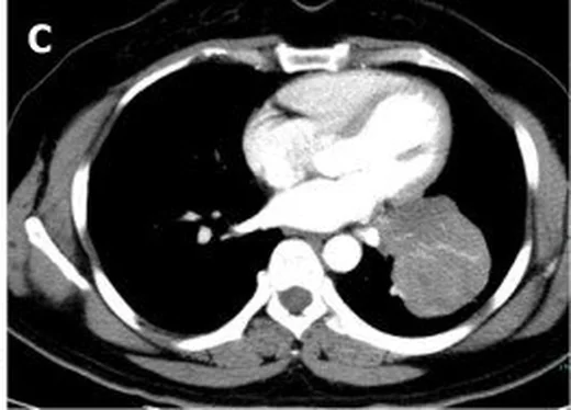
Aynı düzeyde mediasten penceresine geçince parankim ayrıntısı azalırken hiler damarlar ve lenf düğümleri belirginleşir; şüpheli yuvarlak yapının hangi pencerede parankim nodülü, hangi pencerede damar veya yumuşak doku olarak devam ettiğini karşılaştır.
Normal BT ile kıyasla
Aynı bölgenin, kaynak veri setinde patoloji içermediği belirtilen bir BT incelemesini kesit kesit kaydırın; organ sınırlarını açarak anatomiyi hasta görüntüsüyle karşılaştırın. Küçük rastlantısal bulgular tümüyle dışlanmamıştır.
Kaynak: TotalSegmentator v2.01 veri seti (Wasserthal ve ark., Radiology: Artificial Intelligence 2023), CC BY 4.0, vaka s1278; organ sınırları veri setinin otomatik segmentasyonundan, birleştirilerek sadeleştirildi. Görüntüler 2,5 mm kesit aralığına yeniden örneklendi.
Ayırıcı tanı
- Pulmoner nodül
- akciğer penceresinde parankim içinde ayrı odak olarak kalır ve ince kesitlerde devamlıdır.
- Pulmoner arter kesitinin nodülle ilişkisi, BT’de komşu yapılarla anatomik ilişkiler kapsamında değerlendirilir.
- Hiler lenf düğümleri, kontrastlı toraks BT’de büyümüş lenf düğümleri şeklinde izlenebilir.
- Atelektatik bant, pulmoner nodülle birlikte görülebilen eşlik eden atelektazi bulgularından biridir.
Tuzaklar
- Mediasten penceresinde küçük subsolid nodül silikleşebilir; nodülün atenüasyon ve sınır tanımını akciğer penceresinde yap.
- Kontrastlı görüntüde hiler damarı veya lenf düğümünü pulmoner parankim nodülü diye ölçme; damar devamlılığını iki düzlemde izle.
- BTPA’da pulmoner arterlerde dolum defektleri görülebilir; emboli her zaman doğrudan seçilemeyebilir.
- Önceki incelemelerin kesit kalınlığı ve rekonstrüksiyon algoritması nodül ölçümünü etkileyebilir; seri takipte karşılaştırılabilir teknikler kullanılmalıdır.
Kendini dene
Parankim nodüllerinin boyut, şekil ve sınır tanımlamaları için hangi BT serisi önceliklidir?
Cevabı göster
Kontrastsız ince kesit BT. S4'e göre kontrastsız ince kesit BT, nodülün boyutu, şekli ve sınırları dahil morfolojik özelliklerini kapsamlı şekilde gösterir.
S4 çalışmasında tanımlanan standart akciğer penceresi genişlik (W) ve seviye (L) değerleri hangisidir?
Cevabı göster
W: 1200-1500 HU, L: -600 ila -400 HU. S4 metodolojisinde akciğer penceresi için W 1200-1500 HU ve L -600 ila -400 HU olarak belirtilmiştir.
Bu konunun yer aldığı turlar
İlgili konular
Kaynaklar
Bu sayfadaki 32 cümle ve 2 quiz sorusu aşağıdaki kaynaklardan alıntılarla tek tek karşılaştırıldı; 7 cümle bu karşılaştırmada düzeltildi (2026-10-08).
- Glomus Tumors of Uncertain Malignant Potential Arising in the Endobronchus and Lung Parenchymapmc.ncbi.nlm.nih.gov
- Congenital pulmonary airway malformation mimicking lung cancer in adults: a pitfall in oncologic diagnosispmc.ncbi.nlm.nih.gov
- Septic arthritis caused by Nocardia asiatica : a case report and narrative reviewpmc.ncbi.nlm.nih.gov
- Evaluation of benign and malignant pulmonary nodules based on clinical and contrast-enhanced computed tomography data: a multicenter studypmc.ncbi.nlm.nih.gov
- Anatomical-Contextual YOLOv8–YOLOv12 Framework for Pulmonary Nodule Detection in CT: Multi-Organ Learning and Cross-Dataset Validationpmc.ncbi.nlm.nih.gov
- Congenital pulmonary airway malformation mimicking lung cancer in adults: a pitfall in oncologic diagnosis.pubmed.ncbi.nlm.nih.gov
- Incidental Lung Lesions on the CT Component of Oncologic FDG PET/CT: A Pictorial Review of Interpretive Pitfalls and Diagnostic Clues.pubmed.ncbi.nlm.nih.gov
- Image-Domain GAN Denoising for Sn100 kVp Ultra-Low-Dose Chest CT: A Retrospective Paired Image-Quality Study.pubmed.ncbi.nlm.nih.gov
- Case Report: Pulmonary benign metastasizing leiomyoma with mild <sup>18</sup>F-FDG uptake occurring 30 years after hysterectomy.pubmed.ncbi.nlm.nih.gov
- Lung colloid adenocarcinoma: A multidisciplinary diagnostic challenge.pubmed.ncbi.nlm.nih.gov
- ST-Segment Elevation in a Patient With Vocal Cord Cancer: A Great Mimicker.pubmed.ncbi.nlm.nih.gov
- Hypersensitive detection of single millimeter vascular emboli from adhesive in vivo.pubmed.ncbi.nlm.nih.gov
- Thoracic applications of photon-counting CT: where are we after 3 years of clinical implementation?pubmed.ncbi.nlm.nih.gov
- MRI for Lung Cancer Management: Any Closer to Clinical Application?pubmed.ncbi.nlm.nih.gov
- Natural history and clinical characteristics of pulmonary subsolid nodules in pediatric patients: a cohort study.pubmed.ncbi.nlm.nih.gov
- Case Report: Pediatric mediastinal actinomycosis mimicking lymphoma diagnosed by tissue metagenomic next-generation sequencing.pubmed.ncbi.nlm.nih.gov
- Acute mediastinitis, mediastinal granuloma, and fibrosing mediastinitis: a narrative review.pubmed.ncbi.nlm.nih.gov
- Invasive thymoma with rapid response to cisplatin, doxorubicin and cyclophosphamide (PAC) chemotherapy, after an early postoperative recurrence-a case report.pubmed.ncbi.nlm.nih.gov
- Case-matched retrieval improves textual alignment of LLM-generated radiology impressions.pubmed.ncbi.nlm.nih.gov
- Mycotic pulmonary artery aneurysms and septic pulmonary emboli in infective endocarditis: A rare case report and review of imaging findings.pubmed.ncbi.nlm.nih.gov
- Artificial intelligence-based lung nodule detection for pulmonary arteriovenous fistulas on chest computed tomography.pubmed.ncbi.nlm.nih.gov
- Differential diagnosis of benign and malignant pulmonary nodules attached to the interlobar pleura.pubmed.ncbi.nlm.nih.gov
- Left lung combined segmentectomy for lung cancer with displaced left B<sup>1+2</sup>.pubmed.ncbi.nlm.nih.gov
- Deep learning-based super-resolution ultrashort echo time MRI for pulmonary nodule detection and longitudinal assessment in emphysema.pubmed.ncbi.nlm.nih.gov
- Clinicoradiologic features and evolutionary characteristics of pulmonary focal mucinous adenocarcinomas across different density patterns: a retrospective multi-center study.pubmed.ncbi.nlm.nih.gov
- Nodal dissection in thymic surgery: a narrative review of the evolution of practice and evidence in the era of the 9th edition TNM classification (2000-2025).pubmed.ncbi.nlm.nih.gov
- SMARCB1-deficient sinonasal carcinoma in a pediatric patient: Imaging findings and differential diagnosis of a rare entity.pubmed.ncbi.nlm.nih.gov
- POCUS Examination of the Mediastinum in Children: A Simplified and Standardized Protocol for Pulmonary Tuberculosis.pubmed.ncbi.nlm.nih.gov
- Traumatic external jugular vein aneurysm after a kendo thrust (tsuki).pubmed.ncbi.nlm.nih.gov
- Infected splenic artery pseudoaneurysm within a complex splenic cystic lesion: CT angiography pitfalls and surgical correlation.pubmed.ncbi.nlm.nih.gov
- Artificial Intelligence-Assisted Intravascular Imaging in Percutaneous Coronary Intervention: Current Status and Advances.pubmed.ncbi.nlm.nih.gov
- En-Bloc Resection of Stage T4 Non-Small Cell Lung Cancer with Direct Spinal Invasion: Technical Considerations and Comprehensive Literature Review.pubmed.ncbi.nlm.nih.gov
- A diagnostic approach to mediastinal masses in clinical practice.pubmed.ncbi.nlm.nih.gov
- A Patient With Pancoast Tumor Presenting With Cervical Radiculopathy: A Case Report.pubmed.ncbi.nlm.nih.gov
- Application value of high-resolution CT imaging features combined with texture analysis in patients with solitary pulmonary nodules.pubmed.ncbi.nlm.nih.gov
- Phantom-based evaluation of radiomics feature stability for low-dose CT lung cancer screening.pubmed.ncbi.nlm.nih.gov
- Comparative diagnostic accuracy of radiomics, deep learning, and hybrid AI for invasiveness stratification of pulmonary ground-glass nodules: a systematic review and meta-analysis.pubmed.ncbi.nlm.nih.gov
- Features of low-dose CT-detected lung nodules: individuals who never smoked vs. who smoke(d) in a Chinese general population.pubmed.ncbi.nlm.nih.gov
- Tumour Localisation Technologies in Colorectal Cancer Surgery: A Scoping Review of Marking and Detection Methods.pubmed.ncbi.nlm.nih.gov
- Clinical TNM Lung Cancer Staging: A Diagnostic Algorithm with a Pictorial Review.pubmed.ncbi.nlm.nih.gov
- Contrast-Enhanced CT of the Coelom in a Female Bald Eagle (<i>Haliaeetus leucocephalus</i>): Development of a Preliminary CT-Based Anatomical Segmentation Workflow.pubmed.ncbi.nlm.nih.gov
- Texture analysis improves lung-tissue segmentation on high-resolution computed tomography in COVID-19.pubmed.ncbi.nlm.nih.gov
- Development of a Radiologic Nomogram to Predict Invasiveness in Pulmonary Pure Ground-Glass Opacities: Analysis of the GORDON Cohort.pubmed.ncbi.nlm.nih.gov
- A predictive model for fusion gene positivity in treatment-naive advanced non-small cell lung cancer.pubmed.ncbi.nlm.nih.gov
- Machine learning-driven PET-CT and clinical pathology model for predicting mediastinal lymph node metastasis in non-small cell lung cancer: a retrospective cohort study.pubmed.ncbi.nlm.nih.gov
- Diagnostic accuracy of computer-aided detection (CAD)-derived solid component proportion and mean CT attenuation for predicting invasiveness in pulmonary part-solid nodules: a retrospective study.pubmed.ncbi.nlm.nih.gov
- Synergistic fusion of a multilevel visual transformer in CNN for variable-length volumetric radiographic data analysis and content-based retrieval.pubmed.ncbi.nlm.nih.gov
- Pitfall of Wide Wedge Resection: Risk of Overlooking Surgical Margin Shortage.pubmed.ncbi.nlm.nih.gov
- Subacute invasive pulmonary aspergillosis following breast cancer treatment: A one-year diagnostic odyssey.pubmed.ncbi.nlm.nih.gov
- Deep learning-assisted 3D CT angiography for supply-vessel localization in inflammatory peripheral pulmonary artery pseudoaneurysms.pubmed.ncbi.nlm.nih.gov
- A generative adversarial network model for improved three-dimensional mapping of pulmonary arteries and veins from non-contrast computed tomography in sublobar resection planning.pubmed.ncbi.nlm.nih.gov
- Retrospective Evaluation of an AI-Based Computer-Aided Detection Algorithm for Lung Cancer Detection on Cardiac CT: A Multicenter Study.pubmed.ncbi.nlm.nih.gov
- Multisystem Sarcoidosis Presenting as Severe Hypercalcemic Crisis and Generalized Lymphadenopathy Mimicking Hematological Malignancy: A Case Report.pubmed.ncbi.nlm.nih.gov
- CT Imaging Features of Pulmonary Sarcoidosis: Typical and Atypical Radiological Features and Their Differential Diagnosis.pubmed.ncbi.nlm.nih.gov
- The Role of [18F]FDG PET Imaging for the Assessment of Pulmonary Lymphangitic Carcinomatosis: A Comprehensive Narrative Literature Review.pubmed.ncbi.nlm.nih.gov
- Coexistence of Autoantibodies against the Golgi Complex and Ro52 Antigen in a Patient with Nonspecific Interstitial Pneumonia.pubmed.ncbi.nlm.nih.gov
- Photon-counting detector computed tomography in thoracic oncology: revolutionizing tumor imaging through precision and detail.pubmed.ncbi.nlm.nih.gov
- Multi-Institutional Evaluation of Digital Tomosynthesis, Dual-Energy Radiography, and Conventional Chest Radiography for the Detection and Management of Pulmonary Nodules.pubmed.ncbi.nlm.nih.gov
- Spontaneous bilateral external jugular venous aneurysm: A series of two case reports.pubmed.ncbi.nlm.nih.gov
- Spontaneous Coronary Artery Dissection: A Case Series and Review of the Literature.pubmed.ncbi.nlm.nih.gov
- Pulmonary MALT Lymphoma has variable features on CT.pubmed.ncbi.nlm.nih.gov
- Utility of Dual-Energy Computed Tomography in Clinical Conundra.pubmed.ncbi.nlm.nih.gov
- Magnetic resonance imaging for congenital lung malformations.pubmed.ncbi.nlm.nih.gov
- The Role of Imaging in Portal Vein Thrombosis: From the Diagnosis to the Interventional Radiological Management.pubmed.ncbi.nlm.nih.gov
- Factors influencing the outcome of volumetry tools for pulmonary nodule analysis: a systematic review and attempted meta-analysis.pubmed.ncbi.nlm.nih.gov
- A starting guide to root ecology: strengthening ecological concepts and standardising root classification, sampling, processing and trait measurements.pubmed.ncbi.nlm.nih.gov
- IASLC Multidisciplinary Recommendations for Pathologic Assessment of Lung Cancer Resection Specimens After Neoadjuvant Therapy.pubmed.ncbi.nlm.nih.gov
- Evaluation of the efficacy of different lung biopsy modalities in solitary pulmonary nodules.pubmed.ncbi.nlm.nih.gov
- International Society of Paediatric Surgical Oncology (IPSO) Surgical Practice Guidelines.pubmed.ncbi.nlm.nih.gov
- Best Practice Recommendations for Infertility Management.pubmed.ncbi.nlm.nih.gov
- Lipoid Pneumonia: HRCT and MRI Spectrum, Diagnostic Pitfalls, and Imaging-Based Diagnostic Workflow.pubmed.ncbi.nlm.nih.gov
- Revealing hidden material composition via physics-induced deep learning for precision clinical diagnosis.pubmed.ncbi.nlm.nih.gov
- Non-thrombotic pulmonary emboli: imaging findings and differential diagnoses.pubmed.ncbi.nlm.nih.gov
- Diagnosis of Mucormycosis: Current Situation, Challenges and Future Prospects.pubmed.ncbi.nlm.nih.gov
- The New Era of Pulmonary Hypertension: The Dawn of Disease Modification & Therapeutic Modalities.pubmed.ncbi.nlm.nih.gov
- From Magnetic Moment to Magnetic Particle Imaging: A Comprehensive Review on MPI Technology, Tracer Design and Biological Applications.pubmed.ncbi.nlm.nih.gov
- Solid Indeterminate Nodules with a Radiological Stability Suggesting Benignity: A Texture Analysis of Computed Tomography Images Based on the Kurtosis and Skewness of the Nodule Volume Density Histogram.pubmed.ncbi.nlm.nih.gov
- Narrative review of chest wall ultrasound: a practical approach.pubmed.ncbi.nlm.nih.gov
- Clinical, laboratory, pathological, and radiological characteristics and prognosis of patients with pulmonary salivary gland-type tumors.pubmed.ncbi.nlm.nih.gov
- Image-domain deep learning denoising for low-dose chest CT on a single 128-slice CT platform: a retrospective image-quality assessment.pubmed.ncbi.nlm.nih.gov
- Application of an improved YOLOv5s-based deep learning model for automated detection of pulmonary adenocarcinoma <i>in situ</i> and minimally invasive adenocarcinoma.pubmed.ncbi.nlm.nih.gov
- Evaluation of deep-learning image reconstruction for chest CT examinations at two different dose levels.pubmed.ncbi.nlm.nih.gov
- Glomus Tumors of Uncertain Malignant Potential Arising in the Endobronchus and Lung Parenchyma.pubmed.ncbi.nlm.nih.gov
- Analysis of the clinicopathological characteristics of pulmonary sclerosing pneumocytoma (PSP).pubmed.ncbi.nlm.nih.gov
- Typical carcinoid tumor originating from the tracheal carina managed with carinal resection and neo-carinal bronchoplastic reconstruction: a case report.pubmed.ncbi.nlm.nih.gov
- Case Report: Chronic eosinophilic pneumonia with acute-like presentation and diagnostic challenges.pubmed.ncbi.nlm.nih.gov
- Management of pneumonia by the emergency radiologist.pubmed.ncbi.nlm.nih.gov
- An old retained sharp intrapulmonary foreign body: a case report and literature review.pubmed.ncbi.nlm.nih.gov
- Clinical and computed tomography (CT) features of nodular pulmonary histoplasmosis.pubmed.ncbi.nlm.nih.gov
- Comparative analysis of two-dimensional and three-dimensional machine learning models for predicting the degree of invasion in lung non-mucinous adenocarcinomas presenting as subsolid nodules.pubmed.ncbi.nlm.nih.gov
- CT Features of Granulomatous-Lymphocytic Interstitial Lung Disease (GLILD): The "Kebab Sign" as a Marker to Support Differential Diagnosis.pubmed.ncbi.nlm.nih.gov
- Pulmonary lymphangitic carcinomatosis as an unusual presentation of renal cell carcinoma: a case report and a brief literature review.pubmed.ncbi.nlm.nih.gov
- Radiologist review at the time of lung nodule referrals saves clinic appointments and fast tracks investigations: results of the outpatient respiratory nodule management (Ornament) study.pubmed.ncbi.nlm.nih.gov
- Pulmonary nodules with explosive calcification were finally diagnosed as lung adenocarcinom: A case report.pubmed.ncbi.nlm.nih.gov
- Lung imaging methods: indications, strengths and limitations.pubmed.ncbi.nlm.nih.gov
- Stepwise diagnostic algorithm for high-attenuation pulmonary abnormalities on CT.pubmed.ncbi.nlm.nih.gov
- Radiation segmentectomy for hepatocellular carcinoma: redefining the limits of curative-intent therapy.pubmed.ncbi.nlm.nih.gov
- Quantitative imaging features of pulmonary artery involvement in Takayasu arteritis on computed tomography pulmonary angiography in patients with and without pulmonary hypertension.pubmed.ncbi.nlm.nih.gov
- Quantifying the Resolution Ceiling of Prototype-Based Few-Shot Segmentation on Sub-Centimetre Pulmonary Nodules: A Training-Free Geometric Analysis.pubmed.ncbi.nlm.nih.gov
- AI-accelerated 3D gradient echo versus ultrashort echo time MRI for lung nodule detection and measurement.pubmed.ncbi.nlm.nih.gov
- Preoperative and Intraoperative Localization of Small Pulmonary Nodules for Sublobar Resection: Practical Insights into Percutaneous, Bronchoscopic/Robotic, RFID (SuReFInD), and Hybrid-OR CT Workflows.pubmed.ncbi.nlm.nih.gov
- Radiomics and Deep Learning: Bridging Breast Cancer Imaging Phenotypes and Genomic Heterogeneity.pubmed.ncbi.nlm.nih.gov
- Consensus guidelines for the use and interpretation of angiogenesis assays.pubmed.ncbi.nlm.nih.gov
- Agenesis of the left pulmonary artery and cardiac hydatid cysts: A dual rarity.pubmed.ncbi.nlm.nih.gov
- Adult presentation of unilateral pulmonary venous atresia: CT findings in a rare congenital anomaly.pubmed.ncbi.nlm.nih.gov
- Pre-contrast computed tomography reveals distinct characteristics and anatomical distribution of presumably lymphomatous lymph nodes in canine multicentric lymphoma.pubmed.ncbi.nlm.nih.gov
- Cryptococcal infection masquerading as a posterior mediastinal mass on cross sectional imaging: A case report and review of literature.pubmed.ncbi.nlm.nih.gov
- Case Report: Thoracic SMARCA4-deficient undifferentiated tumor with extraocular muscle metastasis: a rare case hidden in diplopia.pubmed.ncbi.nlm.nih.gov
- Giant Breast Angiosarcoma in a 20-Year-Old Young Adult Female: A Case Report and Review of Literature.pubmed.ncbi.nlm.nih.gov
- The first CT-based classification system for pulmonary actinomycosis: correlating imaging patterns with therapeutic strategies and prognosis in a highly misdiagnosed disease.pubmed.ncbi.nlm.nih.gov
- Chest Wall and Diaphragmatic Injuries: Imaging Evaluation and Clinical Considerations.pubmed.ncbi.nlm.nih.gov
- Fleischner Society: Glossary of Terms for Thoracic Imaging.pubmed.ncbi.nlm.nih.gov
- The role of the radiologist in the assessment of thoracic changes after radiotherapy.pubmed.ncbi.nlm.nih.gov
- Subpleural pulmonary nodule marking with patent blue V dye prior to surgical resection.pubmed.ncbi.nlm.nih.gov
- Prospective evaluation of deep learning image reconstruction for Lung-RADS and automatic nodule volumetry on ultralow-dose chest CT.pubmed.ncbi.nlm.nih.gov
Eğitim amaçlıdır; hasta başında tanı ve tedavi kararının yerine geçmez. İçerik yapay zekâ destekli bir süreçle güncel literatüre göre cümle cümle kaynaklarla karşılaştırıldı, ancak uzman radyolog incelemesinden geçmedi. Klinik kararlarda güncel kılavuzları, kurum protokollerini ve radyoloji raporunu esas alın.