Özet
Plevral lenfoma, çoğu kez sistemik lenfomanın torasik tutulumudur; piyotoraksla ilişkili lenfoma (PAL) ise kronik ampiyem boşluğu komşuluğunda gelişen, EBV ile ilişkili diffüz büyük B hücreli lenfoma alt tipidir. BT, PAL için eski plevral boşluğun kenarındaki yeni yumuşak doku kitlesini, nekrozu ve komşu yapılara uzanımı gösterir; tek başına histolojik tanı koymaz. Kronik plevral kalınlaşma veya sıvı içindeki yeni kitleyi yalnız enfeksiyonun alevlenmesi saymak, altta gelişen maligniteyi geciktirebilir. Lenfadenopati, akciğer ve göğüs duvarı tutulumu da hastalığın yayılım haritasını değiştirir.
Faz ve pencere
- Kontrastsız tanısal
- Kronik, sıklıkla kalsifiye ampiyem boşluğunun yanında yumuşak doku dansitesinde plevral veya göğüs duvarı kitlesi; kitlenin içindeki düşük dansiteli nekroz, komşu akciğer opasitesi, lenf düğümleri ve kaburga korteksindeki yıkım aranır. Bu seri kitleyi ve kemik destrüksiyonunu saptar; canlı tümör ile nekrotik merkezi kontrastlanma üzerinden ayırt etmez.
- Venöz tanısal
- İntravenöz kontrastlı BT, PAL kitlesindeki nekrotik merkez çevresinde kontrastlanan solid bileşeni ve ekstraplevral uzanımı göstermeye yardımcı olur; aynı incelemede komşu yapılarla ilişkisini de değerlendirin.
Önerilen pencereler G genişlik, M merkez; değerler Hounsfield birimi (HU)
- MediastenG 350 / M +50
- AkciğerG 1500 / M −600
- KemikG 1800 / M +400
BT bulguları
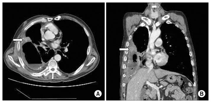
- Kronik ampiyem kenarında yeni kitle — plevral sıvı veya kalınlaşmış plevra boyunca, özellikle boşluğun dış yan duvarında gelişen solid yumuşak doku.
- Heterojen kitle içi — PAL'de düşük dansiteli nekrotik merkez ile çevresindeki daha solid doku birlikte bulunabilir.
- Ekstraplevral uzanım — plevral kaynaklı kitle interkostal yumuşak dokuyu ve göğüs duvarını aşabilir.
- Kaburga korteksinde destrüksiyon — komşu kemiğin erozyonunu kemik penceresinde doğrulayın; kronik sklerozdan farklıdır.
- Akciğere doğrudan yayılım — plevral kitle ile komşu parankim arasında silinen sınır veya parankim içi uzanım.
- Kalsifiye plevral kabuğun içe itilmesi — kitle eski empiyem duvarına bitişikse plevral kalsifikasyonun yer değiştirmesini izleyin.
- Lenfoma yayılımı — mediastinal/hiler lenf düğümleri, başka plevral odaklar ve akciğer infiltrasyonu için tüm toraksı tarayın.
- PAL genellikle kronik piyotoraks kavitesinin kenarında eksantrik, lentiküler veya hilal biçimli plevral yumuşak doku kitlesi olarak görülür; kitle heterojen olabilir ve nekroz içerebilir.
Ölçütler ve sınıflamalar
- WHO-HAEM5: Kronik inflamasyonla ilişkili diffüz büyük B hücreli lenfoma (CI-DLBCL)
- Piyotoraksla ilişkili lenfoma (PAL), WHO-HAEM5'te EBV ile ilişkili CI-DLBCL'nin prototipik biçimidir; EBV-pozitif DLBCL ile aynı sınıflandırma başlığı değildir.
Normalde
Normal parietal plevra ince bir çizgi olarak izlenir; kaburga korteksi kesintisizdir ve ampiyem boşluğu çevresinde yumuşak doku kitlesi bulunmaz. Şüpheli dokuyu kalsifiye plevral kabuğun, interkostal kasların ve karşı hemitorakstaki plevranın görünümüyle karşılaştırın; fokal kabarıklık, nekroz ve kemik korteksinde kayıp normal varyasyon değildir.
Normal BT ile kıyasla
Aynı bölgenin, kaynak veri setinde patoloji içermediği belirtilen bir BT incelemesini kesit kesit kaydırın; organ sınırlarını açarak anatomiyi hasta görüntüsüyle karşılaştırın. Küçük rastlantısal bulgular tümüyle dışlanmamıştır.
Kaynak: TotalSegmentator v2.01 veri seti (Wasserthal ve ark., Radiology: Artificial Intelligence 2023), CC BY 4.0, vaka s1278; organ sınırları veri setinin otomatik segmentasyonundan, birleştirilerek sadeleştirildi. Görüntüler 2,5 mm kesit aralığına yeniden örneklendi.
Ayırıcı tanı
- Kronik ampiyem
- plevral sıvı ve kalın/kalsifiye kabuk görülebilir; yeni solid, nekrotik kitle veya kaburga yıkımı malignite şüphesini artırır.
- Plevral mezotelyoma
- yaygın nodüler rind, fissür ve mediastinal plevra tutulumu lenfomadan çok mezotelyomayı düşündürür.
- Plevral metastaz
- çoklu nodüller veya bilinen primer tümörle eş zamanlı başka metastazlar ön planda olabilir.
- Göğüs duvarı sarkomu
- merkezî plevral boşluk ilişkisi yerine kemik veya yumuşak dokudan dışa büyüme odağı aranır.
- Tüberküloz ampiyemi
- kalın plevral kabuk, kalsifikasyon ve fistül yapabilir; yeni fokal solid kitle ayrıca araştırılmalıdır.
Tuzaklar
- Ampiyemde plevral kalınlaşma ve kalsifikasyon, tüberküloz plörezisinde ise komşu ekstraplevral yağ dansitesinde artış görülebilir; yeni kitleyi önceki incelemelerle karşılaştırın.
- PAL nekrozu, plevral enfeksiyon koleksiyonu gibi görünebilir; solid periferik doku ile kaburga veya göğüs duvarı invazyonunu ayrıca arayın.
- Kitleyi yalnız akciğer parankiminde aramak plevral kökeni kaçırabilir; eski ampiyem kavitesinin tüm çevresini ve interkostal uzanımı izleyin.
- PAL'de BT bulguları tanıyı düşündürür; tanısal değerlendirme için görüntüleme eşliğinde biyopsi ve patolojik inceleme gerekebilir.
Kendini dene
Yıllar önce tüberküloz için yapay pnömotoraks uygulanmış hastada kalsifiye ampiyem kenarında nekrotik kitle ve kaburga yıkımı var. En olası tanı?
Cevabı göster
Piyotoraksla ilişkili lenfoma. Kronik piyotoraks duvarına bitişik yeni solid kitle, nekroz ve göğüs duvarı invazyonu PAL için karakteristik bir bağlam oluşturur. Mezotelyoma genellikle yaygın rind yapar; soliter fibröz tümör iyi sınırlı plevral kitledir; ampiyem tek başına yeni solid tümör dokusunu açıklamaz.
BT'de tek taraflı plevral efüzyon, plevral kalınlaşma veya kitle ve fissür kalınlaşması görülen; ilerleyince akciğeri saran plevral rind oluşturabilen en olası malignite nedir?
Cevabı göster
Malign mezotelyoma. Malign mezotelyomada tek taraflı efüzyon, plevral kalınlaşma veya kitle ve fissür kalınlaşması görülebilir; ilerleyen hastalık akciğeri saran plevral rind oluşturabilir.
İlgili konular
Kaynaklar
Bu sayfadaki 40 cümle ve 2 quiz sorusu aşağıdaki kaynaklardan alıntılarla otomatik olarak karşılaştırıldı; 4 cümle bu karşılaştırmada düzeltildi (2026-10-09).
- Tumor and tumorlike conditions of the pleura and juxtapleural region: review of imaging findingspmc.ncbi.nlm.nih.gov
- Imaging Features of Various Benign and Malignant Tumors and Tumorlike Conditions of the Pleura: A Pictorial Reviewpmc.ncbi.nlm.nih.gov
- Large B-Cell Lymphomas in the 5th Edition of the WHO-Classification of Haematolymphoid Neoplasms—Updated Classification and New Conceptspmc.ncbi.nlm.nih.gov
- [Pyothorax-associated lymphoma with the expression of Epstein-Barr virus latent genes].pubmed.ncbi.nlm.nih.gov
- Case 322: Pyothorax-associated Lymphoma.pubmed.ncbi.nlm.nih.gov
- Primary Malignant Lymphoma Originating from the Chest Wall without Preceding Pleural Disease.pubmed.ncbi.nlm.nih.gov
- Imaging Features of Various Benign and Malignant Tumors and Tumorlike Conditions of the Pleura: A Pictorial Review.pubmed.ncbi.nlm.nih.gov
- Pyothorax-associated lymphoma: imaging findings.pubmed.ncbi.nlm.nih.gov
- Primary diffuse large B-cell lymphoma as a chest-wall mass: A case report.pubmed.ncbi.nlm.nih.gov
- A case report on the effect of rituximab on pyothorax-associated lymphoma.pubmed.ncbi.nlm.nih.gov
- The management of pyothorax-associated lymphoma using 18F-FDG PET/CT.pubmed.ncbi.nlm.nih.gov
- Pyothorax-associated lymphoma: complete remission achieved by chemotherapy alone.pubmed.ncbi.nlm.nih.gov
- Usefulness of diffusion-weighted magnetic resonance imaging-guided biopsy: pyothorax-associated lymphoma.pubmed.ncbi.nlm.nih.gov
- F-18 FDG PET/CT findings in two patients with pyothorax-associated lymphoma.pubmed.ncbi.nlm.nih.gov
- Usefulness of (18)F-FDG positron emission tomography/computed tomography for the diagnosis of pyothorax-associated lymphoma: A report of three cases.pubmed.ncbi.nlm.nih.gov
- Spinal Cord Compression due to Pyothorax-Associated Lymphoma: A Rare Presentation of EBV-Positive Diffuse Large B-Cell Lymphoma.pubmed.ncbi.nlm.nih.gov
- Primary Chest Wall Malignant Lymphoma in a Patient Without a History of Tuberculosis or Chronic Pyothorax: A Case Report With a Review of the Literature.pubmed.ncbi.nlm.nih.gov
- Diffuse Large B-Cell Lymphoma Arising in Postpneumonectomy Cavities: Report of Two Cases and Literature Review.pubmed.ncbi.nlm.nih.gov
- Imaging Findings of Thoracic Extranodal Lymphoma: A Pictorial Essay.pubmed.ncbi.nlm.nih.gov
- Three cases of esophagorespiratory fistula following neoadjuvant DCF therapy for resectable esophageal cancer.pubmed.ncbi.nlm.nih.gov
- Primary Pleural Epithelioid Angiosarcoma Presenting with Recurrent Haemorrhagic Pleural Effusions.pubmed.ncbi.nlm.nih.gov
- Mature anterior mediastinal teratoma presenting with trichoptysis: a case report.pubmed.ncbi.nlm.nih.gov
- Isolated pulmonary lymphangitic carcinomatosis as the initial manifestation of renal cell carcinoma.pubmed.ncbi.nlm.nih.gov
- Localised malignant mesothelioma presenting as an esophageal mass: An unusual manifestation.pubmed.ncbi.nlm.nih.gov
- Management of pneumonia by the emergency radiologist.pubmed.ncbi.nlm.nih.gov
- Case Report: Necrotizing pulmonary destruction and fatal gangrene: the imperative of early recognition.pubmed.ncbi.nlm.nih.gov
- Common and Rare Radiological Findings of Pulmonary and Thoracic Hydatid Cysts.pubmed.ncbi.nlm.nih.gov
- Case Report: A case of tuberculous empyema causing rupture of the diaphragm was misdiagnosed as diaphragmatic hernia.pubmed.ncbi.nlm.nih.gov
- Case Report: Pulmonary benign metastasizing leiomyoma with mild <sup>18</sup>F-FDG uptake occurring 30 years after hysterectomy.pubmed.ncbi.nlm.nih.gov
- Evaluation of benign and malignant pulmonary nodules based on clinical and contrast-enhanced computed tomography data: a multicenter study.pubmed.ncbi.nlm.nih.gov
- Imaging of synchronous multifocal renal masses: a case report.pubmed.ncbi.nlm.nih.gov
- Clinical TNM Lung Cancer Staging: A Diagnostic Algorithm with a Pictorial Review.pubmed.ncbi.nlm.nih.gov
- Pleural Mesothelioma Diagnosis for the Pulmonologist: Steps Along the Way.pubmed.ncbi.nlm.nih.gov
- Identification of CT Features to Differentiate Pulmonary Sarcoma from Carcinoma.pubmed.ncbi.nlm.nih.gov
- Epithelioid Angiosarcoma Presenting With Bilateral Hemothorax and Hemoptysis: A Case Report.pubmed.ncbi.nlm.nih.gov
- Blasto-ing through the chest wall: Empyema necessitans due to pulmonary blastomycosis.pubmed.ncbi.nlm.nih.gov
- Fibrin-associated large B-cell lymphoma arising within a hepatic cystic lesion: a case report.pubmed.ncbi.nlm.nih.gov
- Cavity-based lymphomas: challenges and novel concepts. A report of the 2022 EA4HP/SH lymphoma workshop.pubmed.ncbi.nlm.nih.gov
- Primary Pulmonary B-Cell Lymphoma: A Review and Update.pubmed.ncbi.nlm.nih.gov
- Role of thoracostomy in the management of postoperative empyema following lung resection: a narrative review of surgical strategies, evolving techniques, and outcomes.pubmed.ncbi.nlm.nih.gov
- Single-Position Minimally Invasive Retropleural Asymmetric Vertebral Column Resection and Percutaneous Pedicle Screw Fixation in the Lateral Position for Congenital Kyphoscoliosis.pubmed.ncbi.nlm.nih.gov
- Transsternal bronchial closure: a narrative review.pubmed.ncbi.nlm.nih.gov
- Lymphangiomatosis with pulmonary, mediastinal, and spinal involvement in a pediatric patient: a case report.pubmed.ncbi.nlm.nih.gov
- Tumor and tumorlike conditions of the pleura and juxtapleural region: review of imaging findings.pubmed.ncbi.nlm.nih.gov
- Imaging characteristics of pleural tumours.pubmed.ncbi.nlm.nih.gov
- The expert consensus document on minimally invasive upper lobectomy for lung cancer in China.pubmed.ncbi.nlm.nih.gov
- Free Pericardial Fat Pad Patch Repair Without Parenchymal Approximation for Severe Intraoperative Air Leak: A Technique to Optimize Limited Graft Volume.pubmed.ncbi.nlm.nih.gov
- The first CT-based classification system for pulmonary actinomycosis: correlating imaging patterns with therapeutic strategies and prognosis in a highly misdiagnosed disease.pubmed.ncbi.nlm.nih.gov
- Fundamentals of diagnostic ultrasonography in sheep and goat medicine: a comprehensive illustrated overview.pubmed.ncbi.nlm.nih.gov
- Overview of treatment related complications in malignant pleural mesothelioma.pubmed.ncbi.nlm.nih.gov
- International Harmonization of Nomenclature and Diagnostic Criteria (INHAND): Non-proliferative and Proliferative Lesions of the Non-human Primate ( M. fascicularis ).pubmed.ncbi.nlm.nih.gov
- Invasive thymoma with rapid response to cisplatin, doxorubicin and cyclophosphamide (PAC) chemotherapy, after an early postoperative recurrence-a case report.pubmed.ncbi.nlm.nih.gov
- Peritoneal Metastasis in Advanced Lung Adenocarcinoma Harboring EGFR Exon 19 Deletion/T790M with STRN::ALK Fusion and EGFR Kinase Domain Duplication: Two Case Reports and a Narrative Literature Summary.pubmed.ncbi.nlm.nih.gov
- Systematic review of PET imaging features and clinical applications in pulmonary NUT carcinoma.pubmed.ncbi.nlm.nih.gov
- Cytokeratin-Positive Pleomorphic Chest Wall Sarcoma Mimicking MPNST: A Case Report.pubmed.ncbi.nlm.nih.gov
- Lung ultrasound and the interstitial syndrome in tuberculosis endemic regions: illustrated insights from diverse clinical settings.pubmed.ncbi.nlm.nih.gov
- Comparison of Radiography and Ultrasound for Diagnosis of Rib Fractures in Canine Cadavers.pubmed.ncbi.nlm.nih.gov
- Case Report: Retained gallstone migrating to the chest wall after cholecystectomy presenting as chronic abscess, sinus tract, and inflammatory pseudotumor.pubmed.ncbi.nlm.nih.gov
- PET-CT in Clinical Adult Oncology: II. Primary Thoracic and Breast Malignancies.pubmed.ncbi.nlm.nih.gov
- Beyond the learning curve: a review of complex cases in robotic thoracic surgery.pubmed.ncbi.nlm.nih.gov
- A Rare Entity in Context: Breast Implant-Associated Anaplastic Large Cell Lymphoma Compared With Common Lymphoma Subtypes.pubmed.ncbi.nlm.nih.gov
- Cytologic Diagnosis of Large B-Cell Lymphoma: Diagnostic Approach and Differential Diagnosis.pubmed.ncbi.nlm.nih.gov
- Prognostic Value of the CALLY Index in Diffuse Large B-Cell Lymphoma: Linking Inflammation, Nutrition, and Tumor Biology.pubmed.ncbi.nlm.nih.gov
- Pulmonary <i>Nocardia paucivorans</i> Infection Mimicking Lung Cancer in an Immunocompetent Patient: A Case Report.pubmed.ncbi.nlm.nih.gov
- Early Identification of Children at Risk for Complicated Parapneumonic Pleural Effusion: Development of a Prediction Model.pubmed.ncbi.nlm.nih.gov
- Multidetector CT Findings and Differential Diagnoses of Malignant Pleural Mesothelioma and Metastatic Pleural Diseases in Korea.pubmed.ncbi.nlm.nih.gov
- CT, MRI and PET imaging in peritoneal malignancy.pubmed.ncbi.nlm.nih.gov
- Anaplastic Lymphoma Kinase Positive Aggressive Adult Lung Primary Inflammatory Myofibroblastoma Tumor: A Case and Literature Review.pubmed.ncbi.nlm.nih.gov
- Suspected Paramalignant Pleural Effusion as the Primary Presentation of Disseminated Cholangiocarcinoma in a Cat.pubmed.ncbi.nlm.nih.gov
- Incidental Lung Lesions on the CT Component of Oncologic FDG PET/CT: A Pictorial Review of Interpretive Pitfalls and Diagnostic Clues.pubmed.ncbi.nlm.nih.gov
- Askin Tumor Presents with Ewing Sarcoma of the Femur in Young Female Patient: Case Report and Literature Review.pubmed.ncbi.nlm.nih.gov
- First case description of a highly aggressive thoracopulmonary primitive neuroectodermal tumor in a Moroccan adult male.pubmed.ncbi.nlm.nih.gov
- A case of complicated parapneumonic pleural effusion caused by paragonimus infection in a child was successfully treated by intrapleural injection of urokinase: a case report and literature review.pubmed.ncbi.nlm.nih.gov
- Deepening Understanding of the Clinical Features and Diagnostic Approaches to Anti-Interferon-Gamma Autoantibody Associated Adult-Onset Immunodeficiency in the Last 20 Years: A Case Report and Literature Review.pubmed.ncbi.nlm.nih.gov
- Pulmonary manifestations in hyper IgE syndrome: A case series and review of Indian literature.pubmed.ncbi.nlm.nih.gov
- Pleural Neoplasms-What Could MRI Change?pubmed.ncbi.nlm.nih.gov
- Patterns of injury and violence in Yaoundé Cameroon: an analysis of hospital data.pubmed.ncbi.nlm.nih.gov
- Clinical phenotypes of secondary spontaneous pneumothorax (SSP) in different underlying lung diseases: a retrospective study and literature review on the clinical heterogeneity in this single entity.pubmed.ncbi.nlm.nih.gov
- Biocompatibility of a novel lung sealant based on functionalized polyoxazolines in an ovine model of parenchymal lung injury.pubmed.ncbi.nlm.nih.gov
- COVID-19-Associated Pulmonary Mucormycosis.pubmed.ncbi.nlm.nih.gov
- Retrospective Review Is the Best View: A Primer for Residents and Radiologists on Missed Findings in Chest Radiography.pubmed.ncbi.nlm.nih.gov
- Intraoperative Contrast-Enhanced Ultrasonography (Io-CEUS) to Detect Missed Lung Nodules During Robot-Assisted Thoracoscopic Surgery.pubmed.ncbi.nlm.nih.gov
- Long-term outcomes and patterns of failure in patients with thymic malignancies treated with surgery and radiation therapy.pubmed.ncbi.nlm.nih.gov
- Comparative Interpretation of CT and Standard Radiography of the Pleura.pubmed.ncbi.nlm.nih.gov
- Congenital pulmonary airway malformation mimicking lung cancer in adults: a pitfall in oncologic diagnosis.pubmed.ncbi.nlm.nih.gov
- Case Report: Delayed appearance of the ablation margin after CT-guided microwave ablation of a solid pulmonary nodule.pubmed.ncbi.nlm.nih.gov
- Pathologic findings and causes of death of stranded cetaceans in the Canary Islands (2006-2012).pubmed.ncbi.nlm.nih.gov
- Rare case of primary extranodal marginal zone lymphoma of the thorax.pubmed.ncbi.nlm.nih.gov
- State-of-the-Art Mediastinal Staging in Non-Small-Cell Lung Cancer: Integration of Combined Endosonographic Techniques with Updated IASLC TNM 9th Classification.pubmed.ncbi.nlm.nih.gov
- The 9th Edition of the UICC TNM Classification of Malignant Tumours: Updates and Rationale for Change.pubmed.ncbi.nlm.nih.gov
- Pyothorax-associated lymphoma mimicking chronic expanding hematoma.pubmed.ncbi.nlm.nih.gov
- Radiographic findings useful for diagnosis of primary chest wall lymphoma without preceding pleural disease: A case report.pubmed.ncbi.nlm.nih.gov
- Probable Pleural-Based Pulmonary Tuberculoma Mimicking Lung Malignancy: A Case Report.pubmed.ncbi.nlm.nih.gov
- Radiologist's Guide to the Ninth Edition TNM Staging System for Quantitative and Qualitative Assessment of Pleural Mesothelioma.pubmed.ncbi.nlm.nih.gov
- Positron emission tomography/computed tomography in risk-factor-negative young female with malignant pleural mesothelioma: A case report and review of literature.pubmed.ncbi.nlm.nih.gov
- Plasmablastic Richter Transformation of Chronic Lymphocytic Leukemia With Aberrant Epithelial Marker Expression Presenting as a Pleural Effusion: A Diagnostic Challenge.pubmed.ncbi.nlm.nih.gov
- Bilateral primary adrenal lymphoma presenting with chylothorax: imaging findings and diagnostic challenges.pubmed.ncbi.nlm.nih.gov
- Cavitary Pulmonary Adenocarcinoma in a Young Adult With a History of Treated Hodgkin Lymphoma: A Case Report.pubmed.ncbi.nlm.nih.gov
- First-Line Treatment of Advanced Thoracic SMARCA4-Deficient Undifferentiated Tumor: A Case Report and Review of the Literature.pubmed.ncbi.nlm.nih.gov
- Rare case of primary pleural lymphoma presenting with pleural effusion.pubmed.ncbi.nlm.nih.gov
- Cellular Plasticity in Malignant Transformation: Mesothelial Cells.pubmed.ncbi.nlm.nih.gov
- Antifungal Management in Patients with Hematological Malignancies: From Primary Prophylaxis to Empirical and Diagnosis-Driven Treatmentpubmed.ncbi.nlm.nih.gov
- Epidemiological Features, Etiopathogenesis, Diagnostic Evaluation, and Management Protocol of Splenic Tuberculosis: A Systematic Review.pubmed.ncbi.nlm.nih.gov
- EBV-Positive Lymphoproliferations of B- T- and NK-Cell Derivation in Non-Immunocompromised Hosts.pubmed.ncbi.nlm.nih.gov
- Fibrin-Associated Large B-Cell Lymphoma of Prosthetic Aortic Valve and Aortic Root Graft.pubmed.ncbi.nlm.nih.gov
- Diagnosis of pyogranulomatous and neutrophilic lymphadenitis in 72 cats presenting to a referral hospital: with a focus on nine cats with steroid-responsive lymphadenitis.pubmed.ncbi.nlm.nih.gov
- Retrospective study of 540 cats with respiratory diseases in Japan (2003-2020).pubmed.ncbi.nlm.nih.gov
Eğitim amaçlıdır; hasta başında tanı ve tedavi kararının yerine geçmez, tıbbi tavsiye değildir. Metinler yapay zekâ desteğiyle yazıldı ve otomatik bir adımda kaynak alıntılarıyla karşılaştırıldı; bu bir tıbbi doğrulama değildir ve içerik uzman radyolog incelemesinden geçmedi. Klinik kararlarda güncel kılavuzları, kurum protokollerini ve radyoloji raporunu esas alın.