Özet
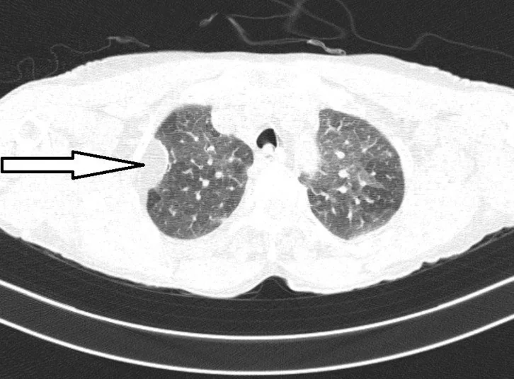
Plevral lipom, çoğu kez başka nedenle çekilen toraks BT'sinde saptanan, plevra veya ekstraplevral yağ dokusuyla ilişkili iyi huylu bir kitle görünümüdür. Kontrastsız BT, makroskopik yağ içeriğini ve lezyonun göğüs duvarı, plevra ve akciğerle ilişkisini göstererek karakterizasyonda temel rol oynar. Yağ içeren kitlede kalın septa, nodüler yumuşak doku veya komşu yapıya infiltrasyon liposarkom kuşkusunu artırır; BT her olguda histolojik ayrımı kesinleştiremez. Agresif bileşeni gözden kaçırmak malign tümörün yanlışlıkla benign kabul edilmesine yol açabilir.
Faz ve pencere
- Kontrastsız tanısal
- Kontrastsız toraks BT'sinde homojen makroskopik yağ çoğunlukla yaklaşık −50 ile −150 HU aralığında ölçülür; düzgün sınırlı kitle göğüs duvarıyla geniş/obtüz açı yapıp akciğer ve damarları içe doğru itebilir. Lipom için iyotlu kontrast gerekmez; kontrastlı inceleme mevcutsa belirgin kontrastlanan yumuşak doku nodülü veya kalın septa ayrıca kuşkulu kabul edilir.
Önerilen pencereler G genişlik, M merkez; değerler Hounsfield birimi (HU)
- AkciğerG 1500 / M −600
- MediastenG 350 / M +50
- Yumuşak dokuG 400 / M +40
BT bulguları
- Homojen makroskopik yağ — plevraya veya göğüs duvarına komşu, düzgün sınırlı lezyonda negatif HU değerleri lipomatöz içeriği destekler.
- Obtüz göğüs duvarı açısı — kitlenin akciğer parankiminden çok plevral/ekstraplevral kökenli olduğunu düşündürür; tek başına kesin lokalizasyon ölçütü değildir.
- Plevraya geniş tabanla oturan kitle plevral köken lehinedir.
- Akciğerin içe itilmesi — komşu parankim ve damarların kitle kenarından uzaklaşması dıştan basıyı gösterir.
- İnce fibröz septalar — lipom içinde görülebilir; kalın, düzensiz ya da nodüler yumuşak dokuya dönüşen septal yapı daha kuşkuludur.
- Kontrastlanan nodüler bileşen — yağ dışı solid odak ve heterojen kitle görünümü liposarkom olasılığını artırır.
- İnvaziv sınır — göğüs duvarı kasına, kaburgaya veya komşu mediastinal yapılara uzanım benign lipom görünümünü desteklemez.
- Homojen makroskopik yağ içeren lezyon lipomayı destekler; iyi diferansiye liposarkomda fibröz septalar lipomdakilerden daha geniş ve nodüler olabilir, görüntüleme ayrımı her zaman kesinleştirmediğinden kesin tanı için histolojik değerlendirme gerekir.
Ölçütler ve sınıflamalar
- Makroskopik yağ atenüasyonu
- −50 ila −150 HU
- Genel yağlı tümör boyutu (plevraya özgü değil)
- Ekstraplevral lipomatöz tümörler için sistematik derlemede ALT/WDL ile ilişkili sık görüntüleme özelliklerinden biri ≥110 mm boyuttur; bu eşik plevraya özgü değildir.
Normalde
Kesitsel görüntülemede kitlenin parietal plevrayla bağlantısını ve akciğere bası yapıp yapmadığını değerlendirin; bu bulgular kökeni belirlemeye yardımcı olabilir.
Normal BT ile kıyasla
Aynı bölgenin, kaynak veri setinde patoloji içermediği belirtilen bir BT incelemesini kesit kesit kaydırın; organ sınırlarını açarak anatomiyi hasta görüntüsüyle karşılaştırın. Küçük rastlantısal bulgular tümüyle dışlanmamıştır.
Kaynak: TotalSegmentator v2.01 veri seti (Wasserthal ve ark., Radiology: Artificial Intelligence 2023), CC BY 4.0, vaka s1278; organ sınırları veri setinin otomatik segmentasyonundan, birleştirilerek sadeleştirildi. Görüntüler 2,5 mm kesit aralığına yeniden örneklendi.
Ayırıcı tanı
- Lipomatoz, birden fazla lipomun bulunduğu ve tek bir lezyonla sınırlı olmayan yağlı kitle tablosudur.
- İyi diferansiye liposarkom
- yağ içerse de kalın düzensiz septa, nodüler non-adipoz odak ve infiltratif sınır gösterebilir.
- Teratom
- yağla birlikte kalsifikasyon ve sıvı-yumuşak doku bileşenleri içerebilir; ön mediastinal yerleşim ayırt ettirir.
- Plevral efüzyon içinde de iyi sınırlı yağ atenüasyonlu nodüller pleural lipomatozis ile ilişkili olabilir; bu nedenle efüzyon içindeki yağlı odaklar plevral lipom ayırıcı tanısında yer alabilir.
Tuzaklar
- Negatif HU değerleri tek başına lipom tanısını kesinleştirmez; plevral liposarkomda da yağ atenüasyonlu alanlar görülebilir ve bu radyolojik özellikler özgül değildir.
- Plevral tabanlı bir kitle akciğer içi kitle sanılabilir; koronal ve sagittal rekonstrüksiyonlarda obtüz açı, ekstraplevral yağ ve akciğerin itilme yönünü birlikte izleyin.
- Benign lipomlar ince septa içerebilir; malignite kuşkusunu tek septaya değil, kalın/nodüler septalara, kontrastlanan yumuşak dokuya ve invazyona dayandırın.
- Plevral lipomun kendiliğinden liposarkoma dönüştüğü varsayımı desteklenmez; asıl sorun, baştan farklı histolojideki yağlı malign tümörün lipom sanılmasıdır.
Kendini dene
Göğüs duvarına obtüz açıyla oturan, homojen −100 HU kitle akciğeri içe itiyor. En olası tanı nedir?
Cevabı göster
Plevral lipom. Homojen makroskopik yağ, düzgün sınır ve dıştan akciğer basısı lipomu destekler. Efüzyon sıvı atenüasyonundadır; teratomda ek doku/kalsifikasyon bileşenleri beklenir. Kalın nodüler septa veya invazyon görülseydi liposarkom daha güçlü bir olasılık olurdu.
Yağ içeren plevral kitlede malignite kuşkusunu en çok hangi eşlikçi görünüm artırır?
Cevabı göster
Kontrastlanan nodüler septa. Kalın veya nodüler yumuşak doku septaları ve kontrastlanan non-adipoz bileşen liposarkom lehine uyarıcıdır. Düzgün kapsül, homojen yağ ve komşu akciğerin itilmesi daha çok benign kitle davranışıdır; histolojik doğruluk yine görüntülemeyle garanti edilemez.
Kaynaklar
Bu sayfadaki 38 cümle ve 2 quiz sorusu aşağıdaki kaynaklardan alıntılarla otomatik olarak karşılaştırıldı; 6 cümle bu karşılaştırmada düzeltildi (2026-10-09).
- Pleural lipomatosis: An often-forgotten intrathoracic tumorpmc.ncbi.nlm.nih.gov
- Primary pleural liposarcoma: A rare entitypmc.ncbi.nlm.nih.gov
- Imaging of the pleura.pubmed.ncbi.nlm.nih.gov
- Pleural lipoma: a non-surgical lesion?pubmed.ncbi.nlm.nih.gov
- Parietal pleura lipoma: a rare intrathoracic tumor.pubmed.ncbi.nlm.nih.gov
- A rare case of huge intrathoracic lipomatosis filling the right pleural cavity removed by open thoracic surgery: A case report.pubmed.ncbi.nlm.nih.gov
- Primary pleural liposarcoma combined spindle cell lipoma of the lung.pubmed.ncbi.nlm.nih.gov
- Intrathoracic lipoma of the chest wall that appeared relatively rapidly and could be resected and diagnosed by minimally invasive thoracoscopic surgery: A case report.pubmed.ncbi.nlm.nih.gov
- A case of pleural lipoma evaluated with multi-imaging methods.pubmed.ncbi.nlm.nih.gov
- Surgical resection of pleural lipoma after 9 years of serial imaging.pubmed.ncbi.nlm.nih.gov
- Huge Intrathoracic Lipoma Occupying the Right Hemithorax.pubmed.ncbi.nlm.nih.gov
- Epipericardial Fat Necrosis in COVID-19-A Case Report.pubmed.ncbi.nlm.nih.gov
- Mediastinal Lymphangioma of the Adult: Case Report and Literature Review.pubmed.ncbi.nlm.nih.gov
- Lung colloid adenocarcinoma: A multidisciplinary diagnostic challenge.pubmed.ncbi.nlm.nih.gov
- Valvular and Outflow Tract Metastases From Germ Cell Tumors: Contrasting Left-Sided Embolism and Right-Sided Silence.pubmed.ncbi.nlm.nih.gov
- Pericardial Diseases: International Position Statement on New Concepts and Advances in Multimodality Cardiac Imaging.pubmed.ncbi.nlm.nih.gov
- A pictorial essay of thoracic wall diseases: multiple pathologies in the same anatomical site.pubmed.ncbi.nlm.nih.gov
- Comments and Illustrations of the European Federation of Societies for Ultrasound in Medicine Guidelines: Benign Pleura Lesions (Benign Pleura Thickening, Lesions and Masses)-What Can Be Seen on Transthoracic Ultrasound?pubmed.ncbi.nlm.nih.gov
- Imaging of duodenal lipoma, case report of a rare tumor of the gastro intestinal tract.pubmed.ncbi.nlm.nih.gov
- Sarcomas of fat and bone: a case report.pubmed.ncbi.nlm.nih.gov
- Spinal lesions: a comprehensive radiologic overview.pubmed.ncbi.nlm.nih.gov
- Mesenchymal Tumors of the Breast: Fibroblastic/Myofibroblastic Lesions and Other Lesions.pubmed.ncbi.nlm.nih.gov
- Probable Pleural-Based Pulmonary Tuberculoma Mimicking Lung Malignancy: A Case Report.pubmed.ncbi.nlm.nih.gov
- Metastatic Carotid Body Paraganglioma Following Three Decades of Indolent Growth: A Case Report.pubmed.ncbi.nlm.nih.gov
- Intrapulmonary solitary fibrous tumor: clinicopathological analysis of 29 cases with emphasis on morphological spectrum and immunohistochemical profile.pubmed.ncbi.nlm.nih.gov
- Endobronchial lipoma presenting as a fat-attenuation lesion causing left upper lobar bronchial obstruction: A case report.pubmed.ncbi.nlm.nih.gov
- Case Report: Primary cardiac atypical lipoblastoma in an adolescent female.pubmed.ncbi.nlm.nih.gov
- Prenatally detected primary renal teratoma in an infant: A case report in Vietnam.pubmed.ncbi.nlm.nih.gov
- Acute Traumatic Aortic Injury: What the Radiologist Needs to Know.pubmed.ncbi.nlm.nih.gov
- Clinical TNM Lung Cancer Staging: A Diagnostic Algorithm with a Pictorial Review.pubmed.ncbi.nlm.nih.gov
- Tips and Tricks in Thoracic Radiology for Beginners: A Findings-Based Approach.pubmed.ncbi.nlm.nih.gov
- Primary Chest Wall Glomus Tumor Resected by Video-Assisted Thoracoscopic Surgery: A Case Report with 10-Year Follow-up.pubmed.ncbi.nlm.nih.gov
- Role of thoracostomy in the management of postoperative empyema following lung resection: a narrative review of surgical strategies, evolving techniques, and outcomes.pubmed.ncbi.nlm.nih.gov
- Surgical Repair of Morgagni Hernia With Liver Herniation and Rib Fractures in an Adult Following Rhinovirus-induced Coughing.pubmed.ncbi.nlm.nih.gov
- Application value of high-resolution CT imaging features combined with texture analysis in patients with solitary pulmonary nodules.pubmed.ncbi.nlm.nih.gov
- Case Report: Successful conversion surgery following chemo-antiangiogenic therapy in a lung adenocarcinoma patient with active rheumatoid arthritis and rare EGFR mutation after immunotherapy-triggered flare.pubmed.ncbi.nlm.nih.gov
- Analysis of risk factors for prolonged postoperative chest tube drainage after uniportal video-assisted thoracoscopic surgery pulmonary resection.pubmed.ncbi.nlm.nih.gov
- Computed tomography and magnetic resonance imaging features of struma ovarii: A retrospective case series.pubmed.ncbi.nlm.nih.gov
- Benign Chest Wall Lipoblastoma in a 21-month-old Child: A Literature Review.pubmed.ncbi.nlm.nih.gov
- Ovarian Low-Grade Mucinous Tumor Combined with Pseudo-Meigs' Syndrome: Report of Two Cases and Literature Review.pubmed.ncbi.nlm.nih.gov
- Echoes of a forgotten lesion: Bilateral lung masses and spontaneous pneumothorax: A clinicopathologic conference.pubmed.ncbi.nlm.nih.gov
- Imaging Diagnosis-Peritoneal Splenosis in a Dog: Computed Tomographic Findings.pubmed.ncbi.nlm.nih.gov
- Cosmic Signs in Radiology: A Pictorial Review.pubmed.ncbi.nlm.nih.gov
- Cytokeratin-Positive Pleomorphic Chest Wall Sarcoma Mimicking MPNST: A Case Report.pubmed.ncbi.nlm.nih.gov
- First case description of a highly aggressive thoracopulmonary primitive neuroectodermal tumor in a Moroccan adult male.pubmed.ncbi.nlm.nih.gov
- Management of Breast Phyllodes Tumors: A Grade-Stratified Surgical and Reconstructive Strategy.pubmed.ncbi.nlm.nih.gov
- Symptomatic pleural lipoma: a case report from Saudi Arabia.pubmed.ncbi.nlm.nih.gov
- Mediastinal Lipoma: An Unusual Cause of Progressive Dyspnea.pubmed.ncbi.nlm.nih.gov
- Effect of surgical management for anterior mediastinal large mass: a retrospective cohort study.pubmed.ncbi.nlm.nih.gov
- Primary liposarcoma of the pericardium: report of a rare case with clinicopathological features and imaging insights.pubmed.ncbi.nlm.nih.gov
- Solitary fibrous tumor: an updated review.pubmed.ncbi.nlm.nih.gov
- Large extrapleural hematoma in a patient with Stanford type B acute aortic dissection: a case report.pubmed.ncbi.nlm.nih.gov
- Lipothorax Following Autologous Fat Grafting to the Breast.pubmed.ncbi.nlm.nih.gov
- Multidisciplinary collaborative treatment of a rare case of chronic hemoptysis: diagnostic and therapeutic insights from a 7-year clinical course.pubmed.ncbi.nlm.nih.gov
- Retrospective Review Is the Best View: A Primer for Residents and Radiologists on Missed Findings in Chest Radiography.pubmed.ncbi.nlm.nih.gov
- Thoracic joint abnormalities and impaired thoracic motion in idiopathic pleuroparenchymal fibroelastosis: an exploratory study using inspiratory and expiratory CT.pubmed.ncbi.nlm.nih.gov
- Restricted Diffusion on Diffusion-Weighted MRI in a Cat With Orbital Lymphoma and Presumptive Pulmonary Lymphoma.pubmed.ncbi.nlm.nih.gov
- Lipomatosis From Prolonged Use of Corticosteroids in Ethiopia: A Case Report.pubmed.ncbi.nlm.nih.gov
- The diagnostic value of ultrasound-guided percutaneous core needle biopsy of musculoskeletal soft tissue lesions.pubmed.ncbi.nlm.nih.gov
- Long-term safety and effectiveness of the "OptEase" vena cava filter.pubmed.ncbi.nlm.nih.gov
- Hybrid eclipse: a case of isolated renal tuberculosis with an unusual infectious-inflammatory-neoplastic triad.pubmed.ncbi.nlm.nih.gov
- General anesthesia with endotracheal intubation in Madelung's Disease.pubmed.ncbi.nlm.nih.gov
- Multiple surgical excision for bilateral multiple mediastinal liposarcoma: a case report.pubmed.ncbi.nlm.nih.gov
- Giant primary mediastinal well-differentiated liposarcoma with bilateral thoracic extension causing complete left lung collapse.pubmed.ncbi.nlm.nih.gov
- Rare Late Cardiac Involvement Following Resected Primary Limb Dedifferentiated Liposarcoma: A Case Report.pubmed.ncbi.nlm.nih.gov
- Giant mature mediastinal teratoma mimicking a pericardial cyst.pubmed.ncbi.nlm.nih.gov
- Rapidly growing anterior mediastinal teratoma in a young adult female: successful resection via robotic-assisted thoracoscopic surgery.pubmed.ncbi.nlm.nih.gov
- Giant Mediastinal Teratoma in Infancy: An Uncommon Cause of Respiratory Compromise.pubmed.ncbi.nlm.nih.gov
- A 37-Year-Old Man With Pleuritic Chest Pain.pubmed.ncbi.nlm.nih.gov
- Comparative Interpretation of CT and Standard Radiography of the Pleura.pubmed.ncbi.nlm.nih.gov
- Radiological review of pleural tumors.pubmed.ncbi.nlm.nih.gov
- Spatial non-uniformity of liver virtual non-contrast attenuation on dual-layer spectral CT: Impact of body habitus and peripheral right-lobe error.pubmed.ncbi.nlm.nih.gov
- Understanding Photon-Counting CT: Physics, Detector Technology, and Image Reconstructions.pubmed.ncbi.nlm.nih.gov
- Plaque-Adapted Virtual Monoenergetic Image Selection for Carotid Plaque Visualization Using Photon-Counting CT.pubmed.ncbi.nlm.nih.gov
- Management of pneumonia by the emergency radiologist.pubmed.ncbi.nlm.nih.gov
- Exploring Extrapleural Hematoma After Ultrasound-Guided Paravertebral Block: A Clinical Case Report.pubmed.ncbi.nlm.nih.gov
- Malignant Pleural Mesothelioma CT Imaging: How to Measure It Correctly?pubmed.ncbi.nlm.nih.gov
- Automated Deep Learning Detection of Hepatic Steatosis on Noncontrast Computed Tomography Scans and Discrepancy With Scan Reports.pubmed.ncbi.nlm.nih.gov
- The diagnostic value of virtual unenhanced spectral CT images in acute pancreatitis: a comparison with true unenhanced images.pubmed.ncbi.nlm.nih.gov
- Ultrasound elastography in pediatric care: bridging innovation and noninvasive diagnostics.pubmed.ncbi.nlm.nih.gov
- Imaging of Thoracic Wall Abnormalities.pubmed.ncbi.nlm.nih.gov
- Multidetector CT Findings and Differential Diagnoses of Malignant Pleural Mesothelioma and Metastatic Pleural Diseases in Korea.pubmed.ncbi.nlm.nih.gov
- Solitary fibrous tumor of the ischioanal fossa-a multidisciplinary approach to management with radiologic-pathologic correlation.pubmed.ncbi.nlm.nih.gov
- Primary Synovial Sarcoma of the Prostate: A Case Report and Review of the Literature.pubmed.ncbi.nlm.nih.gov
- Pancreatic Tail Leiomyosarcoma With Hepatic Metastases: Diagnostic and Therapeutic Challenges: A Case Report.pubmed.ncbi.nlm.nih.gov
- Primary Chest Wall Ewing Sarcoma With Diaphragmatic Invasion in an Adult: En-Bloc Resection and Polypropylene Mesh Reconstruction.pubmed.ncbi.nlm.nih.gov
- Sequential EBUS-Guided Fine Needle Aspiration and Transbronchial Cryobiopsy for the Diagnosis of a Giant Mediastinal Lipoma: A Novel Intraoperative Pivot Strategy.pubmed.ncbi.nlm.nih.gov
- Advanced imaging techniques and artificial intelligence in pleural diseases: a narrative review.pubmed.ncbi.nlm.nih.gov
- Unlocking the Potential of Chest MRI: Strategies for Establishing a Successful Practice.pubmed.ncbi.nlm.nih.gov
- Well-Differentiated Liposarcoma of the Spermatic Cord Involving Testicular Parenchyma.pubmed.ncbi.nlm.nih.gov
- Rare co-occurrence of parosteal lipoma and growing osteochondroma: A case report.pubmed.ncbi.nlm.nih.gov
- Retroperitoneal Masses Decoded: A Practical Multidisciplinary Diagnostic and Surgical Strategy Integrating Radiology and Pathology.pubmed.ncbi.nlm.nih.gov
- Surgical resection of a >50 cm recurrent retroperitoneal spindle cell sarcoma complicated by pulmonary embolism: a case report.pubmed.ncbi.nlm.nih.gov
- Fat-containing soft-tissue tumors: Imaging findings and pathologic correlation.pubmed.ncbi.nlm.nih.gov
- MRI findings for differentiating benign and malignant soft tissue tumors: a systematic review-part 2: key imaging findings.pubmed.ncbi.nlm.nih.gov
- Use of radiologic imaging to differentiate lipoma from atypical lipomatous tumor/well-differentiated liposarcoma: Systematic review.pubmed.ncbi.nlm.nih.gov
- Analysis of MRI features correlated with TNM staging in thymic epithelial tumors.pubmed.ncbi.nlm.nih.gov
- Primary thymic papillary adenocarcinoma with a rare CD5/CD117-negative and PAX8/WT1-positive immunophenotype: A case report.pubmed.ncbi.nlm.nih.gov
- Perioperative Decision-Making for a Solitary Fibrous Tumor of the Pleura: Lessons from Two Cases and Current Evidence.pubmed.ncbi.nlm.nih.gov
- Deep Learning Reconstruction Enhances Lung Cancer CT Imaging.pubmed.ncbi.nlm.nih.gov
- Giant mediastinal lipoma with bilateral hemithoracic extension in a young woman: a case report.pubmed.ncbi.nlm.nih.gov
- Identification of CT Features to Differentiate Pulmonary Sarcoma from Carcinoma.pubmed.ncbi.nlm.nih.gov
- Endobronchial epithelioid leiomyoma (leiomyoblastoma) causing complete right-lung atelectasis: A case report.pubmed.ncbi.nlm.nih.gov
- Extranodal nasal-orbital communicating lesions NK/T cell lymphoma with ocular symptoms as the initial manifestation misdiagnosed as sinusitis and orbital cellulitis: a case report and literature review.pubmed.ncbi.nlm.nih.gov
- Pulmonary angiosarcoma in an HIV-positive patient presenting with hemoptysis and multisystem involvement: Report of a rare case.pubmed.ncbi.nlm.nih.gov
- Local fluid application and needle insertion in gravity-dependent areas to reduce the risk of pneumothorax during lung nodule localization: a single-center observational study.pubmed.ncbi.nlm.nih.gov
- Diagnosis and management of pleural mesothelioma: three unique cases and review of the literature.pubmed.ncbi.nlm.nih.gov
- Case Report: Spontaneous rupture of an internal thoracic artery aneurysm: a rare life-threatening emergency and Its therapeutic challenges.pubmed.ncbi.nlm.nih.gov
- The role of thoracoplasty in modern thoracic surgery: a narrative review with three illustrative cases.pubmed.ncbi.nlm.nih.gov
- Application of postmortem imaging modalities in cases of sudden death due to cardiovascular diseases-current achievements and limitations from a pathology perspective : Endorsed by the Association for European Cardiovascular Pathology and by the International Society of Forensic Radiology and Imaging.pubmed.ncbi.nlm.nih.gov
- State of the Art: Lung Cancer Staging Using Updated Imaging Modalities.pubmed.ncbi.nlm.nih.gov
- Role of Interventional Radiology in the Management of Non-aortic Thoracic Trauma.pubmed.ncbi.nlm.nih.gov
Eğitim amaçlıdır; hasta başında tanı ve tedavi kararının yerine geçmez, tıbbi tavsiye değildir. Metinler yapay zekâ desteğiyle yazıldı ve otomatik bir adımda kaynak alıntılarıyla karşılaştırıldı; bu bir tıbbi doğrulama değildir ve içerik uzman radyolog incelemesinden geçmedi. Klinik kararlarda güncel kılavuzları, kurum protokollerini ve radyoloji raporunu esas alın.