Özet
Renal arter diseksiyonunda intimal yırtık kanı damar duvarına veya yalancı lümene yönlendirerek gerçek lümeni daraltabilir; eşlik eden tromboz distal perfüzyonu bozabilir. MDCT’de renal arter diseksiyonu intimal flep veya tromboze yalancı lümene bağlı gerçek lümen stenozuyla, renal infarkt ise parankimal perfüzyon defektiyle gösterilebilir. İnce flep, eksantrik mural hematom, yalancı lümen ya da ani dal kesilmesi doğrudan damar işaretleridir. Damar lezyonunun atlanması böbrek iskemisinin kaynağını ve olası iki taraflı/ana arter tutulumunu gözden kaçırabilir.
Faz ve pencere
- BTA tanısal
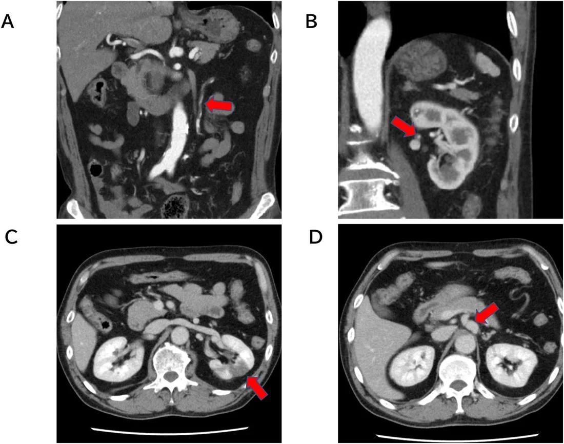
- Arteriyel fazlı ince kesit BT'de renal arter boyunca intimal flep, tromboze yalancı lümen ve bu duruma bağlı gerçek lümen daralması izlenir.
- Nefrografik
- Nefrografik parankim fazında diseksiyonun beslediği segmentte kama biçimli parankimal perfüzyon defekti görülebilir.
Önerilen pencereler: Anjiyo (G 600 / M 150), Batın (G 400 / M 50).
BT bulguları
- Renal arter lümeni içinde intimal flep, gerçek ve yalancı lümen sınırını belirten çizgisel bir yapı olarak izlenebilir.
- Renal arter duvarı boyunca uzanan eksantrik düşük atenüasyonlu alan, tromboze olmuş yalancı lümen veya intramural hematomu düşündürebilir.
- Yalancı lümen tromboze olduğunda renal arter duvarı boyunca eksantrik düşük atenüasyonlu bir alan izlenir.
- Trombüs, renal arterde dolum defekti veya damarın ani sonlanması şeklinde görünür; distal dal opasifikasyonu azalabilir.
- İlgili dalın beslediği parankimde kama biçimli perfüzyon defekti renal infarkt ile uyumludur.
- Renal arter diseksiyonuna fibromüsküler displazi eşlik edebilir.
Ölçütler ve sınıflamalar
- BT’de renal arter diseksiyonu lehine doğrudan bulgular
- BT’de tanı, renal arterde intimal flep görülmesine veya tromboze yalancı lümeni düşündüren eksantrik mural düşük atenüasyona dayanır.
Normalde
Normal renal arter BTA’da aortadan çıkan ana gövde düzenli çapta ve homojen kontrast dolumuyla segmental dallara ayrılır; lümen içinde çizgisel flep veya duvar boyunca eksantrik dolum kusuru bulunmaz. Damar lezyonu değerlendirilirken karşı böbrek ve aort ile karşılaştırma yapılmalı, çok düzlemli rekonstrüksiyonlarda damar yolu takip edilmelidir.
Normal BT ile kıyasla
Aynı bölgenin, kaynak veri setinde patoloji içermediği belirtilen bir BT incelemesini kesit kesit kaydırın; organ sınırlarını açarak anatomiyi hasta görüntüsüyle karşılaştırın. Küçük rastlantısal bulgular tümüyle dışlanmamıştır.
Kaynak: TotalSegmentator v2.01 veri seti (Wasserthal ve ark., Radiology: Artificial Intelligence 2023), CC BY 4.0, vaka s0115; organ sınırları veri setinin otomatik segmentasyonundan, birleştirilerek sadeleştirildi. Görüntüler 2,5 mm kesit aralığına yeniden örneklendi.
Ayırıcı tanı
- Renal arter embolisinde MDCT’de intra-arteriyel dolum defekti görülebilir; diseksiyon lehine intimal flep veya tromboze yalancı lümene ait eksantrik mural düşük atenüasyon aranır.
- Fibromüsküler displazi
- damar duvarında karakteristik 'string-of-beads' (boncuk dizisi) görünümü oluşturur; akut intimal flep veya mural hematom oluşturmaz.
- Renal arter anevrizması, takip görüntülemesinde renal arterde anevrizmal değişiklik olarak izlenebilir.
- Aort diseksiyonunun renal artere uzanımı
- intimal flep aortada da izlenir ve renal ostiuma uzanabilir; tüm aorta ve ana dallar taranmalıdır.
- Vazospazm
- damarda düzgün, geçici daralma oluşturabilir; damar duvarında flep, mural hematom veya yalancı lümen yoktur.
Tuzaklar
- Küçük intimal flep parsiyel hacim etkisiyle kaybolabilir; renal arter boyunca aksiyel kesitlerin yanında koronal ve oblik rekonstrüksiyonları da incele.
- Tromboze yalancı lümen, eksantrik düşük atenüasyonlu mural trombüs şeklinde görünebilir; gerçek lümen daralması ve flebin uzantıları dikkatle taranmalıdır.
- Parankimde infarkt görüp renal arterleri incelemeyi bırakma; diseksiyon ince, ana gövdeye sınırlı veya segmental dal düzeyinde olabilir.
- Tek taraflı damar lezyonu saptansa bile karşı renal arter ve aortayı kontrol et; yaygın diseksiyon/arteriyopati eşlik edebilir.
Kendini dene
BTA'da renal arter lümeninde intimal flep ve distal parankimde kama biçimli hipoenhans alan var. Tanı?
Cevabı göster
Renal arter diseksiyonu. Renal arterde intimal flep diseksiyon için MDCT tanı ölçütüdür; parankimal perfüzyon defekti ise renal infarkt bulgusudur ve kama biçimli olabilir.
Akut renal infarktta MDCT'de renal arter içi dolum defekti doğrudan hangi nedeni destekler?
Cevabı göster
Renal arter embolisi. MDCT'de intra-arteriyel dolum defekti, çalışmada emboli için tanımlanan tek doğrudan işarettir. Bu sorunun açıklamasında diseksiyonun tanısal ölçütlerini ayrıca sıralamak gerekmez; emboli seçeneğini destekleyen doğrudan bulgu arter içi dolum defektidir.
İlgili konular
- Renal infarkt
- Akut renal kortikal nekroz
- Renal ven trombozu
- Renal arter anevrizması veya psödoanevrizması
Kaynaklar
Bu sayfadaki 35 cümle ve 2 quiz sorusu aşağıdaki kaynaklardan alıntılarla otomatik olarak karşılaştırıldı; 21 cümle bu karşılaştırmada düzeltildi (2026-10-09).
- Isolated Renal Artery Dissection: A Systematic Review of Case Reportspmc.ncbi.nlm.nih.gov
- Spontaneous Renal Artery Dissection as a Cause of Acute Renal Infarction: Clinical and MDCT Findingspmc.ncbi.nlm.nih.gov
- Stent-assisted balloon dilatation of chronic aortic dissection.pubmed.ncbi.nlm.nih.gov
- Acute renal infarction: a single center experience.pubmed.ncbi.nlm.nih.gov
- Qualitative and Quantitative Assessments of Blood Flow on Tears in Type B Aortic Dissection With Different Morphologies.pubmed.ncbi.nlm.nih.gov
- Multiple Re-entry Closures After TEVAR for Ruptured Chronic Post-dissection Thoraco-abdominal Aortic Aneurysm.pubmed.ncbi.nlm.nih.gov
- Aorta thrombosis diagnosed due to acute myocardial infarct.pubmed.ncbi.nlm.nih.gov
- [Vascular bypass grafting combined with endovascular aortic repair for treatment of aortic dilatation disease].pubmed.ncbi.nlm.nih.gov
- [Endovascular repair of stanford type B aortic dissections with severe complications].pubmed.ncbi.nlm.nih.gov
- Acute aortic occlusion due to false-lumen expansion after repair of abdominal aortic rupture in type B acute aortic dissection.pubmed.ncbi.nlm.nih.gov
- Diagnosis and Treatment of Aortic Saddle Embolism.pubmed.ncbi.nlm.nih.gov
- A Successful Endovascular Technique for Complete False Lumen Thrombosis in Chronic Abdominal Aortic Dissection.pubmed.ncbi.nlm.nih.gov
- False Lumen Stent-Grafts for Repair of Postdissection Aortic Aneurysms.pubmed.ncbi.nlm.nih.gov
- A floating mural thrombus in the ascending aorta can cause multiorgan infarction.pubmed.ncbi.nlm.nih.gov
- Identification of early complications following pancreas and renal transplantation using contrast enhanced ultrasound (CEUS)--first results.pubmed.ncbi.nlm.nih.gov
- Bilateral Renal Artery Thrombosis With Distal Abdominal Aortic Dissection as the First Presentation of Takayasu Arteritis.pubmed.ncbi.nlm.nih.gov
- Renal artery inferior vena cava arteriovenous fistula complicated by renal infarction after difficult inferior vena cava filter retrieval: A case report.pubmed.ncbi.nlm.nih.gov
- Ten tips for the diagnosis and management of fibromuscular dysplasia.pubmed.ncbi.nlm.nih.gov
- Successful Endovascular Treatment of Traumatic Renal Artery Thrombosis and Dissection Associated with Liver Injury: A Case Report.pubmed.ncbi.nlm.nih.gov
- Localized renal artery vasculopathy: avoiding misdiagnosis between inflammatory arteritis and isolated renal artery dissection.pubmed.ncbi.nlm.nih.gov
- CT-Centered Multimodality Imaging of Arterial Wall Fragility in Acute Aortic Syndromes: A Narrative Review of Imaging Markers and Clinical Implications.pubmed.ncbi.nlm.nih.gov
- Too Young, Too Silent: Painless Stanford Type A Aortic Dissection in a Young Adult.pubmed.ncbi.nlm.nih.gov
- Craniocervical arterial dissections with cerebral infarction: a five-case series with diagnostic application of vessel wall MRI.pubmed.ncbi.nlm.nih.gov
- A Diagnostic Pitfall in the Emergency Department -Aortic Dissection Masquerading as Acute Paraplegia: A Case Report.pubmed.ncbi.nlm.nih.gov
- Bilateral Renal Infarction Associated With Left Renal Artery Thrombosis and Predominant Left Renal Infarction: A Case Report.pubmed.ncbi.nlm.nih.gov
- Evolving cerebrovascular pathology in fibromuscular dysplasia leading to aneurysmal subarachnoid hemorrhage.pubmed.ncbi.nlm.nih.gov
- Modernizing Aortic Dissection Classification in the Era of Endovascular and Hybrid Repair: Implications for Radiology Reporting.pubmed.ncbi.nlm.nih.gov
- Splenic artery fenestration - a previously undescribed arterial variant.pubmed.ncbi.nlm.nih.gov
- Isolated Infrarenal Aortic Dissection Presenting With Predominant Neurological Symptoms Without Limb Ischemia: A Case Report.pubmed.ncbi.nlm.nih.gov
- Computed tomography of renal emergencies: A comprehensive diagnostic guide for radiology residents.pubmed.ncbi.nlm.nih.gov
- Imaging of renal emergencies: Review of infectious, hemorrhagic, vascular, and traumatic etiologies.pubmed.ncbi.nlm.nih.gov
- Acute renal artery infarction secondary to dysfibrinogenemia.pubmed.ncbi.nlm.nih.gov
- Acute aortic dissection masquerading as large bowel obstruction: a case report.pubmed.ncbi.nlm.nih.gov
- "One-Stop" Simultaneous TEVAR and Renal Denervation for Stanford Type B Aortic Dissection Complicated by Refractory Hypertension.pubmed.ncbi.nlm.nih.gov
- Transfemoral TAVR in Chronic Type A Aortic Dissection and Severe Aortic Stenosis.pubmed.ncbi.nlm.nih.gov
- A Rare Case of Spontaneous Dissection of Left Internal Mammary Artery Graft Presenting With Acute Coronary Syndrome.pubmed.ncbi.nlm.nih.gov
- Clinical outcomes of external fenestration TEVAR in Stanford type B aortic dissection involving the left subclavian artery: a retrospective study.pubmed.ncbi.nlm.nih.gov
- Comparative Efficacy of Integrated Stent-Graft versus Conventional Covered Stent-Graft Alone in Type B Aortic Dissection: A Retrospective Study Focusing on Aortic Remodeling and Branch Perfusion.pubmed.ncbi.nlm.nih.gov
- Non-contrast microvascular flow imaging in abdominal ultrasound: a pictorial essay.pubmed.ncbi.nlm.nih.gov
- Recurrent spontaneous coronary artery dissection, diagnostic issues, and management: a case report.pubmed.ncbi.nlm.nih.gov
- Direct balloon dilatation angioplasty for intracranial atherosclerotic stenosis-related acute ischemic stroke: a single-arm retrospective study of technical feasibility and safety.pubmed.ncbi.nlm.nih.gov
- Adult Hepatic Infarction for Internists: A Practical Diagnostic and Management Pathway to Avoid Misdiagnosis, Unnecessary Drainage, and Delayed Vascular Recognition.pubmed.ncbi.nlm.nih.gov
- Acute Renal Infarction as an Initial Manifestation of Factor V Leiden Mutation: A Case Report.pubmed.ncbi.nlm.nih.gov
- Arteriopathy of the Internal Mammary Artery: Implications for Patients With Spontaneous Coronary Artery Dissection.pubmed.ncbi.nlm.nih.gov
- Fibromuscular Dysplasia and Cerebrovascular Dissection: Insights From a Multicenter Cohort.pubmed.ncbi.nlm.nih.gov
- Severe celiac trunk stenosis following blunt trauma in a patient with diabetic ketoacidosis: a diagnostic dilemma of traumatic injury versus median arcuate ligament syndrome.pubmed.ncbi.nlm.nih.gov
- Case Report: Ischemic stroke revealing carotid fibromuscular dysplasia with multiple metabolic disturbances in a 42-year-old man.pubmed.ncbi.nlm.nih.gov
- Bilateral internal carotid artery fibromuscular dysplasia identified during acute ischemic stroke evaluation: A case report.pubmed.ncbi.nlm.nih.gov
- Fused right S-shaped kidney incidentally detected on multidetector CT during staging of periampullary adenocarcinoma: A case report.pubmed.ncbi.nlm.nih.gov
- Extreme Variability of the Kidney Hilar Architecture: A Radioanatomical Map to Guide Surgical Approaches.pubmed.ncbi.nlm.nih.gov
- Contemporary Challenges and Strategies for Diagnosing and Managing in Type 2 Myocardial Infarction: A Narrative Review.pubmed.ncbi.nlm.nih.gov
- Diagnostic Challenges in Takotsubo Syndrome: Bridging Mimics, Mechanisms, and Management.pubmed.ncbi.nlm.nih.gov
- CT Imaging Features of Acute Aortic Syndrome: A Case Series.pubmed.ncbi.nlm.nih.gov
- Prevalence of aortic findings among patients with multifocal fibromuscular dysplasia (FMD) seen at a specialised referral centre.pubmed.ncbi.nlm.nih.gov
- Navigating Diagnostic Uncertainty: A Case Report on Anesthetic Management of Mechanical Thrombectomy in a Patient With Occult Fibromuscular Dysplasia.pubmed.ncbi.nlm.nih.gov
- Clinical characteristics, diagnostic modalities, and therapeutic strategies of spontaneous renal artery dissection: A systematic review and diagnostic analysis.pubmed.ncbi.nlm.nih.gov
- Case Report: Acute Testicular Pain as the Initial Manifestation of a Ruptured Common Iliac Artery Aneurysm.pubmed.ncbi.nlm.nih.gov
- Staged visceral revascularization prior to central aortic repair in acute type A aortic dissection complicated by mesenteric malperfusion: a case report.pubmed.ncbi.nlm.nih.gov
- "Left Main" Pattern Electrocardiogram: A Diagnostic Trap for Cardiologists.pubmed.ncbi.nlm.nih.gov
- Type 2 Myocardial Infarction: Navigating Diagnostic Pathways and Therapeutic Crossroads Between Invasive and Conservative Strategies.pubmed.ncbi.nlm.nih.gov
- Contralateral vertebral artery dissection after an unprecedented 14-year interval with a persistent intimal flap: A case report.pubmed.ncbi.nlm.nih.gov
- Incidence, classification, healing patterns, and vascular remodeling of radial artery dissection assessed by OCT.pubmed.ncbi.nlm.nih.gov
- Unmasking nonspecific pain - clinical clues and diagnostic challenges in acute aortic syndromes: Case series.pubmed.ncbi.nlm.nih.gov
- Applications of 3D Printing and Virtual Modeling in the Assessment of Visceral and Renal Artery Aneurysms.pubmed.ncbi.nlm.nih.gov
- Mechanisms of aortic dissection: From pathological changes to experimental and <i>in silico</i> models.pubmed.ncbi.nlm.nih.gov
- Severe arterial hypertension, a silent complication of Williams-Beuren syndrome: A case report with literature review.pubmed.ncbi.nlm.nih.gov
- "Severe Aortic Regurgitation as the Initial Manifestation of Takayasu Arteritis in a Young Woman: A Case Report".pubmed.ncbi.nlm.nih.gov
- Case report: pheochromocytoma presenting as acute ST-segment elevation myocardial infarction without coronary artery abnormalities.pubmed.ncbi.nlm.nih.gov
- Diagnosis, Perioperative Management, and Surgical Treatment of Suspected Functional Pheochromocytoma in Two Dogs: Surgical Complexity in the Absence of Planned Venotomy.pubmed.ncbi.nlm.nih.gov
- Arterial-Venous Balance in the Human Renal Vasculature: Renal-Aortic and Renal-Caval Ratios and Factors on Contrast-Enhanced Computed Tomography.pubmed.ncbi.nlm.nih.gov
- Renal Artery Stenting for Traumatic Dissection.pubmed.ncbi.nlm.nih.gov
- Detecting Fibromuscular Dysplasia-Related Renal Artery Stenosis: CTA or Doppler US?pubmed.ncbi.nlm.nih.gov
- Risk factors of postoperative complications after F/B-TEVAR for aortic arch lesions: a multicenter retrospective analysis.pubmed.ncbi.nlm.nih.gov
- Total endovascular repair of a post-TEVAR Crawford extent II thoracoabdominal aortic aneurysm with concomitant renal artery aneurysm: a case report.pubmed.ncbi.nlm.nih.gov
- Intravascular ultrasound-mediated stent-assisted coil embolization and follow-up of a large renal artery aneurysm with ostial stenosis in a solitary kidney.pubmed.ncbi.nlm.nih.gov
- The New Era of Pulmonary Hypertension: The Dawn of Disease Modification & Therapeutic Modalities.pubmed.ncbi.nlm.nih.gov
- Assessing Venous Congestion in Acute and Chronic Heart Failure: A Review of Splanchnic, Cardiac and Pulmonary Ultrasound: Part 1: Conventional B-Mode, Colordoppler, and Vexus Protocol.pubmed.ncbi.nlm.nih.gov
- Aortic Dissection With Pseudoaneurysm Formation Masquerading as Pulmonary Embolism After Transcatheter Aortic Valve Replacement.pubmed.ncbi.nlm.nih.gov
- Atrial Fibrillation Versus Nonatrial Fibrillation Coronary Embolism: A Multicenter Study.pubmed.ncbi.nlm.nih.gov
- Venoarterial extracorporeal membrane oxygenation in adults with cardiogenic shock.pubmed.ncbi.nlm.nih.gov
- Diagnostic Role of Multi-Detector Computed Tomography in Acute Mesenteric Ischemia.pubmed.ncbi.nlm.nih.gov
- MDCT Imaging of Non-Traumatic Thoracic Aortic Emergencies and Its Impact on Diagnosis and Management-A Reappraisal.pubmed.ncbi.nlm.nih.gov
- Spontaneous Isolated Superior Mesenteric Artery Dissection Presenting as Chronic Postprandial Abdominal Pain: Conservative Management of a Yun Type IIb Case.pubmed.ncbi.nlm.nih.gov
- Society for Cardiovascular Magnetic Resonance 2024 Cases of SCMR case series.pubmed.ncbi.nlm.nih.gov
- Pulmonary artery dissection with patent ductus arteriosus presenting with differential cyanosis: a case report and review of literature.pubmed.ncbi.nlm.nih.gov
- Case of Aortic Dissection Diagnosed by Suprasternal Notch Point-of-Care Ultrasound (POCUS) in a 73-Year-Old Man With Chest and Lower Extremity Pain.pubmed.ncbi.nlm.nih.gov
- Endovascular Fenestration Using a Coaxial Wingman Catheter Through Preshaped Guiding Catheter for Revascularization of Abdominal Visceral Branches Originating From False Lumen in Aortic Dissection.pubmed.ncbi.nlm.nih.gov
- Left Main Spontaneous Coronary Artery Dissection in a Young Woman With Migraine and Triptan Use.pubmed.ncbi.nlm.nih.gov
- A Case of High-Risk Acute Stanford Type B Aortic Dissection in a Right-Sided Aortic Arch Presenting With Transient Hemiparesis.pubmed.ncbi.nlm.nih.gov
- Endovascular repair of a complicated acute type B aortic dissection in a pediatric patient with visceral and lower-extremity malperfusion.pubmed.ncbi.nlm.nih.gov
- A rare case report of vascular Ehlers-Danlos syndrome as a pulsatile neck mass with an internal carotid artery aneurysm.pubmed.ncbi.nlm.nih.gov
- Trauma-triggered coronary embolism causing extensive anterolateral ST-segment elevation myocardial infarction in a young man with a mechanical aortic valve: a case report.pubmed.ncbi.nlm.nih.gov
- Hidden in Plain Sight: Unmasking a Pseudoaneurysm Without Contrast.pubmed.ncbi.nlm.nih.gov
- Stanford Type B Aortic Dissection in an Adult Patient With Unrepaired Coarctation of the Aorta.pubmed.ncbi.nlm.nih.gov
Eğitim amaçlıdır; hasta başında tanı ve tedavi kararının yerine geçmez, tıbbi tavsiye değildir. Metinler yapay zekâ desteğiyle yazıldı ve otomatik bir adımda kaynak alıntılarıyla karşılaştırıldı; bu bir tıbbi doğrulama değildir ve içerik uzman radyolog incelemesinden geçmedi. Klinik kararlarda güncel kılavuzları, kurum protokollerini ve radyoloji raporunu esas alın.