Özet
Aksesuar renal arter, ana renal arterden ayrı köken alan ve böbreğe ek kan sağlayan damardır; erken dallanmada ise ana arter hilusa ulaşmadan dallara ayrılır. BT anjiyografi (BTA), arter sayısını, aortik veya iliak kökenleri, böbreğe giriş kutbunu, dallanma biçimini ve lümen darlığı olup olmadığını ameliyat öncesi haritalamaya yarar. Damarların sayısını veya alt kutbu besleyen dalı atlamak donör nefrektomisi, transplantasyon ya da renal cerrahi planını etkileyebilir. Yetersiz arteriyel opasifikasyon küçük aksesuar arterlerin izlenememesine veya darlık değerlendirmenin sınırlanmasına yol açabilir.
Faz ve pencere
- Arteriyel faz tanısal
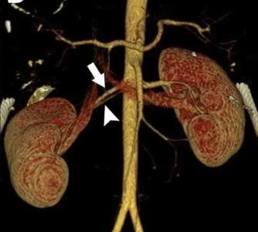
- Arteriyel faz ince kesitlerde ana renal arterden ayrı aortik/iliak çıkışlı aksesuar damar, hilusa eşlik eden arter veya doğrudan üst/alt kutba giren polar arter olarak izlenir; ana arterin erken dalı ostiumdan kısa mesafede ayrılır. Lümen opasifikasyonu darlık, ostial plak ve dal anatomisini değerlendirmeyi sağlar.
Önerilen pencereler: Anjiyo (G 600 / M 150), Batın (G 400 / M 50).
BT bulguları
- Aortadan ana renal arter ostiumundan ayrı çıkan ve böbreğe giren ek arter saptanır; daha aşağıda iliak arter kaynaklı damar da görülebilir.
- Aksesuar hilar arter ana renal arterle birlikte hilustan parankime girer; polar/aberran arter böbreğe doğrudan üst veya alt kutuptan ulaşabilir.
- Her damarın kökeni, seyri, çapı ve böbreğe giriş bölgesi aksiyel kesit ve çok düzlemli rekonstrüksiyonlarda izlenebilir.
- Ana renal arter hilusa ulaşmadan dallara ayrılıyorsa erken/pre-hilar dallanma vardır; ilk dalın ostiuma uzaklığı ölçülüp bildirilir.
- Segmental arterlerin parankim içi dağılımı ve olası kutup beslenmesi cerrahi açıdan anlamlı damar haritasını tamamlar.
- Fokal lümen daralması varsa ostium ve proksimal arter boyunca plak, duvar düzensizliği ve distal dalların dolumu incelenir.
- İnce kalibrasyonlu alt kutup arteri, üst kutup arteri veya çoklu ana arter dalları ayrı ayrı sayılmalı; yalnız ana damar görüntüsüyle yetinilmemelidir.
Ölçütler ve sınıflamalar
- Erken (prehilar) renal arter dallanması
- İlk dal renal arter ostiumundan ≤1,5 cm uzaklıkta çıkar
Normalde
Tipik renal arter anatomisinde her böbrek çoğunlukla aortadan çıkan tek ana renal arterle beslenir; ana arter hilusa kadar izlenir ve ardından segmental dallara ayrılır. BTA'da kıyas için her renal arter ostiumunu aort boyunca sayın, aksesuar köken bulunup bulunmadığını ve ana arterin ilk dalının hilusa göre yerini gösterin; varyant damar da normal arter gibi kontrastlanıp parankime ulaşır.
Normal BT ile kıyasla
Aynı bölgenin, kaynak veri setinde patoloji içermediği belirtilen bir BT incelemesini kesit kesit kaydırın; organ sınırlarını açarak anatomiyi hasta görüntüsüyle karşılaştırın. Küçük rastlantısal bulgular tümüyle dışlanmamıştır.
Kaynak: TotalSegmentator v2.01 veri seti (Wasserthal ve ark., Radiology: Artificial Intelligence 2023), CC BY 4.0, vaka s0115; organ sınırları veri setinin otomatik segmentasyonundan, birleştirilerek sadeleştirildi. Görüntüler 2,5 mm kesit aralığına yeniden örneklendi.
Ayırıcı tanı
- Renal arter stenozu, renal arter varyantlarından ayrı bir anatomik sorun olarak değerlendirilir.
- Renal ven varyantı
- venöz fazda dolan ve böbrekten vena kavaya dönen damardır; BTA'da arterden farklı zamanlamada opaklaşır ve akım yönü/bağlantısı ayrıdır.
- Segmental renal arter dalı
- ana renal arterden ayrıldıktan sonra böbrek içinde dallanır; ayrı aortik/iliak kökenli aksesuar arter değildir.
Tuzaklar
- Arteriyel opasifikasyon yetersizse küçük polar damar fark edilmeyebilir veya dar görünebilir; değerlendirmeyi damar fazı ve görüntü kalitesiyle sınırlayın.
- Aksiyel tek kesitte aksesuar damar çok kısa görünebilir; aortadan çıkışını böbreğe girişine dek koronal/sagittal MPR ve damar izleme ile takip edin.
- Erken dalı aksesuar arter olarak saymayın: ayrı ostiumdan çıkıp çıkmadığını aort ve ana renal arter kökenlerini izleyerek ayırın.
- Erken bifurkasyon, küçük damar çapı ve yüksek dereceli stenozu ayrı anatomik bulgular olarak değerlendirin.
Kendini dene
BTA'da ana renal arterden ayrı bir aortik ostiumdan çıkan ince damar doğrudan alt kutba ulaşıyor. Bu anatomi nasıl adlandırılır?
Cevabı göster
Aksesuar polar renal arter. Ayrı ostiumdan çıkan ve doğrudan kutba ulaşan damar aksesuar polar renal arterdir. Erken dallanan renal arterde tek arter gövdesi hilusa ulaşmadan bölünür; aksesuar renal arter ise ayrı kökenli ek bir damardır.
Ana renal arter tek ostiumdan çıkıyor; ilk dal ostiuma yakın ayrılıyor. Varyantı adlandırmak için hangi ölçüm gerekir?
Cevabı göster
Ostium-ilk dal uzaklığı. Erken dallanma, ana renal arter ostiumundan ilk dalın çıktığı yere olan mesafeyle tanımlanır; kaynağın kullandığı eşik 1.5 cm'dir. Kaynağa göre erken dallanma, ostiumdan 1,5 cm içinde gerçekleşen dallanmadır.
Kaynaklar
Bu sayfadaki 36 cümle ve 2 quiz sorusu aşağıdaki kaynaklardan alıntılarla otomatik olarak karşılaştırıldı; 5 cümle bu karşılaştırmada düzeltildi (2026-10-09).
- Renal transplantation and variations in blood supply: a narrative reviewpmc.ncbi.nlm.nih.gov
- Extreme Variability of the Kidney Hilar Architecture: A Radioanatomical Map to Guide Surgical Approachespmc.ncbi.nlm.nih.gov
- Ambulatory Blood Pressure Characteristics and Target Organ Effects in Hypertensive Children With Renal Artery Variationspmc.ncbi.nlm.nih.gov
- Multidetector Computed Tomography Angiography for Preoperative Anatomical and Functional Assessment of Living Renal Donors: Correlation With Surgical Findings and Renal Scintigraphypmc.ncbi.nlm.nih.gov
- Association of renal artery variations with aggressive pathological features and poor prognosis in clear-cell renal cell carcinoma: a computed tomography-based three-dimensional reconstruction studypmc.ncbi.nlm.nih.gov
- Dual-energy computed tomography angiography for evaluating the renal vascular variants.pubmed.ncbi.nlm.nih.gov
- Diagnostic accuracy of 64-MDCT angiography in the preoperative evaluation of renal vessels and compared with laparotomy findings in living donor kidney.pubmed.ncbi.nlm.nih.gov
- Role of multidetector-row computed tomography in evaluation of living renal donors.pubmed.ncbi.nlm.nih.gov
- Variations in Branching Pattern of Renal Artery in Kidney Donors Using CT Angiography.pubmed.ncbi.nlm.nih.gov
- [Three-dimensional spatial measurement versus conventional CT planning in laparoscopic partial nephrectomy for renal tumors].pubmed.ncbi.nlm.nih.gov
- Implications of renal artery anatomy for endovascular repair using fenestrated, branched, or parallel stent graft techniques.pubmed.ncbi.nlm.nih.gov
- Quantitative analysis of renal arterial variations affecting the eligibility of catheter-based renal denervation using multi-detector computed tomography angiography.pubmed.ncbi.nlm.nih.gov
- Multidetector computed tomography angiography study of the renal arterial vasculature anatomy and its variations in a Bulgarian adult population.pubmed.ncbi.nlm.nih.gov
- Fenestrated and Branched Endovascular Aortic Repair of Thoracoabdominal Aortic Aneurysm With More Than 4 Target Visceral Vessels due to Renovisceral Arterial Anatomical Variations: Feasibility and Early Results.pubmed.ncbi.nlm.nih.gov
- Peri-hilar branching pattern and variations of the renal artery among Indian kidney donors using pre-operative computed tomography angiography: an anatomical study and review.pubmed.ncbi.nlm.nih.gov
- Evaluation of Vascular Structures of Living Donor Kidneys by Multislice Computed Tomography Angiography before Transplant Surgery: Is Arterial Phase Sufficient for Determination of Both Arteries and Veins?pubmed.ncbi.nlm.nih.gov
- Diagnostic performance of non-contrast quiescent-interval slice-selective (QISS) magnetic resonance angiography for evaluation of the renal arterial vasculature compared to computed tomography angiography (CTA) as reference standard.pubmed.ncbi.nlm.nih.gov
- Exploring Anatomical Variations and Potential Associations between the Upper Urinary Tract and Renal Vasculature: Findings from 160-Slice CT Imaging in Northern Nigeria.pubmed.ncbi.nlm.nih.gov
- Prevalence and patterns of renal vascular variations among potential kidney donors: a Computed Tomography Angiography (CTA) study in Sudan.pubmed.ncbi.nlm.nih.gov
- Renal transplantation and variations in blood supply: a narrative review.pubmed.ncbi.nlm.nih.gov
- A partially duplicated left ovarian vein bisected by the left renal artery: Hypothesized embryologic origins and clinical implications.pubmed.ncbi.nlm.nih.gov
- Morphology and morphometry of renal arteries in Indian cadavers.pubmed.ncbi.nlm.nih.gov
- Left Renal Vein Pseudo-Fenestration.pubmed.ncbi.nlm.nih.gov
- Fused right S-shaped kidney incidentally detected on multidetector CT during staging of periampullary adenocarcinoma: A case report.pubmed.ncbi.nlm.nih.gov
- Low- versus high-concentration iodine contrast media for coronary computed tomography angiography and dynamic stress computed tomography perfusion: a study protocol for a prospective randomized trial.pubmed.ncbi.nlm.nih.gov
- Back-Table Repair of Parenchymal and Collecting System Injury of a Living Donor Kidney.pubmed.ncbi.nlm.nih.gov
- Simultaneous Occurrence of Two Renal Artery Variations in a Single Potential Kidney Donor.pubmed.ncbi.nlm.nih.gov
- Spontaneous bacteremia and spontaneous bacterial peritonitis share similar prognosis in patients with cirrhosis: a cohort study.pubmed.ncbi.nlm.nih.gov
- Long-term safety and effectiveness of the "OptEase" vena cava filter.pubmed.ncbi.nlm.nih.gov
- High-affinity alphavbeta3 integrin targeted optical probe as a new imaging biomarker for early atherosclerosis: initial studies in Watanabe rabbits.pubmed.ncbi.nlm.nih.gov
- Radiologic Interventions for Gastrointestinal Bleeding.pubmed.ncbi.nlm.nih.gov
- Cannabis-Associated Spontaneous Coronary Artery Dissection in a Community Hospital: A Case-Based Focused Literature Review.pubmed.ncbi.nlm.nih.gov
- The Weston Score in coronary artery calcification among patients with End-Stage Renal Disease.pubmed.ncbi.nlm.nih.gov
- Ambulatory Blood Pressure Characteristics and Target Organ Effects in Hypertensive Children With Renal Artery Variations.pubmed.ncbi.nlm.nih.gov
- Extreme Variability of the Kidney Hilar Architecture: A Radioanatomical Map to Guide Surgical Approaches.pubmed.ncbi.nlm.nih.gov
- Coexistence of Autoantibodies against the Golgi Complex and Ro52 Antigen in a Patient with Nonspecific Interstitial Pneumonia.pubmed.ncbi.nlm.nih.gov
- Visceral trauma: principles of management and role of embolotherapy.pubmed.ncbi.nlm.nih.gov
- An Overview of Vascular Compression Syndromes and Associations with Autonomic Dysfunction: A Review.pubmed.ncbi.nlm.nih.gov
- From congenital variants to critical clues: A radiologic review of inferior vena cava pathologies.pubmed.ncbi.nlm.nih.gov
- Duplicated Inferior Vena Cava: A Case Study With Clinical Implications.pubmed.ncbi.nlm.nih.gov
- Deep cervical lymphatic-venous anastomosis attenuates cognitive dysfunction and biomarker abnormalities in severe Alzheimer's disease: A prospective single-arm study.pubmed.ncbi.nlm.nih.gov
- The Perception of the Diaphragm with Ultrasound: Always There Yet Overlooked?pubmed.ncbi.nlm.nih.gov
- Personalized Management of Malignant and Non-Malignant Ectopic Mediastinal Thyroid: A Proposed 10-Item Algorithm Approach.pubmed.ncbi.nlm.nih.gov
- Staged hybrid repair of a symptomatic post-traumatic aortic aneurysmatic lesion in a patient with late diagnosed connective tissue disorder.pubmed.ncbi.nlm.nih.gov
- Hyper-rapid progression in Salmonella-associated mycotic aortic aneurysms: a narrative review.pubmed.ncbi.nlm.nih.gov
- Computed tomography of renal emergencies: A comprehensive diagnostic guide for radiology residents.pubmed.ncbi.nlm.nih.gov
- Association of renal artery variations with aggressive pathological features and poor prognosis in clear-cell renal cell carcinoma: a computed tomography-based three-dimensional reconstruction study.pubmed.ncbi.nlm.nih.gov
- Venous reconstruction in living donor liver transplantation: lessons learned from a new national program in a resource-limited setting.pubmed.ncbi.nlm.nih.gov
- Pediatric Renovascular Hypertension: Diagnosis and Management.pubmed.ncbi.nlm.nih.gov
- Emerging risk biomarkers in cardiovascular diseases and disorders.pubmed.ncbi.nlm.nih.gov
- Multimodal imaging in neuro-ophthalmology.pubmed.ncbi.nlm.nih.gov
- Quantitative Cerebrovascular Analysis for Improved Prediction of Post-Stroke Complications.pubmed.ncbi.nlm.nih.gov
- Three-Station Non-Contrast MR Angiography of the Lower Extremities Using Standard and Centric Fresh Blood Imaging.pubmed.ncbi.nlm.nih.gov
- Application of postmortem imaging modalities in cases of sudden death due to cardiovascular diseases-current achievements and limitations from a pathology perspective : Endorsed by the Association for European Cardiovascular Pathology and by the International Society of Forensic Radiology and Imaging.pubmed.ncbi.nlm.nih.gov
- MDCT Imaging of Non-Traumatic Thoracic Aortic Emergencies and Its Impact on Diagnosis and Management-A Reappraisal.pubmed.ncbi.nlm.nih.gov
- Ten things to know about ten imaging studies: A preventive cardiology perspective ("ASPC top ten imaging").pubmed.ncbi.nlm.nih.gov
- Enhancing procedural decision making with cone beam CT in renal artery embolization.pubmed.ncbi.nlm.nih.gov
- Complications Related to Transarterial Treatment of Hepatocellular Carcinoma: A Comprehensive Review.pubmed.ncbi.nlm.nih.gov
- Spectral Computed Tomography Angiography in Visceral Artery Aneurysms: Technical Principles and Clinical Applications.pubmed.ncbi.nlm.nih.gov
- Polyarteritis nodosa with testicular involvement: a rare case report highlighting the role of nuclear imaging and angiography in diagnosis.pubmed.ncbi.nlm.nih.gov
- Individualized strategy of treatment for ruptured abdominal aortic aneurysm using causal inference model: a retrospective observational study.pubmed.ncbi.nlm.nih.gov
- Unmasking the Apex: Multimodality Imaging for the Evaluation of Left Ventricular Apical Obliteration.pubmed.ncbi.nlm.nih.gov
- Perforator-Sparing Microsurgical Clipping of Tandem Dominant-Hemisphere Middle Cerebral Artery Aneurysms: Geometry-Guided Reconstruction of a Wide-Neck Bifurcation and Dorsal M1 Fusiform Lesion.pubmed.ncbi.nlm.nih.gov
- International Society of Paediatric Surgical Oncology (IPSO) Surgical Practice Guidelines.pubmed.ncbi.nlm.nih.gov
- Consensus guidelines for the use and interpretation of angiogenesis assays.pubmed.ncbi.nlm.nih.gov
- Intracardiac Echocardiography during Invasive Electrophysiological Procedures: a Scientific Statement of the European Heart Rhythm Association of the ESC, and the European Association of Percutaneous Cardiovascular Interventions of the ESC.pubmed.ncbi.nlm.nih.gov
- Severe Recurrent Hemorrhage of an Oncogenic Vertebrobasilar Junction Aneurysm Following Stent-Assisted Coil Embolization in Metastatic Testicular Choriocarcinoma.pubmed.ncbi.nlm.nih.gov
- Brazilian Society for Angiology and Vascular Surgery guidelines on abdominal aortic aneurysm.pubmed.ncbi.nlm.nih.gov
- Computed tomography and magnetic resonance imaging of congenital thoracic systemic venous anomalies.pubmed.ncbi.nlm.nih.gov
- Pathologic and Radiologic Correlation of Adult Cystic Lung Disease: A Comprehensive Review.pubmed.ncbi.nlm.nih.gov
- The Prevalence of Variations in the Morphology of Renal Arteries in the North Indian Population.pubmed.ncbi.nlm.nih.gov
- Arterial-Venous Balance in the Human Renal Vasculature: Renal-Aortic and Renal-Caval Ratios and Factors on Contrast-Enhanced Computed Tomography.pubmed.ncbi.nlm.nih.gov
- Communicating bronchopulmonary foregut malformations: Revisiting a rare entity.pubmed.ncbi.nlm.nih.gov
- Three-Dimensional Bronchovascular Modelling in Sublobar Pulmonary Resection: A Tool for Personalised Thoracic Surgery.pubmed.ncbi.nlm.nih.gov
- Anatomical structure of pulmonary vessels and bronchi in the right upper lobe based on three-dimensional reconstruction.pubmed.ncbi.nlm.nih.gov
Eğitim amaçlıdır; hasta başında tanı ve tedavi kararının yerine geçmez, tıbbi tavsiye değildir. Metinler yapay zekâ desteğiyle yazıldı ve otomatik bir adımda kaynak alıntılarıyla karşılaştırıldı; bu bir tıbbi doğrulama değildir ve içerik uzman radyolog incelemesinden geçmedi. Klinik kararlarda güncel kılavuzları, kurum protokollerini ve radyoloji raporunu esas alın.