Özet
Derin renal laserasyonun toplayıcı sisteme uzanması, renal pelvis laserasyonu veya üreteropelvik bileşke ayrışması idrar ekstravazasyonuna yol açabilir. Kontrastlı BT'nin gecikmiş ekskretuar görüntüsü, opak idrarın kaliks-pelvis konturundan nerede ayrıldığını ve perirenal koleksiyona yayıldığını gösterir. Parankim fazı parankimal yaralanmayı belirginleştirebilir; idrar ekstravazasyonunu değerlendirmek için ekskretuar faz görüntülemesi gerekir. Süren idrar kaçağı urinoma oluşumuna yol açabilir; urinoma bazı olgularda enfekte olabilir.
Faz ve pencere
- Nefrografik
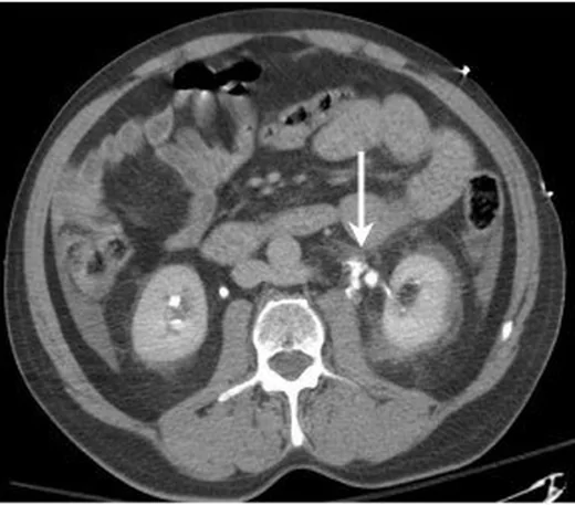
- Derin kortikomedüller yarık, medial hilus çevresinde sıvı ve perirenal hematom saptanır; bu bulgular ekskretuar görüntü ekleme gereğini doğurur.
- Ekskretuar tanısal
- Opak idrarın kaliks, pelvis veya UP bileşke dışına çıktığı çizgisel/serbest kontrast; giderek opaklaşan perirenal koleksiyon ve distal üreterin dolum durumu gösterilir.
Önerilen pencereler G genişlik, M merkez; değerler Hounsfield birimi (HU)
- BatınG 400 / M +50
- Yumuşak dokuG 400 / M +40
BT bulguları
- Gecikmiş görüntüde kaliks veya renal pelvis dışına uzanan kontrastlı idrar ekstravazasyonu.
- Perirenal alandaki başlangıçta hipodens görünen koleksiyona, ekskretuar fazda toplayıcı sistemden kontrastlı idrar geçebilir.
- Medial hilus çevresindeki sıvı ile renal pelvis/UP bileşke arasında izlenebilen ekstravazasyon yolu.
- Derin kortikomedüller laserasyonun renal sinüs ve kaliks sistemine erişmesi.
- UP bileşke ayrışmasında opak renal pelvis ile opaklaşmayan distal üreter arasında kesinti.
- Eşlik eden perirenal hematom, idrar kaçağı ile birlikte yüksek dereceli travmalarda sık görülen bir bulgudur.
- İdrar ekstravazasyonunu değerlendirmek için BT protokolüne ekskretuar faz görüntülemesi eklenir.
Ölçütler ve sınıflamalar
- AAST Böbrek Yaralanma Derecelendirme Ölçeği ile ilişki
- Toplayıcı sisteme uzanan idrar ekstravazasyonlu laserasyon, renal pelvis laserasyonu ve tam üreteropelvik bileşke ayrışması AAST 2018'de derece IV'tür. Derece IV; segmental damar yaralanmalarını, Gerota fasyası dışına aktif kanamayı ve segmental ya da tam böbrek infarktını kapsar; derece V ölçütleri arasında ana renal arter veya ven laserasyonu, renal hilus avülsiyonu, devaskülarize böbrek ve parçalanmış böbrek bulunur.
Normalde
Normal ekskretuar BT'de kontrast kalikslerden renal pelvise, oradan üreter içine devamlı bir lümen boyunca ilerler; çevre perirenal yağ içinde opak idrar birikimi olmaz. Şüpheli alanda kaliks fornikslerinin düzgün sınırını, pelvisin medial konturunu ve distal üreterin opaklaşmasını karşı tarafla kıyasla.
Normal BT ile kıyasla
Aynı bölgenin, kaynak veri setinde patoloji içermediği belirtilen bir BT incelemesini kesit kesit kaydırın; organ sınırlarını açarak anatomiyi hasta görüntüsüyle karşılaştırın. Küçük rastlantısal bulgular tümüyle dışlanmamıştır.
Kaynak: TotalSegmentator v2.01 veri seti (Wasserthal ve ark., Radiology: Artificial Intelligence 2023), CC BY 4.0, vaka s0119; organ sınırları veri setinin otomatik segmentasyonundan, birleştirilerek sadeleştirildi. Görüntüler 2,5 mm kesit aralığına yeniden örneklendi.
Ayırıcı tanı
- Perirenal hematom
- kontrastsız görüntüde kan yoğunluğu vardır ve gecikmiş fazda opak idrar gibi yeni kontrast birikimi göstermez.
- Basit perirenal sıvı
- ekskretuar fazda idrarla devamlı opaklaşmaz ve toplayıcı sistem duvarında kopukluk yoktur.
- Üreter yaralanması
- ekstravazasyon böbrek pelvisinden çok üreter boyunca, yaralanma seviyesindeki kesinti çevresinde belirginleşir.
Tuzaklar
- Gecikmiş fazı çok erken almak, yavaş ekskresyonlu böbrekte kaçak yokmuş izlenimi yaratır; koleksiyonun opaklaşmasını ve distal üreter dolumunu yeterli gecikmede yeniden değerlendir.
- Medial perirenal sıvıyı otomatik olarak hematom sayma; pelvis konturundaki düzensizlikle birlikte ekskretuar serideki kontrast geçişini ara.
- Toplayıcı sisteme uzanan laserasyon tek başına kaçak kanıtı değildir; ekstravazasyon görülmüyorsa sistemin bütünlüğünü ve görüntüleme zamanlamasını belirt.
- Aktif kanama erken faz görüntülerinde kontrast ekstravazasyonu olarak görülebilir ve gecikmiş fazda belirginleşebilir; idrar ekstravazasyonu ise ekskretuar fazda toplayıcı sistem dışına kontrast geçişiyle saptanır.
Kendini dene
Gecikmiş ekskretuar fazda kontrastın renal toplayıcı sistemden perirenal koleksiyona geçtiği görülüyor. Bu bulgu neyi gösterir?
Cevabı göster
İdrar ekstravazasyonu. Ekskretuar fazda toplayıcı sistemden koleksiyona kontrast geçişi idrar ekstravazasyonunu gösterir.
Travma sonrası gecikmiş BT’de renal pelvis opaklaşırken yaralanma distalindeki üreter opaklaşmıyor. Bu bulgu en çok hangi yaralanmayı düşündürür?
Cevabı göster
Tam üreteropelvik bileşke avulsiyonu. Yaralanma distalindeki üreterin opaklaşmaması tam üreteropelvik avulsiyon açısından yüksek derecede şüphelidir.
İlgili konular
Kaynaklar
Bu sayfadaki 36 cümle ve 2 quiz sorusu aşağıdaki kaynaklardan alıntılarla otomatik olarak karşılaştırıldı; 10 cümle bu karşılaştırmada düzeltildi (2026-10-09).
- Computed tomography of renal emergencies: A comprehensive diagnostic guide for radiology residentspmc.ncbi.nlm.nih.gov
- Upper Urinary Tract Clot Obstruction After Delayed Hemorrhage Following Partial Nephrectomy: A Possible Association With Tranexamic Acidpmc.ncbi.nlm.nih.gov
- Dual Challenges: Addressing Post-Traumatic Retroperitoneal Urinoma in the Context of Pyeloureteral Duplicationpmc.ncbi.nlm.nih.gov
- Understanding Air in the Body: A Radiologic Perspective on Emphysematous and Traumatic Conditionspmc.ncbi.nlm.nih.gov
- Renal diseases and blood pressure dysregulation in acute care: Pathophysiology, clinical patternspmc.ncbi.nlm.nih.gov
- Variation in management of pediatric post-traumatic urine leaks.pubmed.ncbi.nlm.nih.gov
- Back-Table Repair of Parenchymal and Collecting System Injury of a Living Donor Kidney.pubmed.ncbi.nlm.nih.gov
- Computed tomography of renal emergencies: A comprehensive diagnostic guide for radiology residents.pubmed.ncbi.nlm.nih.gov
- Conservatively Managed Grade V Blunt Renal Injury.pubmed.ncbi.nlm.nih.gov
- Pseudoaneurysm of the Left Renal Segmental Artery With Delayed Detection After Blunt Trauma.pubmed.ncbi.nlm.nih.gov
- A Presumed Acquired Ureteral Diverticulum Presenting With Hematuria, Urosepsis, and Periureteral Contrast Leak: A Rare Case Report.pubmed.ncbi.nlm.nih.gov
- Urinothorax Secondary to Pyelonephritis With Obstructing Ureteric Calculi: A Rare Cause of Pleural Effusion.pubmed.ncbi.nlm.nih.gov
- Multimodality diagnostic imaging and role of interventional radiology in pancreas transplantation.pubmed.ncbi.nlm.nih.gov
- Human biomarker navigator.pubmed.ncbi.nlm.nih.gov
- Post-operative management of children after lung transplantation.pubmed.ncbi.nlm.nih.gov
- Spontaneous Rupture of the Urinary Bladder in an Elderly Diabetic Male.pubmed.ncbi.nlm.nih.gov
- A 6-year retrospective clinical review of iatrogenic ureteric injuries repaired in a resource-deprived setting.pubmed.ncbi.nlm.nih.gov
- Shortness of Breath: An Unusual Presentation of Bladder Injury. A Case Report and Literature Review of Urinothorax.pubmed.ncbi.nlm.nih.gov
- Time Is Kidney: A Case Study and Literature Review of Bilateral Renal Compartment Syndrome After Blunt Trauma, a Rare Complication.pubmed.ncbi.nlm.nih.gov
- Contrast-enhanced ultrasound (CEUS) in urgent and emergent pediatric conditions: a comprehensive review.pubmed.ncbi.nlm.nih.gov
- Computed tomography of pediatric abdominal trauma: optimizing utilization and enhancing diagnostic interpretation.pubmed.ncbi.nlm.nih.gov
- Vascular and lymphatic complications after renal transplant.pubmed.ncbi.nlm.nih.gov
- Multidetector CT imaging of complications after laparoscopic nephron-sparing surgery.pubmed.ncbi.nlm.nih.gov
- Dual Challenges: Addressing Post-Traumatic Retroperitoneal Urinoma in the Context of Pyeloureteral Duplication.pubmed.ncbi.nlm.nih.gov
- Imaging of an exceptional urinoma complicating a cervical cancer and leading to death: A case report.pubmed.ncbi.nlm.nih.gov
- Retroperitoneal glass foreign body with renal parenchymal involvement after 25 years of silent retention: a case report.pubmed.ncbi.nlm.nih.gov
- Percutaneous nephrostomy: technical aspects and indications.pubmed.ncbi.nlm.nih.gov
- Conventional uroradiology with excretory urography: a forgotten art?pubmed.ncbi.nlm.nih.gov
- Cross-sectional imaging of iatrogenic complications after extracorporeal and endourological treatment of urolithiasis.pubmed.ncbi.nlm.nih.gov
- Spontaneous adrenal hemorrhage after rupture of adenoma: case report and review of 78 cases.pubmed.ncbi.nlm.nih.gov
- Spontaneous perirenal hemorrhage in systemic lupus erythematosus: a rare case report and literature review.pubmed.ncbi.nlm.nih.gov
- Complications of lymphoma in the abdomen and pelvis: clinical and imaging review.pubmed.ncbi.nlm.nih.gov
- Eosinophilic vacuolated tumor of the kidney misdiagnosed as upper tract urothelial carcinoma: a case report and literature review.pubmed.ncbi.nlm.nih.gov
- MR Imaging of the Upper Urinary Tract: Techniques, Diagnostic Performance, and Imaging Biomarkers.pubmed.ncbi.nlm.nih.gov
- ESR Essentials: trauma team and the role of Interventional Radiology-practice recommendations by the Cardiovascular and Interventional Radiological Society of Europe.pubmed.ncbi.nlm.nih.gov
- Management and Outcomes of Blunt Renal Trauma: A Retrospective Analysis from a High-Volume Urban Emergency Department.pubmed.ncbi.nlm.nih.gov
- Case report: traumatic avulsion of the left kidney and spleen with diaphragmatic rupture in a young worker.pubmed.ncbi.nlm.nih.gov
- Pediatric ureteropelvic junction disruption with renal laceration: organ-saving management through delayed diagnosis and surgical repair.pubmed.ncbi.nlm.nih.gov
- The Role of Interventional Radiology in Renal Trauma: A Narrative Review.pubmed.ncbi.nlm.nih.gov
- Use of Contrast-Enhanced Ultrasound in Suspected Traumatic or Spontaneous Renal Injury in Cats: A Case Series.pubmed.ncbi.nlm.nih.gov
- Microfluidics and molecular diagnostics in renal cell carcinoma: advances, challenges, and future directions.pubmed.ncbi.nlm.nih.gov
- Contrast Enhanced Ultrasound Compared with MRI and CT in the Evaluation of Post-Renal Transplant Complications.pubmed.ncbi.nlm.nih.gov
- Radionuclides in nephrourology, Part 2: pitfalls and diagnostic applications.pubmed.ncbi.nlm.nih.gov
- International Society of Paediatric Surgical Oncology (IPSO) Surgical Practice Guidelines.pubmed.ncbi.nlm.nih.gov
- The hallmarks of cancer: relevance to the pathogenesis of polycystic kidney disease.pubmed.ncbi.nlm.nih.gov
- Multiomics Analysis-Based Biomarkers in Diagnosis of Polycystic Ovary Syndrome.pubmed.ncbi.nlm.nih.gov
- Case Report: Acute Testicular Pain as the Initial Manifestation of a Ruptured Common Iliac Artery Aneurysm.pubmed.ncbi.nlm.nih.gov
- Association of Postoperative Drainage Strategy with Persistent Urinary Leakage and Clinical Outcomes Following Percutaneous Nephrolithotomy.pubmed.ncbi.nlm.nih.gov
- Flexible and Navigable Suction Ureteral Access Sheath (FANS) versus conventional stone-pushing technique for impacted upper ureteral stones: a retrospective cohort study.pubmed.ncbi.nlm.nih.gov
- Acute kidney injury in paediatric kidney transplant recipients.pubmed.ncbi.nlm.nih.gov
- Guidelines for Antibiotics Prescription in Critically Ill Patients.pubmed.ncbi.nlm.nih.gov
- CT of Penetrating Abdominopelvic Trauma.pubmed.ncbi.nlm.nih.gov
- Long-term safety and effectiveness of the "OptEase" vena cava filter.pubmed.ncbi.nlm.nih.gov
- Patterns of injury and violence in Yaoundé Cameroon: an analysis of hospital data.pubmed.ncbi.nlm.nih.gov
- Massive bleeding from a Bricker ileal conduit during FOLFIRINOX therapy in noncirrhotic portal hypertension: suspected oxaliplatin-associated sinusoidal injury and presumed irinotecan-associated conduit mucositis: a case report.pubmed.ncbi.nlm.nih.gov
- Imaging of renal pseudotumors in children: a comprehensive review.pubmed.ncbi.nlm.nih.gov
- Symptomatic Parapelvic Cysts in Children: Anatomical and Histological Features, Diagnostic Pitfalls and Urological Management.pubmed.ncbi.nlm.nih.gov
- Obstructed hemivagina with ipsilateral renal and urinary tract anomaly presenting as an unusual cause of acute abdomen: a radiologic perspective.pubmed.ncbi.nlm.nih.gov
- Flexible negative-pressure ureteral sheath lithotripsy compared with mini-PCNL for upper urinary tract stones with moderate-to-severe hydronephrosis: a retrospective clinical study.pubmed.ncbi.nlm.nih.gov
- Tip bendable suction ureteral access sheath versus traditional sheath in retrograde intrarenal stone surgery: an international multicentre, randomized, parallel group, superiority study.pubmed.ncbi.nlm.nih.gov
- The space of Retzius: anatomy, imaging features, and patterns of disease spread.pubmed.ncbi.nlm.nih.gov
- Delayed-Onset Infected Perirenal Hematoma Following Trauma: A Case Report.pubmed.ncbi.nlm.nih.gov
- From imaging to impact: A case of renal arteriovenous malformation.pubmed.ncbi.nlm.nih.gov
- When Stones Escape: A Rare Radiologic Finding of Retroperitoneal Stone Extrusion Following American Association for the Surgery of Trauma (AAST) Grade IV Blunt Renal Trauma in a Chronically Hydronephrotic Kidney.pubmed.ncbi.nlm.nih.gov
- Successful non-operative management of multiple intra-abdominal solid organ injury after blunt abdominal trauma: a case report.pubmed.ncbi.nlm.nih.gov
- Non-operative management for abdominal solidorgan injuries: A literature review.pubmed.ncbi.nlm.nih.gov
- Imaging in renal trauma.pubmed.ncbi.nlm.nih.gov
- Clinical applications of vascular and non-vascular interventional procedures for various urologic diseases: a pictorial essay.pubmed.ncbi.nlm.nih.gov
- Interventional Radiology Management of Genitourinary Trauma: Literature, Current Practice, and Strategies for the Future.pubmed.ncbi.nlm.nih.gov
- Renal relevant radiology: imaging in kidney transplantation.pubmed.ncbi.nlm.nih.gov
- Best Practice Imaging in the Initial Investigation of Paediatric Blunt Abdominal Trauma: A Narrative Review.pubmed.ncbi.nlm.nih.gov
- The Diagnosis and Management of Pediatric Blunt Abdominal Trauma-A Comprehensive Review.pubmed.ncbi.nlm.nih.gov
- Acute kidney injury in urological conditions.pubmed.ncbi.nlm.nih.gov
- The pathogenesis of cerebral small vessel disease and vascular cognitive impairment.pubmed.ncbi.nlm.nih.gov
- ESR Essentials: renal imaging in children-practice recommendations by the European Society of Paediatric Radiology.pubmed.ncbi.nlm.nih.gov
- MDCT of blunt renal trauma: imaging findings and therapeutic implications.pubmed.ncbi.nlm.nih.gov
- Spontaneous Subdiaphragmatic Hemorrhage From an Aneurysm of Inferior Phrenic Artery.pubmed.ncbi.nlm.nih.gov
- Unprovoked Spontaneous Kidney Rupture (Wunderlich's Syndrome) Managed by Renal Artery Embolization.pubmed.ncbi.nlm.nih.gov
- Non-penetrating traumatic psoas muscle hematoma presenting with gross hematuria: a case report.pubmed.ncbi.nlm.nih.gov
- Is There Still a Place for Percutaneous Nephrolithotomy in Current Times?pubmed.ncbi.nlm.nih.gov
- Histotripsy of the Proximal Ureter and Renal Pelvis: Evaluation of Urothelial Injury in a Porcine Survival Model.pubmed.ncbi.nlm.nih.gov
- CIRSE Standards of Practice on Nephrostomy and Ureteric Stent Placement and Exchange.pubmed.ncbi.nlm.nih.gov
- Complications after Nephron-sparing Interventions for Renal Tumors: Imaging Findings and Management.pubmed.ncbi.nlm.nih.gov
- 2023 WSES guidelines for the prevention, detection, and management of iatrogenic urinary tract injuries (IUTIs) during emergency digestive surgery.pubmed.ncbi.nlm.nih.gov
- Optimizing the Diagnosis of Ureteral Complications After Kidney Transplantation Using Retrograde Multidetector Computed Tomography Urography: A Case Series.pubmed.ncbi.nlm.nih.gov
- Divergent outcomes of delayed ureteropelvic junction stenosis after renal cyst sclerotherapy: two case reports.pubmed.ncbi.nlm.nih.gov
- A retroperitoneal urinoma secondary to metastatic urothelial carcinoma mimicking psoas muscle malignancy: Case Report and literature review.pubmed.ncbi.nlm.nih.gov
- Urine Trouble: Forniceal Rupture in the Setting of a Small Urolithic Stone.pubmed.ncbi.nlm.nih.gov
- Successful Closure of Persistent Urinary Fistula After High-Grade Renal Trauma Using N-Butyl-2-Cyanoacrylate: A Case Report.pubmed.ncbi.nlm.nih.gov
- Management of pediatric renal trauma with urinary extravasation: a retrospective study in a single clinical center.pubmed.ncbi.nlm.nih.gov
- Renal Trauma: A 4-Year Retrospective Review of Injury Severity, Treatment Approaches, and Outcomes from a Polish Trauma Center.pubmed.ncbi.nlm.nih.gov
- Solid Organ Injuries: To Irradiate or Operate?pubmed.ncbi.nlm.nih.gov
- Urinary Tract Trauma in Cats: Stabilisation, Diagnosis and Management.pubmed.ncbi.nlm.nih.gov
- Non-Operative Management of Polytraumatized Patients: Body Imaging beyond CT.pubmed.ncbi.nlm.nih.gov
Eğitim amaçlıdır; hasta başında tanı ve tedavi kararının yerine geçmez, tıbbi tavsiye değildir. Metinler yapay zekâ desteğiyle yazıldı ve otomatik bir adımda kaynak alıntılarıyla karşılaştırıldı; bu bir tıbbi doğrulama değildir ve içerik uzman radyolog incelemesinden geçmedi. Klinik kararlarda güncel kılavuzları, kurum protokollerini ve radyoloji raporunu esas alın.