Özet
UPJ obstrüksiyonu, renal pelvis ile proksimal üreter arasındaki geçişin intrinsik darlık, yüksek üreter çıkışı, kıvrılma veya dıştan bası nedeniyle idrar drenajını engellemesidir. Belirti vermeyen hidronefroz insidental bulunabileceği gibi erişkinde ani yan ağrısı, taş veya enfeksiyon araştırılırken de saptanabilir. Kontrastlı BT/BT ürografi genişlemiş pelvis-kaliksleri, daralma seviyesini, distal üreterin kalibrasyonunu, parankim durumunu ve alt pol çapraz damarlarını haritalar; tek başına anatomik genişleme fonksiyonel obstrüksiyonu kesin kanıtlamaz. Gecikmiş tanı tekrarlayan enfeksiyon, taş oluşumu ve etkilenen böbrekte işlev kaybıyla sonuçlanabilir; bu nedenle görüntüleme bulgusu klinik ve gerektiğinde fonksiyonel incelemeyle eşleştirilir.
Faz ve pencere
- Ekskretuar tanısal
- Ekskretuar görüntülerde pelvis ve kaliksler genişlemişken UPJ'de kısa segmentli ani daralma veya yüksek üreter çıkışı görülür; distal üreter genellikle dilate değildir. Kontrastın toplayıcı sistemde gecikmiş geçişi ve parankim incelmesi kronik/ileri etkilenimi destekleyebilir.
- Arteriyel
- Alt pol segmental arterinin veya veninin UPJ önünden/çevresinden geçerek bileşkeye bası ilişkisi multiplanar ya da 3B damar görüntülerinde gösterilebilir; çapraz damar görülmesi tek başına obstrüksiyonun nedeni olduğunu kanıtlamaz.
Önerilen pencereler: Batın (G 400 / M 50), Anjiyo (G 600 / M 150).
BT bulguları
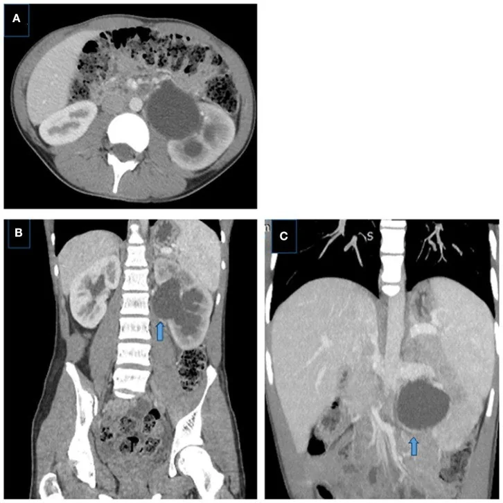
- Renal pelvis ve kalikslerde genişleme; kaliksler küntleşebilir ve parankim korteksi incelmiş olabilir.
- Pelvisin proksimal üretere birleştiği düzeyde ani kalibre değişimi veya kısa dar segment.
- UPJ'nin altında üreterin normal kalibreli olması, geçiş darlığının seviyesini destekler.
- Yüksek üreter insersiyonu, angülasyon veya kıvrılma ile daralan bileşke görünümü.
- Özellikle alt pol segmental arterinin veya veninin UPJ'yi önden çaprazlaması; damar teması klinik nedensellik için tek başına yeterli değildir.
- Uzun süreli basınç etkisinde kortikal incelme, asimetrik nefrogram veya gecikmiş ekskresyon.
- Taş, enfeksiyon bulgusu, geçirilmiş cerrahi veya kitle gibi edinilmiş ikincil nedenlerin araştırılması.
Normalde
Kıyaslamada pelvikaliksiyel sistemin genişleme derecesini, böbrek parankiminin kalınlık ve yapısını, üreter çapını ve varsa çapraz damarı inceleyin.
Normal BT ile kıyasla
Aynı bölgenin, kaynak veri setinde patoloji içermediği belirtilen bir BT incelemesini kesit kesit kaydırın; organ sınırlarını açarak anatomiyi hasta görüntüsüyle karşılaştırın. Küçük rastlantısal bulgular tümüyle dışlanmamıştır.
Kaynak: TotalSegmentator v2.01 veri seti (Wasserthal ve ark., Radiology: Artificial Intelligence 2023), CC BY 4.0, vaka s0541; organ sınırları veri setinin otomatik segmentasyonundan, birleştirilerek sadeleştirildi. Görüntüler 2,5 mm kesit aralığına yeniden örneklendi.
Ayırıcı tanı
- Üreter taşı, BT'de saptanabilir; obstrüksiyona yol açtığında üreterde ve renal kalikslerde genişleme görülebilir.
- Vezikoüreteral reflü, üst üriner sistem genişlemesinin diğer nedenlerinden biridir ve işeme sistoüretrografisiyle dışlanabilir.
- Peripelvik kistler
- renal sinüste sıvı odakları toplayıcı sistemle birleşmez; ekskretuar BT ürografide ayrı kalırlar.
- Geçici/obstrüksiyonsuz pelviektazi
- dilatasyon morfolojik olabilir; BT anatomisi fonksiyonel drenaj gecikmesini tek başına doğrulamaz.
Tuzaklar
- Hidronefroz varlığı tek başına aktif yüksek dereceli obstrüksiyon demek değildir; BT anatomik görünümü gösterir, fonksiyonel doğrulama için renografi gerekebilir.
- Alt pol çapraz damarı obstrüksiyonsuz böbreklerde de görülebilir; damar temasını daralma ve üst sistem genişlemesiyle aynı noktada eşleştirin.
- Tek fazlı incelemede üreter opaklaşmamışsa distal üreterin normal olduğunu kesin varsaymayın; ekskretuar görüntü veya diğer kesitlerle izlenebilirliğini kontrol edin.
Kendini dene
Pelvikaliksiyel sistem genişlemiş, genişleme UPJ düzeyinde sonlanmış ve üreter normal kalibrede. Bu görüntüleme örüntüsü hangi tanıyla uyumludur?
Cevabı göster
UPJ obstrüksiyonu. UPJ tipi obstrüksiyonda pelvikaliksiyel sistem genişleyebilirken üreter normal kalibrede olabilir.
UPJ yakınında alt pol arteri görülüyor; belirgin hidronefroz veya geçiş darlığı yok. En doğru yorum nedir?
Cevabı göster
Damar teması tek başına obstrüksiyonu kanıtlamaz. Çapraz damar, obstrüksiyon olmadan da görülebilir; tek başına obstrüksiyon tanısı koydurmaz.
İlgili konular
- At nalı böbrek
- Renal ektopi ve çapraz ektopi
- Çapraz füzyonlu renal ektopi
- Renal malrotasyon
- Çift toplayıcı sistem
Kaynaklar
Bu sayfadaki 33 cümle ve 2 quiz sorusu aşağıdaki kaynaklardan alıntılarla otomatik olarak karşılaştırıldı; 3 cümle bu karşılaştırmada düzeltildi (2026-10-09).
- Laparoscopic pyeloplasty and isthmusectomy in horseshoe kidney with ureteropelvic junction obstruction: a case report and review of the literaturepmc.ncbi.nlm.nih.gov
- Current Status and Future Perspectives of Robotic‐Assisted Redo Pyeloplasty for Recurrent Ureteropelvic Junction Obstructionpmc.ncbi.nlm.nih.gov
- Horseshoe kidney presenting with features of left ureteropelvic junction obstruction—insights into management: a case report and review of the literaturepmc.ncbi.nlm.nih.gov
- Branchio-oto-renal syndrome: a narrative reviewpmc.ncbi.nlm.nih.gov
- Can We Predict Renal Function Recovery After Pyeloplasty in Pediatrics with Ureteropelvic Junction Obstruction? A Systematic Reviewpmc.ncbi.nlm.nih.gov
- Congenital Anomalies of the Upper Urinary Tract: A Comprehensive Review.pubmed.ncbi.nlm.nih.gov
- Management of ureteropelvic junction obstruction in adults.pubmed.ncbi.nlm.nih.gov
- Multidetector CT of vascular compression syndromes in the abdomen and pelvis.pubmed.ncbi.nlm.nih.gov
- Management of ureteropelvic junction obstruction in children-a roundtable discussion.pubmed.ncbi.nlm.nih.gov
- CT imaging findings of abdominopelvic vascular compression syndromes: what the radiologist needs to know.pubmed.ncbi.nlm.nih.gov
- Robotic Anderson-Hynes dismembered pyeloplasty: initial experience.pubmed.ncbi.nlm.nih.gov
- Is it Always Necessary to Treat an Asymptomatic Hydronephrosis Due to Ureteropelvic Junction Obstruction?pubmed.ncbi.nlm.nih.gov
- Ureteropelvic junction obstruction in children by polar vessels. Is laparoscopic vascular hitching procedure a good solution? Single center experience on 35 consecutive patients.pubmed.ncbi.nlm.nih.gov
- Vascular compression syndromes in the abdomen and pelvis: a concise pictorial review.pubmed.ncbi.nlm.nih.gov
- Hydronephrosis in the course of ureteropelvic junction obstruction: An underestimated problem? Current opinions on the pathogenesis, diagnosis and treatment.pubmed.ncbi.nlm.nih.gov
- Giant Hydronephrosis: A Rare Case Report and Literature Review.pubmed.ncbi.nlm.nih.gov
- Magnetic Resonance Urogram in Pediatric Urology: a Comprehensive Review of Applications and Advances.pubmed.ncbi.nlm.nih.gov
- Combined treatment of ureteropelvic junction obstruction and renal calculi with robot-assisted laparoscopic pyeloplasty and laser lithotripsy in children: Case report and non-systematic review of the literature.pubmed.ncbi.nlm.nih.gov
- Laparoscopic pyeloplasty and isthmusectomy in horseshoe kidney with ureteropelvic junction obstruction: a case report and review of the literature.pubmed.ncbi.nlm.nih.gov
- Acute kidney injury in urological conditions.pubmed.ncbi.nlm.nih.gov
- Matrix Metalloproteinases in Ureteropelvic Junction Obstruction: Their Role in Pathogenesis and Their Use as Clinical Markers.pubmed.ncbi.nlm.nih.gov
- Pelviureteric junction obstruction in adults: A systematic review of the literature.pubmed.ncbi.nlm.nih.gov
- Silent Obstructive Nephrolithiasis Resulting in a Non-functional Kidney.pubmed.ncbi.nlm.nih.gov
- Report-Documented Frequency and Spectrum of Incidental Findings on Emergency Abdominopelvic Computed Tomography: A Retrospective Clinical Data Study from Saudi Arabia.pubmed.ncbi.nlm.nih.gov
- Metastatic Primary Intratesticular Rhabdomyosarcoma in an Adolescent with Rapid Early Response to VAC Chemotherapy: A Case Report and Literature Review of 99 Cases.pubmed.ncbi.nlm.nih.gov
- Robot-Assisted Anderson-Hynes Pyeloplasty for Lower-Moiety Ureteropelvic Junction Obstruction in an Incomplete Duplex Collecting System Presenting as Dietl's Crisis: A Case Report.pubmed.ncbi.nlm.nih.gov
- Primary mucinous adenocarcinoma of the renal pelvis associated with obstructive urolithiasis and giant hydronephrosis.pubmed.ncbi.nlm.nih.gov
- Management of Xanthogranulomatous Pyelonephritis: A Review of Current Evidence and Treatment Strategies and a Proposed Clinical Management Algorithm.pubmed.ncbi.nlm.nih.gov
- Rare Causes of Hydronephrosis in Adults and Diagnosis Algorithm: Analysis of 100 Cases During 15 Years.pubmed.ncbi.nlm.nih.gov
- Bilateral vascular variations at the renal hilum: a case report.pubmed.ncbi.nlm.nih.gov
- Computed tomography of renal emergencies: A comprehensive diagnostic guide for radiology residents.pubmed.ncbi.nlm.nih.gov
- Managing Ureteropelvic Junction Obstruction in the Young Infant.pubmed.ncbi.nlm.nih.gov
- A case report on atypically presented Congenital Solitary pelvic kidney with neglected Ureteropelvic junction obstruction (UPJO) successfully managed with open Anderson-Hynes pyeloplasty. Case report.pubmed.ncbi.nlm.nih.gov
- Systemic inflammatory response syndrome in pediatric urinary tract infections associated with urolithiasis: a retrospective study of risk factors, pathogens, and antimicrobial resistance at two tertiary hospitals.pubmed.ncbi.nlm.nih.gov
- Intermittent Ureteropelvic Junction Obstruction Can Mimic Abdominal Migraine: A Case Report and Review of the Literature.pubmed.ncbi.nlm.nih.gov
- Fetal Hydronephrosis: A Narrative Review of Diagnostic Pathways, Prognostic Markers, and Management.pubmed.ncbi.nlm.nih.gov
- Interobserver agreement of retrospective renal pelvis measurements in cats with confirmed ureteral obstruction.pubmed.ncbi.nlm.nih.gov
- Advances in NIR-II Fluorescence Laparoscopy for Precision Diagnosis and Theranostics of Urological Oncology.pubmed.ncbi.nlm.nih.gov
- Comparison of external drainage ureteral tube versus double-J catheter placement on subjective comfort and adverse events after percutaneous nephrolithotomy.pubmed.ncbi.nlm.nih.gov
- Multilevel congenital ureteral obstruction in a two-year-old child: Staged reconstruction with neo-implantation and V-Y pyeloplasty.pubmed.ncbi.nlm.nih.gov
- Case Report: Laparoscopic repair of a recurrent, huge, inguinoscrotal vesicoureteral hernia: a case report and review of the literature.pubmed.ncbi.nlm.nih.gov
- Prenatal imaging of upper urinary tract abnormalities: when is MRI useful?pubmed.ncbi.nlm.nih.gov
- Grading of Hydronephrosis: An Ongoing Challenge.pubmed.ncbi.nlm.nih.gov
- A rare presentation of unilateral periureteral renal lymphangiomatosis.pubmed.ncbi.nlm.nih.gov
- Spontaneous combined rupture of a pelvicalyceal cyst into the collector system and retroperitoneal space during the acquisition of computed tomography scan images: a case report.pubmed.ncbi.nlm.nih.gov
- Quantitative Dynamic Contrast-Enhanced Ultrasound Confirms Renal Obstruction: A Feasibility Study.pubmed.ncbi.nlm.nih.gov
- Predictive Factors for Spontaneous Resolution in Primary Obstructive Megaureter: The Impact of Hydronephrosis Severity on Clinical Outcomes.pubmed.ncbi.nlm.nih.gov
- Complex Management of Bilateral Congenital Hydronephrosis in a Pediatric Patient: A Multidisciplinary Approach.pubmed.ncbi.nlm.nih.gov
- A radiological evaluation of crossed-fused ectopic kidney in adult female: Case report and comprehensive review of literature.pubmed.ncbi.nlm.nih.gov
- Dietl Syndrome Misattributed to Gastrointestinal Disease in a Child: A Pediatric Diagnostic Pitfall.pubmed.ncbi.nlm.nih.gov
- Mastering the robot-assisted pyeloplasty learning curve: analysis of operative efficiency, safety, and functional outcomes.pubmed.ncbi.nlm.nih.gov
- Clinical Application of the Computed-Tomography-Based Three-Dimensional Models in the Surgical Planning and Intraoperative Navigation of Ureteropelvic Junction Obstruction Complicated with Renal Calculi.pubmed.ncbi.nlm.nih.gov
- A Case of Hydronephrosis Associated With Horseshoe Kidney Successfully Treated by Retroperitoneoscopic Isthmusectomy Without Pyeloplasty.pubmed.ncbi.nlm.nih.gov
- Divergent outcomes of delayed ureteropelvic junction stenosis after renal cyst sclerotherapy: two case reports.pubmed.ncbi.nlm.nih.gov
- Upper tract urothelial carcinoma arising in a horseshoe kidney with ureteropelvic junction obstruction: a case report.pubmed.ncbi.nlm.nih.gov
- Urinary Ultrasound and Other Imaging for Ureteropelvic Junction Type Hydronephrosis (UPJHN).pubmed.ncbi.nlm.nih.gov
- Laparoscopic Anderson-Hynes pyeloplasty with Butler flap for pelviureteric junction obstruction in a pelvic ectopic kidney presenting as a giant palpable abdominal mass: A case report.pubmed.ncbi.nlm.nih.gov
- Beyond Hydronephrosis: Perirenal Fluid on Screening Ultrasound in Patients With Renal Colic at a Basic Emergency Service.pubmed.ncbi.nlm.nih.gov
- When Stones Escape: A Rare Radiologic Finding of Retroperitoneal Stone Extrusion Following American Association for the Surgery of Trauma (AAST) Grade IV Blunt Renal Trauma in a Chronically Hydronephrotic Kidney.pubmed.ncbi.nlm.nih.gov
- Massive uterine angiomyolipoma mimicking uterine leiomyoma in a 49-year-old woman.pubmed.ncbi.nlm.nih.gov
- Large Uterine Angiolipoleiomyoma Masquerading as an Ovarian Tumor: A Case Report.pubmed.ncbi.nlm.nih.gov
- IgG4-related kidney disease presenting as severe renal failure: Diagnostic challenges of a multimodality imaging approach.pubmed.ncbi.nlm.nih.gov
- Diagnosis and treatment of congenital anterior urethral Valves: a retrospective analysis of 8 cases.pubmed.ncbi.nlm.nih.gov
- Multimodal treatment for failed pyeloplasty: Description of a new technique.pubmed.ncbi.nlm.nih.gov
- Revisiting the Anatomy of the Pediatric Uretero-Pelvic Junction Analysis of Intraoperative Video of Robotic-Assisted Pyeloplasty.pubmed.ncbi.nlm.nih.gov
- Ureter as an Innocent Bystander: Presentation and Management in Unusual Vascular Compression Syndromes.pubmed.ncbi.nlm.nih.gov
- Delayed Rupture of the Renal Collecting System Secondary to a Traumatic Hemorrhage in the Setting of Suspected Chronic Ureteropelvic Junction Obstruction.pubmed.ncbi.nlm.nih.gov
- Accuracy of reduced-dose computed tomography for ureteral stones in emergency department patients.pubmed.ncbi.nlm.nih.gov
- Rare case of spontaneous renal calyx rupture due to vesicoureteral junction stone in a horseshoe kidney.pubmed.ncbi.nlm.nih.gov
- Use of MR Urography in Pediatric Patients.pubmed.ncbi.nlm.nih.gov
- Diagnostic value of combined static-excretory MR Urography in children with hydronephrosis.pubmed.ncbi.nlm.nih.gov
- Imaging Diagnosis of Major Kidney and Urinary Tract Disorders in Children.pubmed.ncbi.nlm.nih.gov
- Point-of-Care Kidney and Genitourinary Ultrasound in Adults: Image Acquisition.pubmed.ncbi.nlm.nih.gov
- Combined Minimally Invasive Treatment of Pyeloureteral Junction Obstruction and Primary Obstructive Megaureter in Children: Case Report and Literature Review.pubmed.ncbi.nlm.nih.gov
- Laparoscopic ureteral anastomosis to a bladder diverticulum for the treatment of postoperative ureteral fistula: a case report.pubmed.ncbi.nlm.nih.gov
- Pneumovesicoscopic ureteral reimplantation for pediatric vesicoureteral reflux: Politano-Leadbetter vs. Cohen technique.pubmed.ncbi.nlm.nih.gov
- Robot-assisted reconstruction using ureteroureterostomy and Lich-Gregoir ureteral reimplantation for complicated duplex kidneys in children.pubmed.ncbi.nlm.nih.gov
- Back-Table Repair of Parenchymal and Collecting System Injury of a Living Donor Kidney.pubmed.ncbi.nlm.nih.gov
- Sonographic Anatomy and Normal Measurements of the Human Kidneys: A Comprehensive Review.pubmed.ncbi.nlm.nih.gov
- A case of mid-ureteral stricture with ipsilateral atrophic kidney in a young adult.pubmed.ncbi.nlm.nih.gov
Eğitim amaçlıdır; hasta başında tanı ve tedavi kararının yerine geçmez, tıbbi tavsiye değildir. Metinler yapay zekâ desteğiyle yazıldı ve otomatik bir adımda kaynak alıntılarıyla karşılaştırıldı; bu bir tıbbi doğrulama değildir ve içerik uzman radyolog incelemesinden geçmedi. Klinik kararlarda güncel kılavuzları, kurum protokollerini ve radyoloji raporunu esas alın.