Özet
Ekstrarenal pelvis, renal sinüs yağ dokusunun dışında daha belirgin görünen, böbreğin benign anatomik varyantıdır. Kontrastsız BT taşların saptanmasını sağlar; BT ürogramın ekskretuar fazında kaliksler ve pelvis iyotlu idrarla dolar. Kontrastsız BT, üreter taşını ve taşla ilişkili obstrüksiyonun ikincil bulgularını değerlendirmeye olanak verir. Ekstrarenal pelvis ve üreter dilatasyonu obstrüksiyon olmadan birlikte görülebilir; bu nedenle pelvis dolgunluğu tek başına obstrüksiyonu kanıtlamaz.
Faz ve pencere
- Kontrastsız tanısal
- BT ürogramda ekstrarenal pelvis ve üreter dilatasyonu görülebilir; bildirilen olguda obstrüksiyon veya ikincil neden saptanmamıştır.
Önerilen pencereler G genişlik, M merkez; değerler Hounsfield birimi (HU)
- BatınG 400 / M +50
BT bulguları
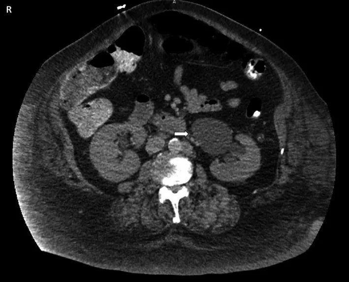
- BT ürogramda ekstrarenal pelvis görünümü, üreter dilatasyonu ile birlikte görülebilir.
- Üreteropelvik bileşke obstrüksiyonunda pelvikaliksiyel sistem değişen derecelerde genişleyebilir.
- Ekstrarenal pelvis ve üreter dilatasyonu, obstrüksiyon olmadan birlikte görülebilir.
- Üreteropelvik bileşke obstrüksiyonunun görüntüleme değerlendirmesinde renal korteksin kalınlığı ve mimarisi incelenir.
- Üreteropelvik bileşke obstrüksiyonunda pelvikaliksiyel sistem genişleyebilirken üreter normal kalibrede olabilir.
- Bir otopsi serisinde sağ ve sol böbrekler arasında toplayıcı sistem morfolojisi bakımından anlamlı fark saptanmamıştır.
- Perinefrik yağ çizgilenmesi, üreter taşına bağlı obstrüksiyonun BT’de değerlendirilebilen ikincil bulgularındandır; tek başına ekstrarenal pelvis tanısını desteklediği kaynaklarda gösterilmemiştir.
Normalde
Bir olgu bildiriminde BT ürogramda ekstrarenal pelvis ve üreter dilatasyonu, obstrüksiyon veya ikincil neden olmaksızın izlenmiştir. Üreter dilatasyonu obstrüksiyon olmadan da görülebilir; bir olgu bildiriminde dilate ekstrarenal pelvis ve üreterlere karşın obstrüksiyon veya ikincil neden saptanmamıştır.
Normal BT ile kıyasla
Aynı bölgenin, kaynak veri setinde patoloji içermediği belirtilen bir BT incelemesini kesit kesit kaydırın; organ sınırlarını açarak anatomiyi hasta görüntüsüyle karşılaştırın. Küçük rastlantısal bulgular tümüyle dışlanmamıştır.
Kaynak: TotalSegmentator v2.01 veri seti (Wasserthal ve ark., Radiology: Artificial Intelligence 2023), CC BY 4.0, vaka s0119; organ sınırları veri setinin otomatik segmentasyonundan, birleştirilerek sadeleştirildi. Görüntüler 2,5 mm kesit aralığına yeniden örneklendi.
Ayırıcı tanı
- Hidronefrozda pelvikaliksiyel sistem değişen derecelerde genişleyebilir; üreteropelvik bileşke obstrüksiyonunda üreter normal kalibrede olabilir.
- Üreteropelvik bileşke obstrüksiyonunda pelvikaliksiyel sistem genişleyebilirken üreter normal kalibrede olabilir.
Tuzaklar
- Pelvisin tek başına geniş görünmesi hidronefroz tanısı koydurmaz; kaliks ve üreter çapı da değerlendirilmelidir.
- Kontrastsız BT taş saptamaya yardımcı olur; kaliks ve pelvisin opaklaşması ise BT ürogramın ekskretuar fazında değerlendirilir.
- Ekstrarenal pelvis görünümüne üreter dilatasyonu eşlik edebilir ve bu birliktelik tek başına obstrüksiyonu göstermez.
- Ekstrarenal pelvis görünümüne üreter dilatasyonu eşlik edebilir; bir olgu bildiriminde bu birliktelikte obstrüksiyon veya ikincil neden saptanmamıştır.
Kendini dene
Bir olgu bildiriminde BT ürogramda ekstrarenal pelvis ve üreterler dilate görülmüş; obstrüksiyon veya ikincil neden saptanmamıştır. Bu olguda hangi sonuç bildirilmiştir?
Cevabı göster
Obstrüksiyonsuz, ikincil nedeni gösterilemeyen dilatasyon. Olgu bildiriminde BT ürogram ekstrarenal pelvis ve üreter dilatasyonunu göstermiş, obstrüksiyon veya ikincil neden saptanmamıştır.
Kontrastsız BT'de üreter taşı saptanan hastada hangi ek değerlendirme yapılabilir?
Cevabı göster
Taşla ilişkili obstrüksiyonun ikincil bulgularını değerlendirmek. Kontrastsız BT üreter içindeki taşı gösterir ve taşla ilişkili obstrüksiyonun ikincil bulgularının değerlendirilmesine olanak verir.
Kaynaklar
Bu sayfadaki 31 cümle ve 2 quiz sorusu aşağıdaki kaynaklardan alıntılarla otomatik olarak karşılaştırıldı; 25 cümle bu karşılaştırmada düzeltildi (2026-10-09).
- Computed Tomography Urography: State of the Art and Beyondpmc.ncbi.nlm.nih.gov
- Bilateral primary nonrefluxing unobstructed megaureter in an adult: a case report and review of the literature.pubmed.ncbi.nlm.nih.gov
- Diagnostic value of combined static-excretory MR Urography in children with hydronephrosis.pubmed.ncbi.nlm.nih.gov
- The evaluation of morphology of renal pelvicalyceal system's and infundibulopelvic anatomy of kidney's lower pole in post-mortem series.pubmed.ncbi.nlm.nih.gov
- [Risk factors for contrast extravasation in renal colic].pubmed.ncbi.nlm.nih.gov
- Stealthy Encroachment: Unraveling an Encounter With Renal Myelolipoma.pubmed.ncbi.nlm.nih.gov
- Pseudodiphallia: a rare kind of diphallia: A case report and literature review.pubmed.ncbi.nlm.nih.gov
- Predictors of Recoverability of Renal Function after Pyeloplasty in Adults with Ureteropelvic Junction Obstruction.pubmed.ncbi.nlm.nih.gov
- Concurrent emphysematous cystitis and emphysematous pyelonephritis in a patient with extrarenal pelvis.pubmed.ncbi.nlm.nih.gov
- Inadvertent placement of an AXIOS TM stent connecting the duodenum to the renal collecting system.pubmed.ncbi.nlm.nih.gov
- Vaginal Agenesis (Mayer-Rokitansky-Kuster-Hauser Syndrome) Presenting with Exertional Dyspnoea, Fatigue and Intermittent Palpitations.pubmed.ncbi.nlm.nih.gov
- Right Iliac Ectopic Kidney With an Extrarenal Pelvis, Aberrant Upper-Pole Arterial Supply, and Multiple Renal Cysts.pubmed.ncbi.nlm.nih.gov
- Hysteroscopic Management of Complete Septate Uterus With Septate Cervix and Longitudinal Vaginal Septum in a Third-Level Mexican Institution: A Case Report.pubmed.ncbi.nlm.nih.gov
- Left Renal Vein Compression Syndrome: Cracking the Nut of Clinical Dilemmas - Three Cases and Review of Literature.pubmed.ncbi.nlm.nih.gov
- Computed tomography findings in xanthogranulomatous pyelonephritis.pubmed.ncbi.nlm.nih.gov
- Lineage infidelity in FH-deficient RCC with secondary somatic alterations: a case report and implications for diagnosis and treatment.pubmed.ncbi.nlm.nih.gov
- Sarcomatoid Renal Cell Carcinoma: Biological Features and Therapeutic Implications-A Narrative Review.pubmed.ncbi.nlm.nih.gov
- Ureterorenoscopic (URS) Lithotripsy and Balloon Dilation Cause Acute Kidney Injury and Distal Renal Tubule Damage: A Prospective Study.pubmed.ncbi.nlm.nih.gov
- My patient has abdominal and flank pain: Identifying renal causes.pubmed.ncbi.nlm.nih.gov
- Comparison between ultrasound and noncontrast helical computed tomography for identification of acute ureterolithiasis in a teaching hospital setting.pubmed.ncbi.nlm.nih.gov
- Granulomatosis with polyangiitis presenting with rare urogenital involvement: a case based review.pubmed.ncbi.nlm.nih.gov
- Enhancing radiology training with GPT-4: Pilot analysis of automated feedback in trainee preliminary reports.pubmed.ncbi.nlm.nih.gov
- Imaging Diagnosis of Major Kidney and Urinary Tract Disorders in Children.pubmed.ncbi.nlm.nih.gov
- Xanthogranulomatous pyelonephritis: A case report and literature review.pubmed.ncbi.nlm.nih.gov
- Radiologic evaluation of the kidney transplant donor and recipient.pubmed.ncbi.nlm.nih.gov
- Multimodality imaging of renal lymphoma and its mimics.pubmed.ncbi.nlm.nih.gov
- Xanthogranulomatous Pyelonephritis: A Narrative Review with Current Perspectives on Diagnostic Imaging and Management, Including Interventional Radiology Techniques.pubmed.ncbi.nlm.nih.gov
- Ultrasonographic features of kidney transplants and their complications: an imaging review.pubmed.ncbi.nlm.nih.gov
- Laparoscopic Anderson-Hynes pyeloplasty with Butler flap for pelviureteric junction obstruction in a pelvic ectopic kidney presenting as a giant palpable abdominal mass: A case report.pubmed.ncbi.nlm.nih.gov
- Fetal Hydronephrosis: A Narrative Review of Diagnostic Pathways, Prognostic Markers, and Management.pubmed.ncbi.nlm.nih.gov
- ESR Essentials: renal imaging in children-practice recommendations by the European Society of Paediatric Radiology.pubmed.ncbi.nlm.nih.gov
- Complete extrarenal migration of a double-J ureteral stent following ureteral stone management: A case report.pubmed.ncbi.nlm.nih.gov
- Urinary Ultrasound and Other Imaging for Ureteropelvic Junction Type Hydronephrosis (UPJHN).pubmed.ncbi.nlm.nih.gov
- A Network of Anomalies Prompting VACTERL Workup in a Trisomy 21 Newborn.pubmed.ncbi.nlm.nih.gov
- Polypoid endometriosis with endometrioid intraepithelial neoplasia mimicking malignancy: a rare case of a postmenopausal pelvic mass.pubmed.ncbi.nlm.nih.gov
- Point-of-Care Kidney and Genitourinary Ultrasound in Adults: Image Acquisition.pubmed.ncbi.nlm.nih.gov
- Persistent Eustachian Valve Causing Abdominal and Pelvic Venous System Dilation in a 44-Year-Old Male: A Case Report.pubmed.ncbi.nlm.nih.gov
- An Uncommon Diagnosis in a Common Presentation: Renal Vein Thrombosis in a Young Adult.pubmed.ncbi.nlm.nih.gov
- Emphysematous conditions of the abdomen and pelvis: pearls and pitfalls.pubmed.ncbi.nlm.nih.gov
- Radiology and Non-operative Management of Emphysematous Infections of the Pancreas, Gallbladder, and Urinary System.pubmed.ncbi.nlm.nih.gov
- Sonographic Anatomy and Normal Measurements of the Human Kidneys: A Comprehensive Review.pubmed.ncbi.nlm.nih.gov
- Complications of lymphoma in the abdomen and pelvis: clinical and imaging review.pubmed.ncbi.nlm.nih.gov
- Standardization of pediatric uroradiological terms: a multidisciplinary European glossary.pubmed.ncbi.nlm.nih.gov
- Prenatal imaging of upper urinary tract abnormalities: when is MRI useful?pubmed.ncbi.nlm.nih.gov
- Symptomatic infundibulopelvic dysgenesis in an adolescent.pubmed.ncbi.nlm.nih.gov
- A rare presentation of unilateral periureteral renal lymphangiomatosis.pubmed.ncbi.nlm.nih.gov
- CT and MRI in Urinary Tract Infections: A Spectrum of Different Imaging Findings.pubmed.ncbi.nlm.nih.gov
- Renal lymphangiomatosis, a rare differential diagnosis for autosomal recessive polycystic kidney disease in pediatric patients.pubmed.ncbi.nlm.nih.gov
- Fetal MRI: abdominal cystic lesions.pubmed.ncbi.nlm.nih.gov
- Use of MR Urography in Pediatric Patients.pubmed.ncbi.nlm.nih.gov
- Quantitative renal magnetic resonance imaging: magnetic resonance urography.pubmed.ncbi.nlm.nih.gov
- Analysis of Diagnosis and Treatment of Active Lupus Enteritis Accompanied by Intestinal Pseudo-Obstruction as the Initial Manifestation: A Case Report and Literature Review.pubmed.ncbi.nlm.nih.gov
- Haematuria: an imaging guide.pubmed.ncbi.nlm.nih.gov
- Radionuclides in nephrourology, Part 2: pitfalls and diagnostic applications.pubmed.ncbi.nlm.nih.gov
- Feline comorbidities: cardiovascular and kidney diseases.pubmed.ncbi.nlm.nih.gov
- The space of Retzius: anatomy, imaging features, and patterns of disease spread.pubmed.ncbi.nlm.nih.gov
- Image Imputation with conditional generative adversarial networks captures clinically relevant imaging features on computed tomography.pubmed.ncbi.nlm.nih.gov
- Renal Sinus Pathologies Depicted by CT Imaging: A Pictorial Review.pubmed.ncbi.nlm.nih.gov
- Current and potential methods to assess kidney structure and morphology in term and preterm neonates.pubmed.ncbi.nlm.nih.gov
- Long-term safety and effectiveness of the "OptEase" vena cava filter.pubmed.ncbi.nlm.nih.gov
- The Hidden Hitchhiker: Retrocaval Ureter Masquerading as Isolated Pelviureteric Junction Obstruction.pubmed.ncbi.nlm.nih.gov
- Sarcoidosis Presenting With Severe Hypercalcemia, Renal Dysfunction, and Bilateral Submandibular Swelling: A Case Report.pubmed.ncbi.nlm.nih.gov
- Percutaneous antegrade-retrograde recanalization (rendezvous technique) of a complete ureteropelvic junction disruption after robotic-assisted partial nephrectomy: A case report.pubmed.ncbi.nlm.nih.gov
- Infantile Homocystinuric Urolithiasis With Staghorn Calculus: A Case Report of Nephron-Sparing Surgical Salvage in an Intraparenchymal Renal Pelvis.pubmed.ncbi.nlm.nih.gov
- Clinical Significance, Differential Diagnosis and Initial Management of Hematuria in Emergency and Outpatient Settings.pubmed.ncbi.nlm.nih.gov
- Ischemic colitis in a child: a case report and narrative review.pubmed.ncbi.nlm.nih.gov
- Contrast-sparing CT using renal-clearable gold nanoclusters for early spatial mapping of renal dysfunction.pubmed.ncbi.nlm.nih.gov
- Point of care renal ultrasonography for the busy nephrologist: A pictorial review.pubmed.ncbi.nlm.nih.gov
- Comparison of secondary signs as shown by unenhanced helical computed tomography in patients with uric acid or calcium ureteral stones.pubmed.ncbi.nlm.nih.gov
- Advances in CT imaging for urolithiasis.pubmed.ncbi.nlm.nih.gov
- Pyeloplasty for pelviureteric junction obstruction in anomalous kidneys: A long-term follow-up experience at a tertiary care centre.pubmed.ncbi.nlm.nih.gov
- Added value of restricted diffusion in acute pyelonephritis: Two cases with different clinical presentations.pubmed.ncbi.nlm.nih.gov
- Imaging of renal pseudotumors in children: a comprehensive review.pubmed.ncbi.nlm.nih.gov
- Outcomes and predictors of postoperative complications in patients undergoing laparoscopic ureterolithotomy: a retrospective analysis.pubmed.ncbi.nlm.nih.gov
- Comparison of the Effects of Laparoscopic Surgery and Traditional Open Surgery on Stone Clearance, Laboratory Indexes and Life Quality in Patients with Renal Calculi.pubmed.ncbi.nlm.nih.gov
- Clinical Approaches and Emerging Therapeutic Horizons in Primary Hyperoxaluria.pubmed.ncbi.nlm.nih.gov
- Examining the Clinical Significance and Embryologic Development of Two Unique Cases of Renal Anatomy.pubmed.ncbi.nlm.nih.gov
- Robot-assisted "Santosh-Post Graduate Institute tubularized flap pyelovesicostomy" in a solitary functioning kidney with giant hydronephrosis: A minimally invasive salvage procedure.pubmed.ncbi.nlm.nih.gov
- Pyonephrosis Ultrasound and Computed Tomography Features: A Pictorial Review.pubmed.ncbi.nlm.nih.gov
- Ultrasonography of acute flank pain: a focus on renal stones and acute pyelonephritis.pubmed.ncbi.nlm.nih.gov
- Prostatic morphometric parameters and ureteral access sheath placement failure during retrograde intrarenal surgery: a retrospective cohort study.pubmed.ncbi.nlm.nih.gov
- Dietl Syndrome Misattributed to Gastrointestinal Disease in a Child: A Pediatric Diagnostic Pitfall.pubmed.ncbi.nlm.nih.gov
- Villous Adenoma of the Renal Pelvis: Clinicopathologic Spectrum and Diagnostic Considerations.pubmed.ncbi.nlm.nih.gov
- Nonoperative Management of Technically Resectable Pancreatic Cancer With Ablative Radiation Therapy.pubmed.ncbi.nlm.nih.gov
- Magnetic Resonance Imaging of the Gastrointestinal Tract: Current Role, Recent Advancements and Future Prospectives.pubmed.ncbi.nlm.nih.gov
- Complicated pyelonephritis associated with chronic renal stone disease.pubmed.ncbi.nlm.nih.gov
- Histology Atlas of the Developing Mouse Urinary System With Emphasis on Prenatal Days E10.5-E18.5.pubmed.ncbi.nlm.nih.gov
- Multiphase CT-derived markers for the characterization of large solid and cystic renal masses by histology and grade.pubmed.ncbi.nlm.nih.gov
- Urogenital Tuberculosis: A Narrative Review and recommendations for diagnosis and treatment.pubmed.ncbi.nlm.nih.gov
- Spilling the beans: an inside scoop on the imaging of renal parenchymal disease.pubmed.ncbi.nlm.nih.gov
- Totally tubeless, tubeless, and tubed percutaneous nephrolithotomy for treating kidney stones.pubmed.ncbi.nlm.nih.gov
- A Rare Case of Primary Follicular Dendritic Cell Sarcoma of the Kidney.pubmed.ncbi.nlm.nih.gov
- Beyond Hydronephrosis: Perirenal Fluid on Screening Ultrasound in Patients With Renal Colic at a Basic Emergency Service.pubmed.ncbi.nlm.nih.gov
- Laparoscopic Management of a Symptomatic Paraduodenal Hernia Complicated by Ureteric Compression: A Case Report.pubmed.ncbi.nlm.nih.gov
- Epidemiology, pathophysiology, and management of uric acid urolithiasis: A narrative review.pubmed.ncbi.nlm.nih.gov
- Perirenal fat stranding on CT: is there an association with bladder outlet obstruction?pubmed.ncbi.nlm.nih.gov
- When Stones Escape: A Rare Radiologic Finding of Retroperitoneal Stone Extrusion Following American Association for the Surgery of Trauma (AAST) Grade IV Blunt Renal Trauma in a Chronically Hydronephrotic Kidney.pubmed.ncbi.nlm.nih.gov
- Hypernatremia secondary to partial urinary tract obstruction: a case report.pubmed.ncbi.nlm.nih.gov
- Cognitive bias in veterinary clinical decision-making: Antemortem misdiagnosis of suspected giant kidney worm (Dioctophyme renale) in Australia.pubmed.ncbi.nlm.nih.gov
- Stage III xanthogranulomatous pyelonephritis with sarcomatoid degeneration.pubmed.ncbi.nlm.nih.gov
- Can resident-performed POCUS and clinical predictors effectively risk-stratify emergency department patients with confirmed renal colic?pubmed.ncbi.nlm.nih.gov
- Kidney-sparing resection of concomitant renal artery and renal vein aneurysms by single-port retroperitoneoscopic surgery: a case report.pubmed.ncbi.nlm.nih.gov
- Imaging urolithiasis: complications and interventions in children.pubmed.ncbi.nlm.nih.gov
- Massive upper tract hematuria following decompression of chronic PUJ obstruction: A rare but clinically significant complication.pubmed.ncbi.nlm.nih.gov
- Paraspinal Extrarenal Wilms Tumor Case Report and Review of Literature.pubmed.ncbi.nlm.nih.gov
- Analysis of factors affecting the upstaging and downstaging of renal cell carcinoma from T1 to T3a and the impact of different surgical approaches on patient prognosis and survival: a multicenter retrospective study.pubmed.ncbi.nlm.nih.gov
- Renal and Genitourinary Ultrasound Evaluation in Emergency and Critical Care: An Overview.pubmed.ncbi.nlm.nih.gov
- MR Imaging of the Upper Urinary Tract: Techniques, Diagnostic Performance, and Imaging Biomarkers.pubmed.ncbi.nlm.nih.gov
- Periaortitis and Retroperitoneal Fibrosis in a Patient With a Normal IgG4 Level: A Case Report and Review of the Literature.pubmed.ncbi.nlm.nih.gov
- Ureter as an Innocent Bystander: Presentation and Management in Unusual Vascular Compression Syndromes.pubmed.ncbi.nlm.nih.gov
- 18 F-FDG PET/CT Manifestations of IgG4-related Disease.pubmed.ncbi.nlm.nih.gov
Eğitim amaçlıdır; hasta başında tanı ve tedavi kararının yerine geçmez, tıbbi tavsiye değildir. Metinler yapay zekâ desteğiyle yazıldı ve otomatik bir adımda kaynak alıntılarıyla karşılaştırıldı; bu bir tıbbi doğrulama değildir ve içerik uzman radyolog incelemesinden geçmedi. Klinik kararlarda güncel kılavuzları, kurum protokollerini ve radyoloji raporunu esas alın.