Özet
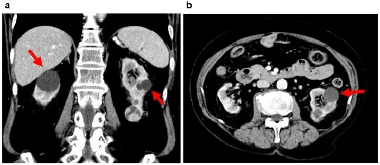
Edinsel kistik böbrek hastalığı, özellikle uzun süreli böbrek yetmezliği ve diyaliz öyküsü olan kişilerde iki böbreğe yayılmış çok sayıda kistle karşımıza çıkar. Kronik böbrek hastalığında kortikal incelme ve skarlaşma görülebilir; BT'de kistler yanında solid lezyonların değerlendirilmesi de önemlidir. Kontrastsız BT kist içeriğinin yüksek atenüasyonunu ve kalsifikasyonu gösterebilir; ancak kontrast madde kullanılmadan kistin solid lezyondan ayrılması zor olabilir. ACKD’deki çok sayıda ve karmaşık kist, eşlikçi renal hücreli karsinomun görüntülemede fark edilmesini güçleştirebilir.
Faz ve pencere
- Kontrastsız tanısal
- Her iki böbrekte farklı boyut ve atenüasyonda kistler görülebilir; kistlerde hemorajik veya proteinöz içerik ve duvar kalsifikasyonu bulunabilir. Kontrastsız BT lezyonların atenüasyonunu gösterir; kontrast madde olmadan kistleri solid lezyonlardan ayırmak zor olabilir.
- Portal tanısal
- Kistler kontrastlı BT’de tipik olarak kontrastlanmaz; kalınlaşmış duvar veya septalarda kontrastlanma malignite kuşkusu doğurur. ACKD ilişkili RCC kontrastlı BT'de solid, kistik veya karma atenüasyonlu görünebilir.
Önerilen pencereler: Batın (G 400 / M 50), Yumuşak doku (G 400 / M 40).
BT bulguları
- Korteks ve medullada iki taraflı, çok sayıda ve boyutları değişken kistik boşluklar; aralarda ince parankim köprüleri kalabilir.
- Böbrek konturları atrofik, düzensiz ya da korunmuş hacimde olabilir; yalnız böbrek boyutu hastalığı dışlamaz.
- Kist içeriği kontrastsız BT'de sıvı atenüasyonunda olabilir; hemorajik veya proteinöz içerik daha yüksek atenüasyon gösterebilir ve bu bulgu tek başına solid bileşen olduğunu göstermez.
- ACKD'de kist içindeki solid bileşen kontrastlı BT'de belirgin kontrastlanma gösterebilir.
- ACKD ilişkili RCC iyi sınırlı ve dışa büyüyen bir kitle şeklinde gelişebilir; kistik ve solid bileşenler aynı lezyonda birlikte bulunabilir.
- Küçük neoplastik odaklar yoğun kist dağılımı içinde saklanabilir; böbreği seri kesitlerde ve çok düzlemli rekonstrüksiyonda taramak gerekir.
- Şüpheli renal kitlenin evrelemesinde kontrastlı BT ile venöz tutulumu değerlendirin.
- ACKD’de böbrekler atrofik veya normal boyutta olabilir ve iki böbrekte çok sayıda mikrokist görülebilir.
Normalde
Normal böbrekte korteks ve medulla kesintisiz, düzenli parankim oluşturur; renal sinüs yağlıdır ve bu alana yayılmış çoklu kistik boşluklar bulunmaz. Aynı kesitte iki böbreğin kalan kortikal dokusunu karşılaştırın; kontrastsız seride yoğun görünen kist içeriğini portal serideki gerçek nodüler doku boyanmasından ayırın.
Normal BT ile kıyasla
Aynı bölgenin, kaynak veri setinde patoloji içermediği belirtilen bir BT incelemesini kesit kesit kaydırın; organ sınırlarını açarak anatomiyi hasta görüntüsüyle karşılaştırın. Küçük rastlantısal bulgular tümüyle dışlanmamıştır.
Kaynak: TotalSegmentator v2.01 veri seti (Wasserthal ve ark., Radiology: Artificial Intelligence 2023), CC BY 4.0, vaka s0541; organ sınırları veri setinin otomatik segmentasyonundan, birleştirilerek sadeleştirildi. Görüntüler 2,5 mm kesit aralığına yeniden örneklendi.
Ayırıcı tanı
- Otozomal dominant polikistik böbrek hastalığı
- böbrekler çoğu kez belirgin büyür ve karaciğer kistleri eşlik edebilir.
- Basit renal kist
- düzgün ince duvarlı, homojen sıvı içerikli ve kontrastlanmayan odaktır.
- ACKD ilişkili renal hücreli karsinom
- kist içinde mural doku veya kistler arasında kontrastlanan solid bileşen oluşturabilir.
- Kanamalı veya proteinöz kist kontrastsız BT'de yüksek atenüasyon gösterebilir; kontrastlı BT'de kistler tipik olarak kontrastlanmaz.
- Renal lenfoma, infiltratif renal kitlelerin ayırıcı tanısında yer alır.
Tuzaklar
- Yoğun kist içeriğini kontrastlanan tümör sanmayın; başlangıç atenüasyonunu kontrast sonrası aynı odakla karşılaştırın.
- Küçük mural nodüller veya duvar kalınlaşmaları, hemorajik/proteinöz içerikle karışabilir; kontrastlı serilerde gerçek vasküler boyanma ayrıştırılmalıdır.
- Kalıtsal polikistik hastalıkla ayrımda yalnız kist sayısına yaslanmayın; böbrek hacmini ve karaciğer kistlerini de görüntü alanına katın.
- Kontrastsız BT’de intrarenal, kontrastlanmayan ve 3 cm veya daha büyük hiperatenuan kitleler Bosniak IIF kapsamında sınıflandırılır.
Kendini dene
Kontrastlı BT'de ACKD ilişkili renal hücreli karsinom hangi atenüasyon paterninde görülebilir?
Cevabı göster
Solid, kistik veya solid-kistik paternlerde. ACKD ilişkili renal hücreli karsinom kontrastlı BT'de solid, kistik veya solid-kistik paternlerde görülebilir.
Kaynağa göre kontrastsız BT'de yüksek atenüasyonlu renal kist içeriği hangi iki durumla ilişkili olabilir?
Cevabı göster
Hemoraji veya yüksek protein içeriği. Kist içindeki kanama veya yüksek protein içeriği kontrastsız BT'de yüksek atenüasyonlu görünüm oluşturabilir.
İlgili konular
Kaynaklar
Bu sayfadaki 36 cümle ve 2 quiz sorusu aşağıdaki kaynaklardan alıntılarla otomatik olarak karşılaştırıldı; 19 cümle bu karşılaştırmada düzeltildi (2026-10-09).
- Diagnostic evaluation of renal masses in chronic kidney disease: a clinical and radiologic challengepmc.ncbi.nlm.nih.gov
- Cystic Kidney Diseases That Require a Differential Diagnosis from Autosomal Dominant Polycystic Kidney Disease (ADPKD)pmc.ncbi.nlm.nih.gov
- Renal Cell Carcinoma in End-Stage Renal Disease: A Review and Updatepmc.ncbi.nlm.nih.gov
- Renal Cell Carcinoma and Kidney Transplantation: A Narrative Reviewpmc.ncbi.nlm.nih.gov
- Imaging of haemodialysis: renal and extrarenal findingspmc.ncbi.nlm.nih.gov
- CT and MR imaging for evaluation of cystic renal lesions and diseases.pubmed.ncbi.nlm.nih.gov
- Diagnosis and Management of Acquired Cystic Kidney Disease and Renal Tumors in ESRD Patients.pubmed.ncbi.nlm.nih.gov
- Imaging Patients with Kidney Failure.pubmed.ncbi.nlm.nih.gov
- Imaging of Kidney Cysts and Cystic Kidney Diseases in Children: An International Working Group Consensus Statement.pubmed.ncbi.nlm.nih.gov
- Cystic renal neoplasms and renal neoplasms associated with cystic renal diseases in adults: cross-sectional imaging findings.pubmed.ncbi.nlm.nih.gov
- Spontaneous perirenal hemorrhage in hemodialysis patient treated with selective embolization: A case series and review of the literature.pubmed.ncbi.nlm.nih.gov
- Diagnostic evaluation of renal masses in chronic kidney disease: a clinical and radiologic challenge.pubmed.ncbi.nlm.nih.gov
- Renal cell carcinoma: links and risks.pubmed.ncbi.nlm.nih.gov
- Acquired Multiple Cysts of the Kidney in Neuroblastoma Survivors.pubmed.ncbi.nlm.nih.gov
- Diagnosis and evaluation of renal cysts.pubmed.ncbi.nlm.nih.gov
- Imaging of haemodialysis: renal and extrarenal findings.pubmed.ncbi.nlm.nih.gov
- Spontaneous perirenal hemorrhage (Wunderlich syndrome): An analysis of 28 cases.pubmed.ncbi.nlm.nih.gov
- Clinical Scenarios in Chronic Kidney Disease: Kidneys' Structural Changes in End-Stage Renal Disease.pubmed.ncbi.nlm.nih.gov
- CT features of acquired cystic kidney disease-associated renal cell carcinoma.pubmed.ncbi.nlm.nih.gov
- Giant Clear Cell Renal Cell Carcinoma With Continuous Tumor Thrombus Extension Into the Left Renal, Gonadal, Ascending Lumbar, and Hemiazygos Veins: A Case Report.pubmed.ncbi.nlm.nih.gov
- Early-Onset Bilateral Clear Cell Renal Cell Carcinoma in a Pediatric Renal Transplant Recipient: A Rare Case Report.pubmed.ncbi.nlm.nih.gov
- A Case of Bilateral Renal Cancer in a Dialysis Patient With Enlarged ACDK Treated With Staged Laparoscopic Nephrectomy.pubmed.ncbi.nlm.nih.gov
- Upper tract urothelial carcinoma mimicking emphysematous pyelonephritis: a diagnostic challenge case report.pubmed.ncbi.nlm.nih.gov
- Renal cell carcinoma of the native kidney in renal transplant recipients: case report and literature review.pubmed.ncbi.nlm.nih.gov
- Imaging Diagnosis of Major Kidney and Urinary Tract Disorders in Children.pubmed.ncbi.nlm.nih.gov
- Cystic lesions and their mimics involving the intrahepatic bile ducts and peribiliary space: diagnosis, complications, and management.pubmed.ncbi.nlm.nih.gov
- Large-scale pancreatic cancer detection via non-contrast CT and deep learning.pubmed.ncbi.nlm.nih.gov
- Imaging Approach to Pulmonary Infections in the Immunocompromised Patient.pubmed.ncbi.nlm.nih.gov
- Primary Neoplasms of the Seminal Vesicles: A Narrative Review of Carcinoma and the Rarer Non-Epithelial and Biphasic Tumours.pubmed.ncbi.nlm.nih.gov
- Acquired immune-mediated thrombotic thrombocytopenic purpura with severe cholestatic liver injury and reversible acute kidney injury after ivonescimab-containing chemoimmunotherapy: a case report and literature review.pubmed.ncbi.nlm.nih.gov
- Atypical Sjögren-Like Presentation as a Paraneoplastic Manifestation of Metastatic Parotid Adenocarcinoma.pubmed.ncbi.nlm.nih.gov
- Cancer predisposition syndromes: an imaging review.pubmed.ncbi.nlm.nih.gov
- CT and MRI features of renal inflammatory myofibroblastic tumor and its differential diagnosis from clear cell renal cell carcinoma and chromophobe renal cell carcinoma: a study of 13 cases from two centers.pubmed.ncbi.nlm.nih.gov
- Zinner Syndrome: A Narrative Review of Imaging Findings with an Illustrative Case Report.pubmed.ncbi.nlm.nih.gov
- The Central Role of Imaging in Renal Cell Carcinoma: A Comprehensive Review of Tumor Aggressiveness, Histology, and Radiomics.pubmed.ncbi.nlm.nih.gov
- Spectral CT and Photon-counting Detector CT: Principles and Applications in Neuroradiology.pubmed.ncbi.nlm.nih.gov
- Multiparametric Abdominal Ultrasound as a Source of Imaging Biomarkers in Predictive and Personalized Medicine: A Narrative Review.pubmed.ncbi.nlm.nih.gov
- Navigating Ovarian-Adnexal Reporting and Data System Magnetic Resonance Imaging (O-RADS MRI): A Review of Its Evolution, Current Advances, and Persistent Challenges in Ovarian Imaging.pubmed.ncbi.nlm.nih.gov
- Xanthogranulomatous pyelonephritis presenting as a mass-like renal lesion mimicking neoplasia in a dog: first documented case and diagnostic considerations.pubmed.ncbi.nlm.nih.gov
- Diagnostic Approaches to Adult Cystic Nephroma: Insights From Two Cases.pubmed.ncbi.nlm.nih.gov
- The SLC-ome of membrane transport: From molecular discovery to physiology and clinical applications.pubmed.ncbi.nlm.nih.gov
- Cystic kidney diseases: many ways to form a cyst.pubmed.ncbi.nlm.nih.gov
- EACR 2025 Congress: Innovative Cancer Science, 16-19 June 2025.pubmed.ncbi.nlm.nih.gov
- Testicular Disorders in Equids: A Case Series.pubmed.ncbi.nlm.nih.gov
- Fundamentals of diagnostic ultrasonography in sheep and goat medicine: a comprehensive illustrated overview.pubmed.ncbi.nlm.nih.gov
- Applications of Optical Coherence Tomography in Optic Nerve Head Diseases: A Narrative Review.pubmed.ncbi.nlm.nih.gov
- Dual-energy computed tomography in children: Technique and clinical applications.pubmed.ncbi.nlm.nih.gov
- Imaging insights into pediatric liver masses: A comprehensive minireview for hepatology practice.pubmed.ncbi.nlm.nih.gov
- Long-term safety and effectiveness of the "OptEase" vena cava filter.pubmed.ncbi.nlm.nih.gov
- Multiphase CT-derived markers for the characterization of large solid and cystic renal masses by histology and grade.pubmed.ncbi.nlm.nih.gov
- Von Hippel-Lindau Syndrome: An Updated Narrative Review for the First-Contact Clinician.pubmed.ncbi.nlm.nih.gov
- In-situ ²²³Ra-doped calcium-alginate composite microspheres: a high-LET and immunoactivating platform for α-particle radioembolization in hepatocellular carcinoma.pubmed.ncbi.nlm.nih.gov
- Multidisciplinary management of spinal metastases: what the radiologist needs to know.pubmed.ncbi.nlm.nih.gov
- 2.5-mm articulated endoluminal forceps using a compliant mechanism.pubmed.ncbi.nlm.nih.gov
- Case Report: Clinical pathological characteristics, diagnosis, and treatment analysis of eosinophilic solid and cystic renal cell carcinoma: experience from a five-case series.pubmed.ncbi.nlm.nih.gov
- Renal Sinus Pathologies Depicted by CT Imaging: A Pictorial Review.pubmed.ncbi.nlm.nih.gov
- Imaging Tips and Tricks in Management of Renal and Urothelial Malignancies.pubmed.ncbi.nlm.nih.gov
- Sonographic Anatomy and Normal Measurements of the Human Kidneys: A Comprehensive Review.pubmed.ncbi.nlm.nih.gov
- Spontaneous Lesions of Endangered Geriatric Julia Creek Dunnarts ( Sminthopsis douglasi , Archer 1979) with Emphasis in Reproductive Pathology.pubmed.ncbi.nlm.nih.gov
- Urologic Diseases Germane to the Medical Renal Biopsy: Review of a Large Diagnostic Experience in the Context of the Renal Architecture and Its Environs.pubmed.ncbi.nlm.nih.gov
- Photon-counting detector CT in oncology: a new era of cancer imaging.pubmed.ncbi.nlm.nih.gov
- Multimodality Imaging Features of Papillary Renal Cell Carcinoma.pubmed.ncbi.nlm.nih.gov
- Autosomal Recessive Polycystic Kidney Diseasepubmed.ncbi.nlm.nih.gov
- Autosomal Dominant Polycystic Kidney Disease Presenting as a Painless Epigastric Mass: A Case Report.pubmed.ncbi.nlm.nih.gov
- A Rare PKHD1 Frameshift Variant Identified in a Prenatally Diagnosed ARPKD Case From a Consanguineous Jordanian Family.pubmed.ncbi.nlm.nih.gov
- Breast cystic echinococcosis with ultrasonographic "cerebral gyrus sign": a case report.pubmed.ncbi.nlm.nih.gov
- When size misleads: CT attenuation and enhancement characteristics predict benignity in a giant ovarian mucinous cystadenoma.pubmed.ncbi.nlm.nih.gov
- Adult renal lymphangioma misdiagnosed as a simple renal cyst: A case report.pubmed.ncbi.nlm.nih.gov
- Diagnostic accuracy of iodine quantification and material density imaging with rapid Kilovoltage-switching DECT for small hyperattenuating renal lesions.pubmed.ncbi.nlm.nih.gov
- Contemporary Review of Clinical Features, Multi-Modality Imaging, and Management of Pericardial Cysts.pubmed.ncbi.nlm.nih.gov
- Invasive Renal Parenchymal Urothelial Carcinoma: A Case Report and Literature Review of Imaging Features and Spectrum of Infiltrative Renal Masses.pubmed.ncbi.nlm.nih.gov
- Primary hepatic angiosarcoma misdiagnosed as hemangioma despite repeated negative biopsies and sclerotherapy: A diagnostic pitfall.pubmed.ncbi.nlm.nih.gov
- Beyond Chordoma: A Comprehensive Review of Sacral Lesions.pubmed.ncbi.nlm.nih.gov
- Imaging in Renal Cell Carcinoma Detection.pubmed.ncbi.nlm.nih.gov
- Joint Consensus Statement of the Indian National Association for Study of the Liver and Indian Radiological and Imaging Association for the Diagnosis and Imaging of Hepatocellular Carcinoma Incorporating Liver Imaging Reporting and Data System.pubmed.ncbi.nlm.nih.gov
- MRI phenotype in renal cancer: is it clinically relevant?pubmed.ncbi.nlm.nih.gov
- Special Issue: EACR 2026 Congress: Innovative Cancer Science, 8-11 June 2026.pubmed.ncbi.nlm.nih.gov
- Autosomal Dominant Polycystic Kidney Disease: A Review.pubmed.ncbi.nlm.nih.gov
- Cholangiocyte biology in primary sclerosing cholangitis and other cholangiopathies: pathogenesis, clinical insights, and experimental tools.pubmed.ncbi.nlm.nih.gov
- Autosomal Dominant Polycystic Kidney Disease-Related Multifocal Renal Cell Carcinoma: A Narrative Iconographic Review.pubmed.ncbi.nlm.nih.gov
- Tuberous Sclerosis Complex: A Case Series from a Romanian Genetics Center and a Review of the Literature.pubmed.ncbi.nlm.nih.gov
- Human biomarker navigator.pubmed.ncbi.nlm.nih.gov
- Tips and Tricks in Thoracic Radiology for Beginners: A Findings-Based Approach.pubmed.ncbi.nlm.nih.gov
- Iatrogenic-related transplant injuries: the role of the interventional radiologist.pubmed.ncbi.nlm.nih.gov
- Renal Lymphangiectasia: An Unusual Mimicker of Cystic Renal Disease - A Case Series and Literature Review.pubmed.ncbi.nlm.nih.gov
- Utilization of immunotherapy as a neoadjuvant treatment in liver transplant recipients with hepatobiliary carcinoma.pubmed.ncbi.nlm.nih.gov
- Open Surgical Management of Renal Cell Carcinoma with Infradiaphragmatic Venous Tumor Thrombus (Mayo Levels 0-III): The Epitome of Surgical Self-Reliance in Urology.pubmed.ncbi.nlm.nih.gov
- Utilization of Immunotherapy as a Neoadjuvant Therapy for Liver Transplant Recipients with Hepatocellular Carcinoma.pubmed.ncbi.nlm.nih.gov
- Initial misdiagnosis of gastritis cystica profunda: Two case reports.pubmed.ncbi.nlm.nih.gov
- Molecular Determinants of Thyroid Cancer Progression: Thyroid Hormone Signaling, the BRAF/MAPK Pathway, and Emerging miRNA Biomarkers.pubmed.ncbi.nlm.nih.gov
- Renal and Genitourinary Ultrasound Evaluation in Emergency and Critical Care: An Overview.pubmed.ncbi.nlm.nih.gov
- CT and MRI in Urinary Tract Infections: A Spectrum of Different Imaging Findings.pubmed.ncbi.nlm.nih.gov
- Xantogranulomatous pyeloneprhritis in children.pubmed.ncbi.nlm.nih.gov
- ESR Essentials: characterisation and staging of adnexal masses with MRI and CT-practice recommendations by ESUR.pubmed.ncbi.nlm.nih.gov
- Dual-energy computed tomography of the abdomen: A reliable trouble-shooter.pubmed.ncbi.nlm.nih.gov
- Radiologic-Pathologic Correlation in Biliary Cystic Neoplasms: Insights From a Single-Centre Experience.pubmed.ncbi.nlm.nih.gov
- Imaging Assessment of Complex Renal Cysts: Comparative Value of Superb Microvascular Imaging and Contrast-Enhanced Ultrasound.pubmed.ncbi.nlm.nih.gov
- Incidental Focal Spleen Lesions: Integrated Imaging and Pattern Recognition Approach to the Differential Diagnosis.pubmed.ncbi.nlm.nih.gov
- Utility of Dual-Energy Computed Tomography in Clinical Conundra.pubmed.ncbi.nlm.nih.gov
- Imaging of Cardiac Masses: An Updated Overview.pubmed.ncbi.nlm.nih.gov
- Adrenal Lesions: A Review of Imaging.pubmed.ncbi.nlm.nih.gov
- International Harmonization of Nomenclature and Diagnostic Criteria (INHAND): Non-proliferative and Proliferative Lesions of the Non-human Primate ( M. fascicularis ).pubmed.ncbi.nlm.nih.gov
- Patterns of injury and violence in Yaoundé Cameroon: an analysis of hospital data.pubmed.ncbi.nlm.nih.gov
- Dialysis-Associated Acquired Cystic Kidney Disease as a Cause of Progressive Renal Hemorrhage in a Patient Receiving Hemodialysis: A Case Report.pubmed.ncbi.nlm.nih.gov
- Acquired cystic disease-associated renal cell carcinoma mimicking gallbladder carcinoma.pubmed.ncbi.nlm.nih.gov
- Congenital biliary abnormalities in adults: a practical CT and MRI guide.pubmed.ncbi.nlm.nih.gov
- Low-grade oncocytic fumarate hydratase-deficient renal cell carcinoma in a pediatric liver transplant recipient: a case report.pubmed.ncbi.nlm.nih.gov
- Primary Renal Mesenchymal Tumors: A Study from North India, Emphasizing Rare Entities and Molecular Profiles.pubmed.ncbi.nlm.nih.gov
- SDH-deficient renal cell carcinoma with intracytoplasmic mucinous material: a case report and literature review.pubmed.ncbi.nlm.nih.gov
- A Rare Case of Motor Neuropathy Secondary to Non-Hodgkin Lymphoma.pubmed.ncbi.nlm.nih.gov
- Primary Small Cell Carcinoma of the Liver Presenting With Acute Liver Failure.pubmed.ncbi.nlm.nih.gov
- 불분명한 소아 복부 종괴: 영상 기반 진단 접근법.pubmed.ncbi.nlm.nih.gov
- Computed Tomographic Characteristics of Feline Renal Cell Carcinoma and Renal Lymphoma: A Comparative Analysis.pubmed.ncbi.nlm.nih.gov
- Unveiling the Invisible: A Case of Canine Diffuse Microscopic Pancreatic Adenocarcinoma With Normal Imaging and Gross Appearance of the Pancreas Complicated by Sterile Peritonitis and Disseminated Intravascular Coagulation.pubmed.ncbi.nlm.nih.gov
- Lung imaging methods: indications, strengths and limitations.pubmed.ncbi.nlm.nih.gov
Eğitim amaçlıdır; hasta başında tanı ve tedavi kararının yerine geçmez, tıbbi tavsiye değildir. Metinler yapay zekâ desteğiyle yazıldı ve otomatik bir adımda kaynak alıntılarıyla karşılaştırıldı; bu bir tıbbi doğrulama değildir ve içerik uzman radyolog incelemesinden geçmedi. Klinik kararlarda güncel kılavuzları, kurum protokollerini ve radyoloji raporunu esas alın.