Özet
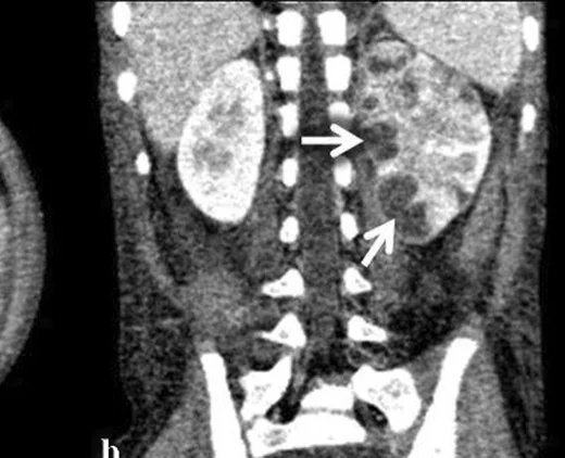
Renal apse, çoğunlukla yükselen üriner enfeksiyonun parankimde sınırlı ve sıvılaşmış odağıdır; enfekte kist de benzer koleksiyon görünümü verebilir. Kontrastlı BT, merkezi kontrastlanmayan düşük atenüasyonlu bir koleksiyon ile çevresinde kontrastlanan kalın duvarı ve komşu perirenal yağ dokusunda kirlenmeyi gösterir. Enfeksiyonlu hastada fokal şişlik kitle sanılabilir; tersine tümör içindeki nekroz da apseyi taklit edebilir. Gazın sadece toplayıcı sistemde mi yoksa parankim ve perirenal alana mı yayıldığı, emfizematoz pielonefrit ile pielit ayrımı yaparak tedavi aciliyetini belirler.
Faz ve pencere
- Kontrastsız
- Kontrastsız BT ile taş ve obstrüksiyon saptanabilir; gaz oluşumu varsa parankim veya toplayıcı sistemde hava yoğunluğunda çok düşük atenüasyonlu odaklar görülür; enfekte böbrekte büyüme ve perirenal yağ kirlenmesi en iyi kontrastlı fazda değerlendirilir. Kontrastsız BT tek başına apse duvarının kontrastlanmasını göstermez.
- Portal tanısal
- Apse, merkezi kontrastlanmayan düşük atenüasyonlu bir koleksiyon ile çevresinde kontrastlanan bir duvardan oluşur; komşu parankimde hipoenhansman, perirenal yağ dokuda kirlenme ve Gerota fasyasında kalınlaşma eşlik edebilir. Apse içi gaz nadiren bulunur ve enfeksiyonu işaret eden güçlü bir bulgudur.
Önerilen pencereler: Batın (G 400 / M 50), Yumuşak doku (G 400 / M 40).
BT bulguları
- İntraparenkimal yuvarlak ya da coğrafi düşük atenüasyonlu sıvı koleksiyonu; merkezinde canlı doku kontrastlanması bulunmaz.
- Çevresel duvar kalın, düzensiz ve kontrastlanan yapıdadır; koleksiyon olgunlaştıkça sınırı daha belirginleşebilir.
- Komşu renal parankim ödemli ve hipoenhans olabilir; apse çevresinde inflamatuvar halo görülebilir.
- Perirenal yağ dokuda çizgilenme ve Gerota fasyasında kalınlaşma ekstrarenal inflamasyon yayılımını destekler.
- Koleksiyon içinde gaz nadirdir ancak enfeksiyon lehine güçlü ipucudur; gazın toplayıcı sistemde mi parankimde mi olduğu belirlenir.
- Enfekte kistte önceden bilinen basit kistin duvarı kalınlaşıp kontrastlanabilir ve içerik heterojenleşebilir.
- Taş, hidronefroz veya toplayıcı sistemde debris eşlik ediyorsa enfeksiyonun obstrüktif kaynağı araştırılır.
Normalde
Normal böbrek korteksi homojen kontrastlanır; perirenal yağ temiz, Gerota fasyası ince, renal pelvis ve kaliksler genişlemesizdir. Aynı seviyede karşı böbreğin parankim yoğunluğunu referans alarak merkezde kontrastlanmayan sıvı boşluğunu, çevresel duvarı ve etrafındaki yağ dokusu reaksiyonunu kıyaslayın.
Normal BT ile kıyasla
Aynı bölgenin, kaynak veri setinde patoloji içermediği belirtilen bir BT incelemesini kesit kesit kaydırın; organ sınırlarını açarak anatomiyi hasta görüntüsüyle karşılaştırın. Küçük rastlantısal bulgular tümüyle dışlanmamıştır.
Kaynak: TotalSegmentator v2.01 veri seti (Wasserthal ve ark., Radiology: Artificial Intelligence 2023), CC BY 4.0, vaka s0541; organ sınırları veri setinin otomatik segmentasyonundan, birleştirilerek sadeleştirildi. Görüntüler 2,5 mm kesit aralığına yeniden örneklendi.
Ayırıcı tanı
- Akut fokal bakteriyel nefrit
- fokal hipoenhansman yapar ancak sıvılaşmış merkez ve olgun çevresel apse duvarı yoktur.
- Kistik RCC
- kontrastlanan mural nodül veya solid septa görülebilir; enfeksiyon öyküsü ve çevre yağ reaksiyonu tek başına yeterli ayrım sağlamaz.
- Renal infarkt
- damar alanına uyan kama biçimli hipoenhansman gösterir; yuvarlak sıvı koleksiyonu ve çevresel inflamatuvar duvar beklenmez.
- Enfekte hidronefroz / piyonefroz
- sıvı ve partiküler artık (debris), genişlemiş renal pelvis-kaliks sistemi içindedir; parankim içi koleksiyon değildir.
- Nekrotik renal tümör
- canlı tümör dokusu merkezdeki nekrozu çevreleyebilir; kitle konturu ve solid bileşenler enfeksiyon bulgularından bağımsız incelenir.
Tuzaklar
- Olgunlaşmamış apse belirgin kapsül oluşturmadan yalnız hipoenhansman yapabilir; odak içindeki sıvılaşmayı ve seri klinik/görüntüleme değişimini göz önüne alın.
- Tümör içi nekroz apseyi taklit edebilir; kontrastlanan mural nodül, düzensiz solid bileşen ve çevre dokulara infiltrasyonu özellikle arayın.
- Toplayıcı sistemdeki hava parankimal gazlı apseyle aynı değildir; gazın kaliks, pelvis veya renal parankimle konum ilişkisini çok düzlemli izleyin.
Kendini dene
Ateşli hastada renal parankimde merkezinde kontrastlanmayan bir koleksiyon, kalın kontrastlanan duvar ve perirenal yağ çizgilenmesi var. En olası tanı nedir?
Cevabı göster
Renal apse. Merkezde sıvılaşma, çevresel kontrastlanan duvar ve perirenal inflamasyon apseyi destekler. İnfarkt kama biçiminde perfüzyon kaybıdır; kistik RCC'de mural solid nodül kuşkuludur, basit kistte kalın kontrastlanan duvar olmaz.
Böbrek içindeki gaz kabarcıkları genişlemiş kaliks ve pelvis lümeniyle devamlılık gösteriyor; parankimde koleksiyon yok. Hangi süreç daha olasıdır?
Cevabı göster
Emfizematoz pielit (enfekte toplayıcı sistem). Gazın genişlemiş toplayıcı sistemde yer alması emfizematoz pielit lehinedir. Parankimal apse gazı renal dokuda ayrı koleksiyon içinde olur; infarkt gaz oluşturmaz, nekrotik RCC'de bu dağılım beklenmez.
Bu konunun yer aldığı turlar
Kaynaklar
Bu sayfadaki 38 cümle ve 2 quiz sorusu aşağıdaki kaynaklardan alıntılarla tek tek karşılaştırıldı; 7 cümle bu karşılaştırmada düzeltildi (2026-10-08).
- Imaging of renal emergencies: Review of infectious, hemorrhagic, vascular, and traumatic etiologiespmc.ncbi.nlm.nih.gov
- CT and MR imaging of cystic renal lesionspmc.ncbi.nlm.nih.gov
- Follow-up of defecation, urination, growth and development in children with Currarino syndrome.pubmed.ncbi.nlm.nih.gov
- Efficacy and safety of flexible and navigable suction ureteral access sheath combined with flexible ureteroscopic lithotripsy versus flexible ureteroscopic lithotripsy alone for infected upper urinary tract stones: a retrospective cohort study.pubmed.ncbi.nlm.nih.gov
- A Case of Perinephric Pancreatic Pseudocyst Secondary to Pancreatitis.pubmed.ncbi.nlm.nih.gov
- Diffuse large B-cell lymphoma mimicking musculoskeletal nontuberculous mycobacterial infection: A fatal diagnostic challenge.pubmed.ncbi.nlm.nih.gov
- The space of Retzius: anatomy, imaging features, and patterns of disease spread.pubmed.ncbi.nlm.nih.gov
- Computed tomography of renal emergencies: A comprehensive diagnostic guide for radiology residents.pubmed.ncbi.nlm.nih.gov
- A Rare Case Report on Dedifferentiated Liposarcoma of the Thigh Mimicking an Abscess With Vascular Features: A Diagnostic Challenge With Fatal Outcome.pubmed.ncbi.nlm.nih.gov
- Inflammatory mass or malignancy? The radiology of fish bone phlegmon: a review of 3 cases.pubmed.ncbi.nlm.nih.gov
- Pyonephrosis Ultrasound and Computed Tomography Features: A Pictorial Review.pubmed.ncbi.nlm.nih.gov
- Bilateral Pneumonia and Emphysematous Pyelonephritis as an Inaugural Presentation of Diabetes: A Case Report and Review of Literature.pubmed.ncbi.nlm.nih.gov
- Complications after Nephron-sparing Interventions for Renal Tumors: Imaging Findings and Management.pubmed.ncbi.nlm.nih.gov
- What every neuropathologist needs to know: Update on neuro infectious disease workups and consultation resources.pubmed.ncbi.nlm.nih.gov
- Gastrointestinal stromal tumors: a comprehensive radiological review.pubmed.ncbi.nlm.nih.gov
- Utility of Contrast-Enhanced Ultrasound in Optimizing Hepatic Abscess Treatment and Monitoring.pubmed.ncbi.nlm.nih.gov
- Venous gas caused by emphysematous pyelonephritis: a case report and review of literature.pubmed.ncbi.nlm.nih.gov
- Clinical, Radiological, and Endoscopic Features of Pancreatic Pseudocyst and Walled-Off Necrosis: How to Diagnose and How to Drain Them.pubmed.ncbi.nlm.nih.gov
- CT and MRI in Urinary Tract Infections: A Spectrum of Different Imaging Findings.pubmed.ncbi.nlm.nih.gov
- Cross-sectional imaging of iatrogenic complications after extracorporeal and endourological treatment of urolithiasis.pubmed.ncbi.nlm.nih.gov
- International Society of Paediatric Surgical Oncology (IPSO) Surgical Practice Guidelines.pubmed.ncbi.nlm.nih.gov
- Arterial and biliary complications after transarterial chemoembolization for hepatocellular carcinoma.pubmed.ncbi.nlm.nih.gov
- Injectable hydrogels for osteomyelitis treatment induce metabolic reprogramming for protection against reinfection.pubmed.ncbi.nlm.nih.gov
- Common and uncommon adult unilateral renal masses other than renal cell carcinoma.pubmed.ncbi.nlm.nih.gov
- Renal Hydatid Cyst: A Report of Two Cases.pubmed.ncbi.nlm.nih.gov
- ESUR female pelvis group approach to cystic female pelvic lesions.pubmed.ncbi.nlm.nih.gov
- Excision of a Giant Bilobed Second Branchial Cleft Cyst: A Case Report.pubmed.ncbi.nlm.nih.gov
- Colorenal Fistula in a Patient With Recurrent Urinary Tract Infections and Urinary Urgency: A Case Report.pubmed.ncbi.nlm.nih.gov
- Management of Xanthogranulomatous Pyelonephritis: A Review of Current Evidence and Treatment Strategies and a Proposed Clinical Management Algorithm.pubmed.ncbi.nlm.nih.gov
- Clinical Characteristics, Microbiological Spectrum, Biomarkers, and Imaging Insights in Acute Pyelonephritis and Its Complicated Forms-A Systematic Review.pubmed.ncbi.nlm.nih.gov
- A Concise Review of the Multimodality Imaging Features of Renal Cell Carcinoma.pubmed.ncbi.nlm.nih.gov
- Soft-Tissue Sarcomas of the Abdomen and Pelvis: Radiologic-Pathologic Features, Part 1-Common Sarcomas: From the Radiologic Pathology Archives.pubmed.ncbi.nlm.nih.gov
- Advances of multidetector computed tomography in the characterization and staging of renal cell carcinoma.pubmed.ncbi.nlm.nih.gov
- Stem cell injections for osteoarthritis of the knee.pubmed.ncbi.nlm.nih.gov
- Volumetric Modulated Arc (Radio) Therapy in Pets Treatment: The "La Cittadina Fondazione" Experience.pubmed.ncbi.nlm.nih.gov
- Contrast-enhanced ultrasound of transplant organs - liver and kidney - in children.pubmed.ncbi.nlm.nih.gov
- Mucinous cystic neoplasms of the liver: current insights into epidemiology, diagnosis, and treatment.pubmed.ncbi.nlm.nih.gov
- Analysis of risk factors for early detection of cystic lung cancer based on CT genomics and its relationship with pathological invasion.pubmed.ncbi.nlm.nih.gov
- Adult Hepatic Infarction for Internists: A Practical Diagnostic and Management Pathway to Avoid Misdiagnosis, Unnecessary Drainage, and Delayed Vascular Recognition.pubmed.ncbi.nlm.nih.gov
- Management of neonatal pyonephrosis in a low-resource setting: a case report.pubmed.ncbi.nlm.nih.gov
- Clinical utility of renal and urinary tract point-of-care ultrasound in the neonatal intensive care unit: insights from a case series.pubmed.ncbi.nlm.nih.gov
- Liposarcoma With Extensive Necrosis Obscuring the Diagnosis in an HIV-Positive Patient: A Potential Diagnostic Pitfall.pubmed.ncbi.nlm.nih.gov
- Imaging diagnosis of cystic intraparenchymal brain neoplasms.pubmed.ncbi.nlm.nih.gov
- Diagnostic imaging and emerging image-guided neurointervention in brain tumors: a narrative review.pubmed.ncbi.nlm.nih.gov
- 불분명한 소아 복부 종괴: 영상 기반 진단 접근법.pubmed.ncbi.nlm.nih.gov
- Mimics and Pitfalls of Primary Ovarian Malignancy Imaging.pubmed.ncbi.nlm.nih.gov
- Approach to Cystic Lesions in the Abdomen and Pelvis, with Radiologic-Pathologic Correlation.pubmed.ncbi.nlm.nih.gov
- Management of Adult Bronchiectasis: Consensus-Based Guidelines of the German Respiratory Society.pubmed.ncbi.nlm.nih.gov
- Multidetector CT imaging of complications after laparoscopic nephron-sparing surgery.pubmed.ncbi.nlm.nih.gov
- Long-term safety and effectiveness of the "OptEase" vena cava filter.pubmed.ncbi.nlm.nih.gov
- The early fogging effect in cerebral infarction - a case report and review of the literature.pubmed.ncbi.nlm.nih.gov
- Computed tomography in the diagnosis of bilateral renal tuberculosis: diagnostic value, limitations, and future directions.pubmed.ncbi.nlm.nih.gov
- Predicting the clinical prognosis of acute ischemic stroke using machine learning: an application of radiomic biomarkers on non-contrast CT after intravascular interventional treatment.pubmed.ncbi.nlm.nih.gov
- Contrast-Enhanced Low-Mechanical-Index Endoscopic Ultrasound for the Evaluation of Focal Liver Lesions.pubmed.ncbi.nlm.nih.gov
- Comments and Illustrations of Ultrasound Findings in Extrapulmonary Tuberculosis Manifestations.pubmed.ncbi.nlm.nih.gov
- Lower extremity infections: Essential anatomy and multimodality imaging findings.pubmed.ncbi.nlm.nih.gov
- Radiological diagnosis of perinephric pathology: pictorial essay 2015.pubmed.ncbi.nlm.nih.gov
- Imaging of renal emergencies: Review of infectious, hemorrhagic, vascular, and traumatic etiologies.pubmed.ncbi.nlm.nih.gov
- Infected splenic artery pseudoaneurysm within a complex splenic cystic lesion: CT angiography pitfalls and surgical correlation.pubmed.ncbi.nlm.nih.gov
- Pyelo-Hepatic Fistula Resulting From a Perinephric Abscess: A Case Report and Literature Review.pubmed.ncbi.nlm.nih.gov
- Severe Splenic Rupture After Minor Blunt Trauma in a Patient With an Epidermoid Splenic Cyst: A Case Report.pubmed.ncbi.nlm.nih.gov
- Abdominal visceral tuberculosis: a malignancy mimic.pubmed.ncbi.nlm.nih.gov
- Endometriosis MR mimickers: T1-hyperintense lesions.pubmed.ncbi.nlm.nih.gov
- Clinical manifestations and immune response to tuberculosis.pubmed.ncbi.nlm.nih.gov
- Multimodality imaging of renal lymphoma and its mimics.pubmed.ncbi.nlm.nih.gov
- Tips and Tricks in Thoracic Radiology for Beginners: A Findings-Based Approach.pubmed.ncbi.nlm.nih.gov
- Neonatal stress hyperglycemia mimicking diabetes mellitus secondary to massive intraparenchymal brain abscess due to escherichia coli sepsis: A case report.pubmed.ncbi.nlm.nih.gov
- Complications and expected imaging findings after endoscopic retrograde cholangiopancreatography.pubmed.ncbi.nlm.nih.gov
- Diagnosis and management of type II endoscopic retrograde cholangiopancreatography-related perforations: a multicenter retrospective study.pubmed.ncbi.nlm.nih.gov
- Diagnostic value of CT enterography combined with inflammatory indicators in active Crohn's disease.pubmed.ncbi.nlm.nih.gov
- Anatomy of a Rescue: Explantation and Reconstruction After Complex Endovascular Aortic Repair-A Systematic Review of Case Reports.pubmed.ncbi.nlm.nih.gov
- Sigmoid colon fistulization of an infected pelvic lymphocele after pelvic lymphadenectomy: a case report.pubmed.ncbi.nlm.nih.gov
- Autopsy case report of rapidly progressive iliopsoas abscess caused by <i>Escherichia coli</i> and <i>Prevotella bivia</i>.pubmed.ncbi.nlm.nih.gov
- Role of CT in Pyelonephritis: A Comprehensive Pictorial Review.pubmed.ncbi.nlm.nih.gov
- Kidney involvement during the course of febrile urinary tract infection.pubmed.ncbi.nlm.nih.gov
- Navigating the diagnostic pitfalls of cystic and solid breast masses: a comparative case report of metaplastic carcinoma and benign lesions.pubmed.ncbi.nlm.nih.gov
- Gynecologic imaging errors and pitfalls: how to avoid misdiagnosis on pelvic MRI.pubmed.ncbi.nlm.nih.gov
Eğitim amaçlıdır; hasta başında tanı ve tedavi kararının yerine geçmez. İçerik yapay zekâ destekli bir süreçle güncel literatüre göre cümle cümle kaynaklarla karşılaştırıldı, ancak uzman radyolog incelemesinden geçmedi. Klinik kararlarda güncel kılavuzları, kurum protokollerini ve radyoloji raporunu esas alın.