Özet
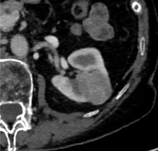
Kromofob renal hücreli karsinom, kortikal kökenli ve diğer RCC alt tipleriyle görüntüleme bulguları örtüşebilen solid bir böbrek tümörüdür. Kontrastlı BT'de kitle genellikle iyi sınırlı ve homojen görünümlüdür; orta düzeyde kontrastlanır ve kontrastlanması normal korteksten ve berrak hücreli RCC'den genellikle daha düşüktür. Merkezi yıldızsı skar veya spoke-wheel kontrastlanma görülebilir ancak onkositomla ortak özelliklerdir ve tanı koydurmaz. Görünümü kesin histoloji gibi yorumlamak benign onkositomla karışıklığa veya tümör yayılımının yetersiz değerlendirilmesine yol açabilir.
Faz ve pencere
- Kontrastsız tanısal
- Kromofob RCC'de BT'de kalsifikasyon görülebilir; ancak kesitsel görüntülemede patognomonik özellik olmadığından kalsifikasyon tek başına histolojik ayrım sağlamaz.
- Arteriyel tanısal
- Kontrastlanma orta düzeydedir ve arteriyel/kortikomedüller fazda bile normal korteksten ve berrak hücreli RCC'den daha düşük yoğunluktadır.
- Nefrografik tanısal
- Nefrografik fazda kromofob kitleler genellikle görece homojendir ve berrak hücreli RCC'den daha az kontrastlanır; tümör içinde merkezi yıldızsı skar görülebilirken nekroz veya kistik değişiklikler nadirdir.
Önerilen pencereler: Batın (G 400 / M 50), Yumuşak doku (G 400 / M 40).
BT bulguları
- Korteksten köken alan, genellikle iyi sınırlı ve ekspansil büyüyen solid renal kitle.
- Kontrastlanma genellikle berrak hücreli RCC'den düşüktür; tümör ile bitişik korteks arasında fazlar boyunca kıyaslama yapılmalıdır.
- Kromofob RCC BT'de genellikle solid bir kitle olarak izlenir; santral skar görülebilir, nekroz ise nadirdir. Kromofob RCC'de BT'de nekroz nadir görülür.
- Merkezi yıldızsı skar kitlelerin yaklaşık yüzde yirmisinde görülebilir; bu bulgu onkositomda da izlenir ve özgül değildir.
- Kitlelerde kalsifikasyon görülebilir (14–34%); ancak tek başına kalsifikasyon alt tipi belirlemez.
- Kromofob RCC genellikle renal parankimle sınırlı kalır; metastaz geliştiğinde karaciğer, akciğer, kemik, adrenal bez ve periton tutulabilir.
Normalde
Normal korteks, kromofob RCC kitlesinden tüm kontrastlı fazlarda daha fazla kontrastlanır. Kitleyi aynı böbreğin sağlam korteksiyle fazlar boyunca karşılaştırın ve olası santral skarın normal renal sinüs yağından farklı, tümör içinde yerleşmiş olduğunu doğrulayın.
Normal BT ile kıyasla
Aynı bölgenin, kaynak veri setinde patoloji içermediği belirtilen bir BT incelemesini kesit kesit kaydırın; organ sınırlarını açarak anatomiyi hasta görüntüsüyle karşılaştırın. Küçük rastlantısal bulgular tümüyle dışlanmamıştır.
Kaynak: TotalSegmentator v2.01 veri seti (Wasserthal ve ark., Radiology: Artificial Intelligence 2023), CC BY 4.0, vaka s0541; organ sınırları veri setinin otomatik segmentasyonundan, birleştirilerek sadeleştirildi. Görüntüler 2,5 mm kesit aralığına yeniden örneklendi.
Ayırıcı tanı
- Onkositom
- merkezi yıldızsı skar ve homojen yapı kromofob RCC ile örtüşebilir; görüntüleme ile kesin ayrım çoğu zaman mümkün değildir.
- Papiller RCC, belirsiz renal kitlelerin görüntüleme ayırıcı tanısında kromofob RCC ile birlikte değerlendirilir.
- Berrak hücreli RCC, kromofob RCC'ye göre genellikle daha fazla kontrastlanır.
- Yağsız anjiyomiyolipom
- makroskopik yağ göstermeyen solid kitle olarak örtüşür; homojen kontrastlanma histoloji ayrımı sağlamaz.
Tuzaklar
- Merkezi yıldızsı skarı onkositom ya da kromofob RCC için özgül kabul etmeyin; her iki lezyonda da benzer şekilde görülebilir.
- Kitle boyutu ve homojenlik iyi huyluluk kanıtı değildir; kromofob RCC büyük ve düzenli sınırlı görünebilir.
- Tek faz kontrastlanma ölçümü alt tip ayrımını zayıflatır; kortikomedüller ve nefrografik görünümü birlikte okuyun.
Kendini dene
BT'de kromofob RCC için santral yıldızsı skar hangi sıklıkta bildirilmiştir ve bu bulgu nasıl yorumlanmalıdır?
Cevabı göster
%19–34; tanı koydurucu değildir. Kaynakta BT'de santral skar %19–34 oranında bildirilmiştir; kromofob RCC için kesitsel görüntülemede patognomonik bulgu yoktur.
Kromofob RCC'de kontrastlanma normal renal kortekse göre nasıldır?
Cevabı göster
Tüm fazlarda daha düşüktür. Kromofob RCC, tüm fazlarda renal korteksten daha az kontrastlanır.
İlgili konular
Kaynaklar
Bu sayfadaki 33 cümle ve 2 quiz sorusu aşağıdaki kaynaklardan alıntılarla otomatik olarak karşılaştırıldı; 15 cümle bu karşılaştırmada düzeltildi (2026-10-09).
- Retro-orbital Metastasis in Chromophobe Renal Cell Carcinoma: A Case Report and Reviewpmc.ncbi.nlm.nih.gov
- Chromophobe renal cell carcinoma – a rare kidney cancer with limited therapy options: a narrative reviewpmc.ncbi.nlm.nih.gov
- Unilateral Renal Castleman Disease Presenting with Acute Abdominal Pain: A Case Report and Literature Reviewpmc.ncbi.nlm.nih.gov
- Chromophobe thyroid carcinoma: a distinct entity associated with TSC gene alterationspmc.ncbi.nlm.nih.gov
- Sarcomatoid Chromophobe Renal Cell Carcinoma: A Case Report and Review of the Literaturepmc.ncbi.nlm.nih.gov
- Review of renal cell carcinoma and its common subtypes in radiology.pubmed.ncbi.nlm.nih.gov
- Image Interpretation: Practical Triage of Benign from Malignant Renal Masses.pubmed.ncbi.nlm.nih.gov
- Unilateral renal oncocytosis and combined hybrid tumor: a case report and review of the literature.pubmed.ncbi.nlm.nih.gov
- Chromophobe renal cell carcinoma with osteosarcoma differentiation: case report and literature review.pubmed.ncbi.nlm.nih.gov
- Renal hybrid oncocytic/chromophobe tumor associated with multiple schwannomas: Case report and literature review.pubmed.ncbi.nlm.nih.gov
- Birt-Hogg-Dubé syndrome with simultaneous hyperplastic polyposis of the gastrointestinal tract: case report and review of the literature.pubmed.ncbi.nlm.nih.gov
- Sarcomatoid Chromophobe Renal Cell Carcinoma: A Case Report and Review of the Literature.pubmed.ncbi.nlm.nih.gov
- Diagnostic accuracy of 99mTc-sestamibi SPECT/CT for detecting renal oncocytomas and other benign renal lesions: a systematic review and meta-analysis.pubmed.ncbi.nlm.nih.gov
- Chromophobe Renal Cell Carcinoma Presenting as a Cystic Renal Mass: Case Report and Review of the Literature.pubmed.ncbi.nlm.nih.gov
- [Circumferential calcifications in cromophobe renal cell carcinoma: Description of a clinical case and review of the literature.].pubmed.ncbi.nlm.nih.gov
- Renal adenocarcinoma presenting as a spontaneous perirenal hematoma in a patient on warfarin therapy--case report and literature review.pubmed.ncbi.nlm.nih.gov
- Metachronous contralateral testicular and bilateral adrenal metastasis of chromophobe renal cell carcinoma: a case report and review of the literature.pubmed.ncbi.nlm.nih.gov
- Eosinophilic solid and cystic renal cell carcinoma: a review of literature focused on radiological findings and differential diagnosis.pubmed.ncbi.nlm.nih.gov
- [The diagnostic and therapeutic challenges of bilateral renal oncocytosis : Illustrative case presentations and a review of the literature].pubmed.ncbi.nlm.nih.gov
- Chromophobe Renal Cell Carcinoma With Multimodal Imaging Features: A Case Report.pubmed.ncbi.nlm.nih.gov
- Renal Oncocytoma: A Case Report and Review of the Literature.pubmed.ncbi.nlm.nih.gov
- Genetic Heterogeneity in Renal Cell Carcinoma: A Case of Concurrent Clear Cell and Papillary Renal Carcinomas.pubmed.ncbi.nlm.nih.gov
- The Central Role of Imaging in Renal Cell Carcinoma: A Comprehensive Review of Tumor Aggressiveness, Histology, and Radiomics.pubmed.ncbi.nlm.nih.gov
- Utility of Tumor Sphericity on CT for Differentiating Fat-Poor Angiomyolipoma from Renal Cell Carcinoma.pubmed.ncbi.nlm.nih.gov
- Imaging features of eosinophilic solid and cystic renal cell carcinoma.pubmed.ncbi.nlm.nih.gov
- Diffusion kurtosis versus diffusion-weighted magnetic resonance imaging in differentiating clear cell renal cell carcinoma and renal angiomyolipoma with minimal fat: a comparative study.pubmed.ncbi.nlm.nih.gov
- Chromophobe renal cell carcinoma with nodal involvement: A rare case report highlighting unusual molecular features.pubmed.ncbi.nlm.nih.gov
- The Role of CT Imaging in Characterization of Small Renal Masses.pubmed.ncbi.nlm.nih.gov
- Chromophobe Renal Cell Carcinoma with Radiologic-Pathologic Correlation.pubmed.ncbi.nlm.nih.gov
- Computed Tomography Appearance of Renal Hybrid Oncocytic/Chromophobe Tumors.pubmed.ncbi.nlm.nih.gov
- IgG4-related retroperitoneal fibrosis mimicking perinephric extension of renal cell carcinoma on CT: a case report.pubmed.ncbi.nlm.nih.gov
- Multiphase CT-derived markers for the characterization of large solid and cystic renal masses by histology and grade.pubmed.ncbi.nlm.nih.gov
- <i>LIMCH1</i> in Cancer: A Context-Dependent Modulator Beyond the Tumor Suppressor-Oncogene Dichotomy.pubmed.ncbi.nlm.nih.gov
- Glomerular filtration rate in patients with renal space-occupying masses using combination of SPECT 99m Tc-DTPA renal dynamic imaging and contrast-enhanced CT.pubmed.ncbi.nlm.nih.gov
- Risk Stratification in Renal Cell Carcinoma: A Narrative Review.pubmed.ncbi.nlm.nih.gov
- Multimodal deep learning model for multiclass classification of renal tumors.pubmed.ncbi.nlm.nih.gov
- Differentiating Fat-Poor Angiomyolipoma from Renal Cell Carcinoma Using Contrast-Enhanced CT.pubmed.ncbi.nlm.nih.gov
- Tumor heterogeneity on contrast-enhanced computed tomography outperforms attenuation ratio in differentiating renal cell carcinoma subtypes: a pathologic correlation study.pubmed.ncbi.nlm.nih.gov
- MR Virtual Biopsy of Solid Renal Masses: An Algorithmic Approach.pubmed.ncbi.nlm.nih.gov
- Exploratory Analysis of the Role of Radiomic Features in the Differentiation of Oncocytoma and Chromophobe RCC in the Nephrographic CT Phase.pubmed.ncbi.nlm.nih.gov
- Role of diffusion-weighted magnetic resonance imaging and apparent diffusion coefficient in subtyping renal cell carcinoma.pubmed.ncbi.nlm.nih.gov
- Rosy Cells with a Rocky Core: Low-Grade Oncocytic Tumor with Osseous Metaplasia.pubmed.ncbi.nlm.nih.gov
- Prognostic Factors for Localized Clear Cell Renal Cell Carcinoma and Their Application in Adjuvant Therapy.pubmed.ncbi.nlm.nih.gov
- Imaging Tips and Tricks in Management of Renal and Urothelial Malignancies.pubmed.ncbi.nlm.nih.gov
- Development and validation of a machine learning-based radiomics model using 45-keV virtual monoenergetic images for differentiating fat-poor angiomyolipoma from clear cell renal cell carcinoma.pubmed.ncbi.nlm.nih.gov
- Imaging of synchronous multifocal renal masses: a case report.pubmed.ncbi.nlm.nih.gov
- CT-Based Radiomics in Renal Tumors: Current Evidence, Methodological Challenges, and Future Perspectives for Precision Oncology.pubmed.ncbi.nlm.nih.gov
- Navigating Recurrence in Clear Cell Renal Cell Carcinoma after Adjuvant Immunotherapy.pubmed.ncbi.nlm.nih.gov
- Circular RNA circEVI5 functions as a miR-433 sponge to promote renal cell carcinoma progression via GBP2-mediated oncogenic signaling.pubmed.ncbi.nlm.nih.gov
- Multimodality Radiological Pictorial Review of Testicular Carcinoma: From Initial Staging to Restaging.pubmed.ncbi.nlm.nih.gov
- Prognostic indicators for survival in renal cell carcinoma with venous thrombus and development of predictive nomograms.pubmed.ncbi.nlm.nih.gov
- ESMO/ASCO Recommendations for a Global Curriculum in Medical Oncology Edition 2023.pubmed.ncbi.nlm.nih.gov
- Reassessing re-TURBT in the modern era: Is it time to move from routine to selective?pubmed.ncbi.nlm.nih.gov
- Development and validation of a machine learning model for predicting distant metastasis in patients with renal cell carcinoma: a population-based study.pubmed.ncbi.nlm.nih.gov
- Venous Tumor Thrombus Level in Renal Cell Carcinoma: Impact on Surgical and Oncologic Outcomes.pubmed.ncbi.nlm.nih.gov
- Exploring the Role of Intracorporeal Ultrasound in Partial Nephrectomies: A Systematic Review.pubmed.ncbi.nlm.nih.gov
- The clinical significance of computed tomography texture features of renal cell carcinoma in predicting pathological T1-3 staging.pubmed.ncbi.nlm.nih.gov
- SEOM SOGUG clinical guideline for treatment of kidney cancer (2022).pubmed.ncbi.nlm.nih.gov
- Predicting Complications After Radical Nephrectomy With Venous Tumor Thrombectomy and Various IVC Reconstruction Approaches: A Simplified Risk Calculator.pubmed.ncbi.nlm.nih.gov
- From congenital variants to critical clues: A radiologic review of inferior vena cava pathologies.pubmed.ncbi.nlm.nih.gov
- Early-Stage Renal Cell Carcinoma: Who Needs Adjuvant Therapy?pubmed.ncbi.nlm.nih.gov
- Primary Renal Mesenchymal Tumors: A Study from North India, Emphasizing Rare Entities and Molecular Profiles.pubmed.ncbi.nlm.nih.gov
- Nasopharyngeal carcinoma with inguinal lymph node metastasis: A case report.pubmed.ncbi.nlm.nih.gov
- Primary Surgery in Metastatic Breast Cancer.pubmed.ncbi.nlm.nih.gov
- Association Between Preoperative Single Dose of Dexamethasone and Postoperative Outcomes in Elderly Patients with Gastric Cancer.pubmed.ncbi.nlm.nih.gov
- The 9th Edition of the UICC TNM Classification of Malignant Tumours: Updates and Rationale for Change.pubmed.ncbi.nlm.nih.gov
- The impact of prevalent primary cancers in melanoma patients on survival.pubmed.ncbi.nlm.nih.gov
- Missed Diagnosis of Clear Cell Renal Cell Carcinoma on Noncontrast Computed Tomography: The Potential Role of Artificial Intelligence in Reducing Radiological Diagnostic Error.pubmed.ncbi.nlm.nih.gov
- The Malignant Imposter: Cystic Renal Oncocytoma Challenging Bosniak Criteria.pubmed.ncbi.nlm.nih.gov
- Photon-Counting CT: Technology, Current and Potential Future Clinical Applications, and Overview of Approved Systems and Those in Various Stages of Research and Development.pubmed.ncbi.nlm.nih.gov
- Adrenal Washout Under Scrutiny: Spectrum, Pitfalls, and Mimics.pubmed.ncbi.nlm.nih.gov
- Renal hemangioblastoma mimicking clear cell renal cell carcinoma: a case report emphasizing contrast-enhanced ultrasound findings.pubmed.ncbi.nlm.nih.gov
- Heterogeneity Analyzed by CT-Based Habitat Analysis for Clinical Management of Cancers: A Narrative Review.pubmed.ncbi.nlm.nih.gov
- A cfDNA fragmentomics classifier for noninvasive differentiation of benign and malignant renal masses.pubmed.ncbi.nlm.nih.gov
- Metastatic mucinous tubular and spindle cell carcinoma of the kidney: Case report and literature review.pubmed.ncbi.nlm.nih.gov
- Extensive Von Meyenburg Complexes Mimicking Diffuse Hepatic Metastases: Diagnostic Role of MRI and Magnetic Resonance Cholangiopancreatography.pubmed.ncbi.nlm.nih.gov
- A case report of a composite pheochromocytoma that occurred 13 years after adrenalectomy for pheochromocytoma.pubmed.ncbi.nlm.nih.gov
- Carcinoma of Unknown Primary Origin Presenting as Rapidly Progressive Multifocal Skeletal Lesions and Fatal Systemic Thrombosis in a Dog: A Case Report.pubmed.ncbi.nlm.nih.gov
- Integrated clinicopathological, genomic, and immunophenotypic landscape of renal tubulocystic oncocytoma.pubmed.ncbi.nlm.nih.gov
- Isoechoic Renal Tumors: A Case Report and Literature Review.pubmed.ncbi.nlm.nih.gov
- A Causal Framework for Making Individualized Treatment Decisions in Oncology.pubmed.ncbi.nlm.nih.gov
- Evaluating Established Roles, Future Perspectives and Methodological Heterogeneity for Wilms' Tumor 1 (WT1) Antigen Detection in Adult Renal Cell Carcinoma, Using a Novel N-Terminus Targeted Antibody (Clone WT49).pubmed.ncbi.nlm.nih.gov
- Advances in MALDI Mass Spectrometry Imaging Single Cell and Tissues.pubmed.ncbi.nlm.nih.gov
- Differentiation of venous thrombus consistency in patients with renal cell carcinoma using volumetric analysis of computed tomography first-order radiomic features.pubmed.ncbi.nlm.nih.gov
- CT and MRI features of renal inflammatory myofibroblastic tumor and its differential diagnosis from clear cell renal cell carcinoma and chromophobe renal cell carcinoma: a study of 13 cases from two centers.pubmed.ncbi.nlm.nih.gov
- CT-Based Radiomics in the Characterization of Solid Renal Tumors: A Systematic Review.pubmed.ncbi.nlm.nih.gov
- Patterns of injury and violence in Yaoundé Cameroon: an analysis of hospital data.pubmed.ncbi.nlm.nih.gov
- Computed tomography of renal emergencies: A comprehensive diagnostic guide for radiology residents.pubmed.ncbi.nlm.nih.gov
- Imaging findings of common benign renal tumors in the era of small renal masses: differential diagnosis from small renal cell carcinoma: current status and future perspectives.pubmed.ncbi.nlm.nih.gov
- Management of patients with atrial tachycardia: a clinical consensus statement of the European Heart Rhythm Association (EHRA) of the ESC, endorsed by the Heart Rhythm Society (HRS), the Asia Pacific Heart Rhythm Society (APHRS), the Latin American Heart Rhythm Society (LAHRS), and the Association for European Paediatric and Congenital Cardiology (AEPC).pubmed.ncbi.nlm.nih.gov
- Cardiac immunity and heart repair: Mechanism, challenge, and future direction.pubmed.ncbi.nlm.nih.gov
- Measuring blood-brain barrier dysfunction: a critical appraisal of fluid biomarkers, in vitro models, in vivo imaging, and post-mortem approaches.pubmed.ncbi.nlm.nih.gov
- A case series: Four cases of urothelial carcinoma with sarcomatoid differentiation.pubmed.ncbi.nlm.nih.gov
- Tumor in the veins: an abdominal perspective with an emphasis on CT and MR imaging.pubmed.ncbi.nlm.nih.gov
- International Society of Paediatric Surgical Oncology (IPSO) Surgical Practice Guidelines.pubmed.ncbi.nlm.nih.gov
- Not all masses are metastases: a diagnostic dilemma resolved - synchronous HER2-positive breast cancer and renal oncocytoma.pubmed.ncbi.nlm.nih.gov
- Chromophobe renal cell carcinoma - a rare kidney cancer with limited therapy options: a narrative review.pubmed.ncbi.nlm.nih.gov
- Application of three-dimensional Superb micro-vascular imaging (3D-SMI) combined with quantitative blood flow analysis in the noninvasive diagnosis of renal tumors.pubmed.ncbi.nlm.nih.gov
- Scientific Status Quo of Small Renal Lesions: Diagnostic Assessment and Radiomics.pubmed.ncbi.nlm.nih.gov
- Diagnostic evaluation of renal masses in chronic kidney disease: a clinical and radiologic challenge.pubmed.ncbi.nlm.nih.gov
- Evaluation of Multimodal MRI Diffusion and Multiphase CT Parameters for Estimating Glomerular Filtration Rate in Patients with Acute Renal Obstruction.pubmed.ncbi.nlm.nih.gov
- Eosinophilic vacuolated tumor of the kidney misdiagnosed as upper tract urothelial carcinoma: a case report and literature review.pubmed.ncbi.nlm.nih.gov
- ESR Essentials: renal imaging in children-practice recommendations by the European Society of Paediatric Radiology.pubmed.ncbi.nlm.nih.gov
- Toward protocol simplification: Deep learning-based image synthesis in three-phase CT urography.pubmed.ncbi.nlm.nih.gov
- Prognostic Factors of Survival in Patients with Surgically Treated Penile Squamous Cell Carcinoma: A Retrospective Cohort Analysis.pubmed.ncbi.nlm.nih.gov
- A 7-cm inflection point: nonlinear association between maximum tumor diameter and capsular invasion risk after primary liver cancer resection.pubmed.ncbi.nlm.nih.gov
- Development and validation of a nomogram for predicting the impact of tumor size on cancer-specific survival of locally advanced renal cell carcinoma: a SEER-based study.pubmed.ncbi.nlm.nih.gov
- Nomograms based on SIRI for predicting postoperative survival outcomes in patients with non-metastatic clear cell renal cell carcinoma.pubmed.ncbi.nlm.nih.gov
- A dynamic nomogram and risk classification for predicting prognosis in renal cell carcinoma with venous tumor thrombus.pubmed.ncbi.nlm.nih.gov
- Beyond Thrombus Level: Increased Surgical Complexity, Nutritional Status, and Postoperative Complications Shape Outcomes in Renal Cell Carcinoma with Venous Tumor Thrombus-A 14-Year Single-Center Experience.pubmed.ncbi.nlm.nih.gov
- The new (version 9) American Joint Committee on Cancer (AJCC) tumor-node-metastasis (TNM) staging protocol for carcinoma of the vulva.pubmed.ncbi.nlm.nih.gov
- Narrative review of the staging classification controversy in stage N3 small cell lung cancer: from the perspective of overlapping Veterans Administration Lung Study Group and International Association for the Study of Lung Cancer definitions.pubmed.ncbi.nlm.nih.gov
- From Anatomical Staging to Precision Prognostication: Evolution of Gallbladder Cancer Staging Across AJCC Editions and the 2025 UICC TNM Update.pubmed.ncbi.nlm.nih.gov
- Clinicopathological-based nomogram prediction and molecular characterization of postoperative recurrence in clinical T1 clear cell renal cell carcinoma.pubmed.ncbi.nlm.nih.gov
- Added value of restricted diffusion in acute pyelonephritis: Two cases with different clinical presentations.pubmed.ncbi.nlm.nih.gov
Eğitim amaçlıdır; hasta başında tanı ve tedavi kararının yerine geçmez, tıbbi tavsiye değildir. Metinler yapay zekâ desteğiyle yazıldı ve otomatik bir adımda kaynak alıntılarıyla karşılaştırıldı; bu bir tıbbi doğrulama değildir ve içerik uzman radyolog incelemesinden geçmedi. Klinik kararlarda güncel kılavuzları, kurum protokollerini ve radyoloji raporunu esas alın.