Özet
Aralıklı hematüri veya irritatif işeme yakınmaları olan erişkinde mesane amiloidozu, ürotelyal tümör görünümünü taklit edebilen nadir bir depo hastalığıdır. BT'de fokal nodüler ya da plak tarzı duvar kalınlaşması, polipoid kitle veya daha yaygın duvar tutulumu görülebilir; görüntüleme tek başına amiloidi kanserden güvenilir biçimde ayıramaz. BT ürografi lezyonun yerini, yayılımını ve üst üriner sistemi göstererek hematüri değerlendirmesine katkı sağlar. Bulguyu yalnız tümör olarak etiketlemek biyopsiyle tanınabilecek taklitçiyi kaçırabilir; amiloid varsayımıyla maligniteyi dışlamamak da önemlidir.
Faz ve pencere
- Kontrastsız
- Duvar içinde ince çizgisel submukozal veya intramural kalsifikasyon varsa seçilir; kalsifikasyon amiloidoz için karakteristik olabilir, fakat yokluğu hastalığı dışlamaz.
- Ekskresyon tanısal
- Mesane yeterince doluyken fokal/plak tarzı kalınlaşma, nodüler veya polipoid kontur ve kontrastlanan duvar lezyonu görülebilir; eşlik eden lümen dolum defekti ile üst üriner sistem de haritalanır. Bu görünüm amiloidozu ürotelyal kanserden ayırmaz; kesin tanı doku incelemesine dayanır.
Önerilen pencereler: Batın (G 400 / M 50), Yumuşak doku (G 400 / M 40).
BT bulguları
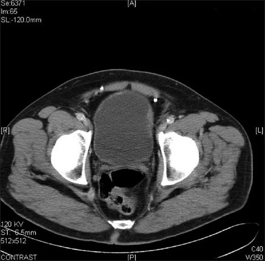
- Fokal eksantrik duvar kalınlaşması — mesane doluyken de devam eden plak veya nodül şeklinde izlenebilir.
- Polipoid intralüminal komponent — kontrastlı incelemede duvarla devamlı ve kontrastlanan yumuşak doku olarak belirir.
- Yaygın duvar tutulumu — tek bir tümör odağından farklı olarak birden fazla duvar segmentinde düzensiz kalınlaşma oluşturabilir.
- Submukozal/intramural çizgisel kalsifikasyon — kontrastsız kesitlerde duvar boyunca ince kalsifiye çizgiler görülebilir; karakteristik olsa da her olguda bulunmaz.
- Lümen dolum defekti — polipoid birikim veya mukozal düzensizlik mesane idrar sütununa taşabilir.
- Üreterovezikal bileşke komşuluğu — tutulum bu bölgede ise distal üreter duvarı ve üst sistemde genişleme eşlik ediyor mu incelenir.
- Perivezikal uzanım ve lenf düğümleri — infiltratif kenar, yağ planı kaybı veya pelvik adenopati varsa eşlik eden malignite olasılığı ayrıca değerlendirilir; bu bulgular amiloid tanısı koydurmaz.
Normalde
Mesane amiloidozunda BT'de duvar kalınlaşması ve kitle lezyonları görülebilir. Mesane amiloidozunda fokal veya diffüz duvar kalınlaşması ve intramural kalsifikasyon görülebilir.
Normal BT ile kıyasla
Aynı bölgenin, kaynak veri setinde patoloji içermediği belirtilen bir BT incelemesini kesit kesit kaydırın; organ sınırlarını açarak anatomiyi hasta görüntüsüyle karşılaştırın. Küçük rastlantısal bulgular tümüyle dışlanmamıştır.
Kaynak: TotalSegmentator v2.01 veri seti (Wasserthal ve ark., Radiology: Artificial Intelligence 2023), CC BY 4.0, vaka s0119; organ sınırları veri setinin otomatik segmentasyonundan, birleştirilerek sadeleştirildi. Görüntüler 2,5 mm kesit aralığına yeniden örneklendi.
Ayırıcı tanı
- Ürotelyal karsinom
- kontrastlanan papiller kitle veya asimetrik duvar kalınlaşması yapar; BT görünümü amiloidozla örtüşebilir ve doku tanısı gerekir.
- Enfeksiyöz sistit
- komplike alt üriner sistem enfeksiyonlarının BT/MR değerlendirmesinde ele alınan durumlardandır.
- Mesane lenfoması
- yaygın veya nodüler duvar kitlesi yapabilir; eşlik eden lenfadenopati ve diğer ekstravezikal hastalık alanları ipucudur.
- Mesane endometriozisi
- çoğunlukla fokal duvar odağıdır; siklik semptomlar ve MR'da kan ürünlerine ait sinyal özellikleri yardımcı olur.
- Mesane pıhtısı
- lümen içinde duvara yapışmayan dolum defekti oluşturabilir; kontrastlanmaz ve pozisyonla yer değiştirebilir.
- Fokal mesane duvarı kalınlaşması amiloidozda görülebilir ve maligniteyi taklit edebilir; BT hastalığı mesane tümörlerinden güvenilir biçimde ayıramaz.
Tuzaklar
- Duvar kalınlaşmasını yalnızca yetersiz distansiyona bağlamayın; iyi dolu komşu kesitlerde aynı fokal nodül sürüyorsa gerçek lezyondur.
- Kalsifikasyon görülmesi tek başına amiloidoz tanısı koşturmaz; schistosomiazis, kronik inflamasyon ve tümörlerde de izlenebilir, kesin tanı için doku örnekleme şarttır.
- BT'de kanserle örtüşen görünüm nedeniyle amiloidoz görüntüleme ile kesinleştirilemez; Congo red boyası ve polarize ışıkta çift kırılma doku tanısını sağlar.
Kendini dene
Hematürili hastada mesane BT'sinde kontrastlanan fokal plak kalınlaşması ve duvar içi çizgisel kalsifikasyon var. BT ile hangisi kesinleştirilebilir?
Cevabı göster
Lezyonun duvar yerleşimi. BT lezyonun yerini, şeklini ve yayılımını gösterir; amiloidoz ile ürotelyal tümörü kesin ayıramaz. Amiloid tipi ve sistemik tutulum ayrıca değerlendirilir, kesin lokal tanı histolojiyle konur.
Mesane duvarında nodüler kontrastlanan lezyon saptanıyor; idrar sitolojisi negatif. En uygun görüntüleme yorumu hangisidir?
Cevabı göster
Amiloid-karsinom ayrımı yapılamaz. Amiloidoz ve karsinom BT'de benzer nodüler ya da polipoid görünüm verebilir; negatif sitoloji depo hastalığını doğrulamaz veya tümörü güvenle dışlamaz. Doku örneklemesi ayrımı sağlar.
İlgili konular
Kaynaklar
Bu sayfadaki 39 cümle ve 2 quiz sorusu aşağıdaki kaynaklardan alıntılarla otomatik olarak karşılaştırıldı; 7 cümle bu karşılaştırmada düzeltildi (2026-10-09).
- Urological manifestations in familial Mediterranean fever excluding renal amyloidosis: a systematic reviewpmc.ncbi.nlm.nih.gov
- Bladder triangle amyloidosis: A case report and literature reviewpmc.ncbi.nlm.nih.gov
- Bladder Amyloidosis With Pulmonary Lesions: A Case of Spontaneous Remission After Transurethral Resectionpmc.ncbi.nlm.nih.gov
- Primary Localized Amyloidosis of the Urinary Bladder Mimicking Urothelial Carcinoma: A Case Reportpmc.ncbi.nlm.nih.gov
- Primary localized bladder amyloidosis with negative postoperative Congo red staining: A case reportpmc.ncbi.nlm.nih.gov
- Primary bladder amyloidosis.pubmed.ncbi.nlm.nih.gov
- Intractable hematuria secondary to systemic amyloidosis with bladder involvement.pubmed.ncbi.nlm.nih.gov
- Primary bladder amyloidosis as a cause of haematuria.pubmed.ncbi.nlm.nih.gov
- Bladder triangle amyloidosis: A case report and literature review.pubmed.ncbi.nlm.nih.gov
- Primary localized amylodoisis of bladder: Is there a need for cystoscopic surveillance?pubmed.ncbi.nlm.nih.gov
- Localized amyloidosis of the upper urinary tract: a case series of three patients managed with reconstructive surgery or surveillance.pubmed.ncbi.nlm.nih.gov
- Bladder Amyloidosis With Pulmonary Lesions: A Case of Spontaneous Remission After Transurethral Resection.pubmed.ncbi.nlm.nih.gov
- Primary Localized Amyloidosis of the Urinary Bladder Mimicking Urothelial Carcinoma: A Case Report.pubmed.ncbi.nlm.nih.gov
- [A Case of Systemic Wild-Type Transthyretin Amyloid (ATTR) Amyloidosis Diagnosed in a Patient with Gross Hematuria].pubmed.ncbi.nlm.nih.gov
- Localized amyloidosis of the prostatic urethra mimicking urothelial carcinoma.pubmed.ncbi.nlm.nih.gov
- Role of computed tomography urography in the clinical evaluation of upper tract urothelial carcinoma.pubmed.ncbi.nlm.nih.gov
- Imaging evaluation of amyloidosis of the urinary tract and retroperitoneum.pubmed.ncbi.nlm.nih.gov
- Primary Amyloidosis of the Urinary Bladder: A Case Report.pubmed.ncbi.nlm.nih.gov
- The Value of Amide Proton Transfer MRI in the Diagnosis of Malignant and Benign Urinary Bladder Lesions: Comparison With Diffusion-Weighted Imaging.pubmed.ncbi.nlm.nih.gov
- Primary bladder amyloidosis.pubmed.ncbi.nlm.nih.gov
- Incidental bladder cancers found on multiparametric MRI of the prostate gland: a single center experience.pubmed.ncbi.nlm.nih.gov
- [Management of isolated urinary bladder amyloidsis].pubmed.ncbi.nlm.nih.gov
- Case Report: Ureteral obstruction reveals rare underlying fibro-inflammatory, infiltrative, and proliferative disorders: report of four cases.pubmed.ncbi.nlm.nih.gov
- Emphysematous conditions of the abdomen and pelvis: pearls and pitfalls.pubmed.ncbi.nlm.nih.gov
- Imaging Tips and Tricks in Management of Renal and Urothelial Malignancies.pubmed.ncbi.nlm.nih.gov
- MR Imaging of the Upper Urinary Tract: Techniques, Diagnostic Performance, and Imaging Biomarkers.pubmed.ncbi.nlm.nih.gov
- Sonographic Anatomy and Normal Measurements of the Human Kidneys: A Comprehensive Review.pubmed.ncbi.nlm.nih.gov
- p53 expression in repair/reactive renal tubular cells: A potential pitfall leading to a false-positive diagnosis of urine cytology.pubmed.ncbi.nlm.nih.gov
- High-affinity alphavbeta3 integrin targeted optical probe as a new imaging biomarker for early atherosclerosis: initial studies in Watanabe rabbits.pubmed.ncbi.nlm.nih.gov
- Society for Cardiovascular Magnetic Resonance 2024 Cases of SCMR case series.pubmed.ncbi.nlm.nih.gov
- Navigating Ovarian-Adnexal Reporting and Data System Magnetic Resonance Imaging (O-RADS MRI): A Review of Its Evolution, Current Advances, and Persistent Challenges in Ovarian Imaging.pubmed.ncbi.nlm.nih.gov
- Benign Lesion of the Common Bile Duct Mimicking a Malignant Tumor: an Entity Commonly Overdiagnosed Clinically.pubmed.ncbi.nlm.nih.gov
- Small Bowel Imaging from Stepchild of Roentgenology to MR Enterography, Part II: The Reliable Disclosure of Crohn's Disease and Non-Inflammatory Small Bowel Disorder Plot through MRI Findings.pubmed.ncbi.nlm.nih.gov
- Imaging for abdominal involvement in amyloidosis.pubmed.ncbi.nlm.nih.gov
- Glomerular disease associated with cancer: a case series of paraneoplastic nephrotic syndrome.pubmed.ncbi.nlm.nih.gov
- Non-functional muscle-invasive bladder paraganglioma-a case report.pubmed.ncbi.nlm.nih.gov
- International Harmonization of Nomenclature and Diagnostic Criteria (INHAND): Non-proliferative and Proliferative Lesions of the Non-human Primate ( M. fascicularis ).pubmed.ncbi.nlm.nih.gov
- Nonproliferative and Proliferative Lesions of the Gastrointestinal Tract, Pancreas and Salivary Glands of the Rat and Mouse.pubmed.ncbi.nlm.nih.gov
- Aseptic Abscess of the Gallbladder Mimicking Gallbladder Carcinoma after Surgery for Ulcerative Colitis.pubmed.ncbi.nlm.nih.gov
- Adenomatous Hyperplasia of the Gallbladder in the Setting of Mirizzi Syndrome, Mimicking Adenocarcinoma of the Gallbladder.pubmed.ncbi.nlm.nih.gov
- Intracellular amyloidosis in peripheral nerve and skeletal muscle.pubmed.ncbi.nlm.nih.gov
- T1 relaxation: Chemo-physical fundamentals of magnetic resonance imaging and clinical applications.pubmed.ncbi.nlm.nih.gov
- Amyloidosis of the renal pelvis presenting as flank pain.pubmed.ncbi.nlm.nih.gov
- An extremely rare case of dermoid cyst of urinary bladder.pubmed.ncbi.nlm.nih.gov
- Fluid transport in the brain.pubmed.ncbi.nlm.nih.gov
- Commentary: an eye on PET quantification.pubmed.ncbi.nlm.nih.gov
- Patterns of injury and violence in Yaoundé Cameroon: an analysis of hospital data.pubmed.ncbi.nlm.nih.gov
- Congenital Anomalies of the Kidney and Urinary Tract in Down Syndrome: Prevalence, Phenotypes, Genetics and Clinical Management.pubmed.ncbi.nlm.nih.gov
- Antenatal hydronephrosis: differential diagnosis, evaluation, and treatment options.pubmed.ncbi.nlm.nih.gov
- Typical and Atypical Imaging Features of Malignant Lymphoma in the Abdomen and Mimicking Diseases.pubmed.ncbi.nlm.nih.gov
- Autoimmunity and Infection in Glomerular Disease.pubmed.ncbi.nlm.nih.gov
- Diagnostic and Prognostic Non-Invasive Markers in Bladder Cancer.pubmed.ncbi.nlm.nih.gov
- Evaluation of total urethral thickness using ultrasonography in cats.pubmed.ncbi.nlm.nih.gov
- Microwave Radar Sensing for Non-Invasive Intra-Abdominal Pressure Monitoring: A Simulation-Based Analysis with Phantom Testing.pubmed.ncbi.nlm.nih.gov
- ESMO / ASCO Recommendations for a Global Curriculum in Medical Oncology Edition 2016.pubmed.ncbi.nlm.nih.gov
- Chronic Ketamine Toxicity Involving both Urinary and Hepatobiliary Systems.pubmed.ncbi.nlm.nih.gov
- Imaging of abdominal and pelvic infections in the cancer patient.pubmed.ncbi.nlm.nih.gov
- Imaging side effects and complications of chemotherapy and radiation therapy: a pictorial review from head to toe.pubmed.ncbi.nlm.nih.gov
- Inflammatory Myofibroblastic Tumor of the Urinary Bladder Presenting as a Radiological cT3 Mass: A Case Report and Narrative Review of 89 Cases.pubmed.ncbi.nlm.nih.gov
- Primary Diffuse Large B-cell Lymphoma of the Urinary Bladder: A Case Report and Narrative Review of Treatment Modalities and Clinical Outcomes.pubmed.ncbi.nlm.nih.gov
- Rare Early Coexistence of Classic Urothelial and Small Cell Carcinoma of the Urinary Bladder.pubmed.ncbi.nlm.nih.gov
- Gynecologic imaging errors and pitfalls: how to avoid misdiagnosis on pelvic MRI.pubmed.ncbi.nlm.nih.gov
- Head-to-head prospective comparison of diagnostic accuracy of preoperative ultrasonography and MRI in patients undergoing surgery for deep endometriosis using #Enzian classification.pubmed.ncbi.nlm.nih.gov
- A Rare Mimicker: Renal Endometriosis and Its Diagnostic Pitfalls-Review of Reported Cases.pubmed.ncbi.nlm.nih.gov
- CT of Penetrating Abdominopelvic Trauma.pubmed.ncbi.nlm.nih.gov
- Pediatric emergency medicine point-of-care ultrasound: summary of the evidence.pubmed.ncbi.nlm.nih.gov
- Contrast-enhanced ultrasound of the pediatric bowel.pubmed.ncbi.nlm.nih.gov
- Inflammatory bowel disease imaging: Current practice and future directions.pubmed.ncbi.nlm.nih.gov
- Research-Relevant Conditions and Pathology of Laboratory Mice, Rats, Gerbils, Guinea Pigs, Hamsters, Naked Mole Rats, and Rabbits.pubmed.ncbi.nlm.nih.gov
- Medical hyperspectral imaging: an updated review of technology advancements and biomedical applications.pubmed.ncbi.nlm.nih.gov
- Case Report: Diagnostic challenges in cervical small cell neuroendocrine carcinoma of a reproductive-age woman.pubmed.ncbi.nlm.nih.gov
- Guidelines on the Use of Therapeutic Apheresis in Clinical Practice-Evidence-Based Approach From the Writing Committee of the American Society for Apheresis: The Tenth Special Issue.pubmed.ncbi.nlm.nih.gov
- Nanoscale Analysis beyond Imaging by Atomic Force Microscopy: Molecular Perspectives on Oncology and Neurodegeneration.pubmed.ncbi.nlm.nih.gov
- Spontaneous bacteremia and spontaneous bacterial peritonitis share similar prognosis in patients with cirrhosis: a cohort study.pubmed.ncbi.nlm.nih.gov
- Case Report: early regression and bladder-intact survival after limited-course immunotherapy-based systemic therapy in two patients with clinically staged bulky muscle-invasive bladder cancer.pubmed.ncbi.nlm.nih.gov
- Clinical and imaging manifestations of metastases from rare sites.pubmed.ncbi.nlm.nih.gov
- Recent advances in three-dimensional ultrasound virtual cystoscopy in modeling and local staging for urothelial carcinoma with histopathological correlation: a cohort prospective study.pubmed.ncbi.nlm.nih.gov
- Long-term safety and effectiveness of the "OptEase" vena cava filter.pubmed.ncbi.nlm.nih.gov
- Does laparoscopic surgery decrease the risk of atrial fibrillation after foregut surgery?pubmed.ncbi.nlm.nih.gov
- Eggshell calcification of the urinary bladder in chronic <i>Schistosoma haematobium</i> infection: A radiologic-pathologic case report.pubmed.ncbi.nlm.nih.gov
- Spontaneus Suburothelial Hemorrhage Associated With Forniceal Rupture and Retroperitoneal Hemorrhage.pubmed.ncbi.nlm.nih.gov
- Molecular, Biochemical, and Bioimaging Markers of MEN Syndromes.pubmed.ncbi.nlm.nih.gov
- X-inactive specific transcript (XIST) can determine sex differences in cardiovascular drug responses: focus on RNA therapeutics.pubmed.ncbi.nlm.nih.gov
- Sentinel Lymph Node Biopsy in Melanoma: Overview and Updates.pubmed.ncbi.nlm.nih.gov
- American Society of Hematology 2026 guidelines on diagnosis of light chain amyloidosis.pubmed.ncbi.nlm.nih.gov
- A Dose-Tailored Anti-Plasma Cell Regimen Lowers the Mortality of Late-Stage Cardiac Amyloidosis.pubmed.ncbi.nlm.nih.gov
- First Report of Lysozyme Amyloidosis with p.F21L/T88N Amino Acid Substitutions in a Russian Family.pubmed.ncbi.nlm.nih.gov
- Primary localised amyloidosis of the urinary bladder: A recurrent and progressive disease.pubmed.ncbi.nlm.nih.gov
- What pulmonologists need to know about extrapulmonary tuberculosis.pubmed.ncbi.nlm.nih.gov
- Primary Localized Vesical Amyloidosis Mimicking Bladder Carcinoma: A Case Report.pubmed.ncbi.nlm.nih.gov
- Management of Immune-Related Adverse Events in Patients Treated With Immune Checkpoint Inhibitor Therapy: American Society of Clinical Oncology Clinical Practice Guideline.pubmed.ncbi.nlm.nih.gov
- Residual Graft Pathology During CABG as a Minimally Invasive Diagnostic Pathway for Transthyretin Amyloidosis.pubmed.ncbi.nlm.nih.gov
- A Potential Approach to Bridge the Diagnostic Gap and Propose a Referral Pathway for Suspected ATTR-CA Based on Incidental Cardiac Uptake in <sup>99m</sup>Tc-MDP Bone Scans: A Pilot Descriptive Study.pubmed.ncbi.nlm.nih.gov
- Clinical Utility of Magnetic Resonance Urethrography in Assessment of Canine Urethra.pubmed.ncbi.nlm.nih.gov
- Encrusted Uropathy: A Comprehensive Overview-To the Bottom of the Crust.pubmed.ncbi.nlm.nih.gov
- Recommended screening and preventive practices for long-term survivors after hematopoietic cell transplantation.pubmed.ncbi.nlm.nih.gov
- The space of Retzius: anatomy, imaging features, and patterns of disease spread.pubmed.ncbi.nlm.nih.gov
- Nanotechnology in healthcare, and its safety and environmental risks.pubmed.ncbi.nlm.nih.gov
- Technology Roadmap of Micro/Nanorobots.pubmed.ncbi.nlm.nih.gov
- How to use intestinal ultrasonography in patients with Crohn disease: its role in the assessment of disease activity and disease monitoring in the era of the treat-to-target strategy.pubmed.ncbi.nlm.nih.gov
- Peritoneal Carcinosis: What the Radiologist Needs to Know.pubmed.ncbi.nlm.nih.gov
- A Survey on AI Techniques for Thoracic Diseases Diagnosis Using Medical Images.pubmed.ncbi.nlm.nih.gov
- Dual-Layer Spectral CT for Advanced Tissue Characterization: Differentiating Bladder Neoplasm from Intraluminal Thrombus-A Case Report.pubmed.ncbi.nlm.nih.gov
- A novel four-step protocol using intracavitary contrast-enhanced ultrasound combined with pelvic floor ultrasound for the diagnosis and preoperative planning of vesicovaginal fistulas.pubmed.ncbi.nlm.nih.gov
- Diagnostic accuracy of <sup>18</sup>F-FDG PET/CT in muscle-invasive bladder cancer: rationale and design of the MIBC-PET study.pubmed.ncbi.nlm.nih.gov
- Sintilimab‑related cystitis/ureteritis in high‑grade endometrial stromal sarcoma: A case report and structured literature review.pubmed.ncbi.nlm.nih.gov
- Granulomatosis with polyangiitis presenting with rare urogenital involvement: a case based review.pubmed.ncbi.nlm.nih.gov
- Acute and chronic complications of cesarean delivery: An update for the general radiologist.pubmed.ncbi.nlm.nih.gov
- Medical emergencies in pediatric blood & marrow transplant and cellular therapies.pubmed.ncbi.nlm.nih.gov
- Epidemiology and risk factors of urolithiasis in the Turkestan region: a case-control study.pubmed.ncbi.nlm.nih.gov
- Urgent considerations on renal immune-related adverse events in oncology practice.pubmed.ncbi.nlm.nih.gov
- Cross-sectional imaging of complicated urinary infections affecting the lower tract and male genital organs.pubmed.ncbi.nlm.nih.gov
- Autonomic nervous system and gastrointestinal manifestations with an emphasis on gastric dysfunction.pubmed.ncbi.nlm.nih.gov
- Ultrasound findings in urogenital schistosomiasis: a pictorial essay.pubmed.ncbi.nlm.nih.gov
- Laparoscopic ureteral anastomosis to a bladder diverticulum for the treatment of postoperative ureteral fistula: a case report.pubmed.ncbi.nlm.nih.gov
Eğitim amaçlıdır; hasta başında tanı ve tedavi kararının yerine geçmez, tıbbi tavsiye değildir. Metinler yapay zekâ desteğiyle yazıldı ve otomatik bir adımda kaynak alıntılarıyla karşılaştırıldı; bu bir tıbbi doğrulama değildir ve içerik uzman radyolog incelemesinden geçmedi. Klinik kararlarda güncel kılavuzları, kurum protokollerini ve radyoloji raporunu esas alın.