Özet
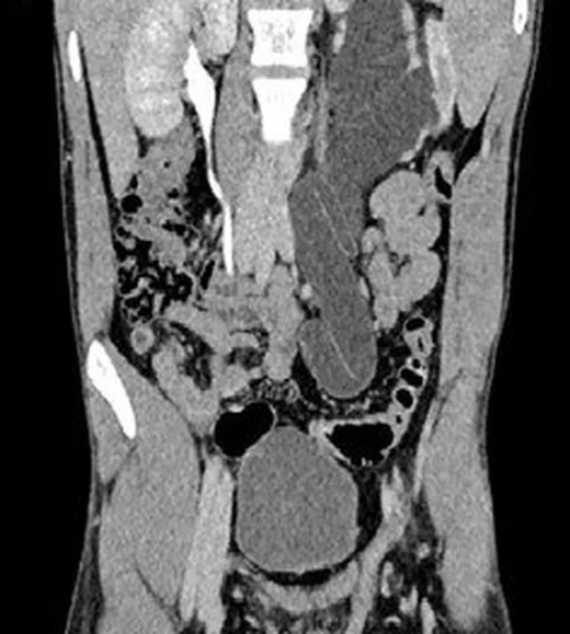
Üreter obstrüksiyonu idrar akımını bozarak üst sistemde genişleme ve böbrek işlevinde etkilenmeye yol açabilir. BT, genişlemenin bittiği geçiş noktasını gösterir ve intraluminal taşı, duvar lezyonunu, çevre dokudan basıyı ya da cerrahi sonrası darlığı araştırır. Tekrarlayan veya nedenleri açıklanamayan üreteral obstrüksiyon vakalarında, nadir tümörlerin dahil olduğu ayırıcı tanı geniş tutulmalı; bazı nadir patolojiler tipik obstrüktif bulguları taklit edebilmektedir.
Faz ve pencere
- BT ürografi tanısal
- BT ürografide hidroureteronefrozun başlangıç düzeyi ve ani veya kademeli daralma gösteren geçiş bölgesi ortaya konur; ayrıca üreter içi dolum defektleri, duvar kalınlaşması ve çevre yapıların etkisi değerlendirilebilir. Tek fazlı kontrastsız inceleme taş için yararlı olsa da yumuşak doku nedeni ve darlığın tüm seyri için yeterli olmayabilir.
- Kontrastsız
- Kalkül, kanama veya ameliyat sonrası klip gibi hiperdens odaklar ve üst sistem dilatasyonu görülebilir; taş kuşkusu varsa bu faz doğrudan nedenin saptanmasına yardım eder.
Önerilen pencereler: Batın (G 400 / M 50), Yumuşak doku (G 400 / M 40).
BT bulguları
- Kaliks, renal pelvis ve üreterin geçiş seviyesinin üzerinde genişlemesi; tek taraflı veya iki taraflı dağılımı belirt.
- Geniş üreterden ince üretere geçişin yeri ve biçimi: kısa ani daralma, uzun segmentli daralma veya kademeli sonlanma.
- Geçiş düzeyinde intraluminal kalkül, kontrastlanan duvar kitlesi, yumuşak doku dolum defekti ya da lümen dışından bası.
- Üreteral duvarda konsantrik kalınlaşma, periüreteral kalınlaşma veya yoğun inflamasyon nedeniyle oluşan çevre yağ planı silkinmesi izlenebilir.
- Cerrahi anastomoz, önceki üreter girişimi, radyasyon alanı veya travma iziyle anatomik olarak örtüşen kısa dar segment.
- Nükleer tıp tetkikleri (MAG3 skintigrafisi) veya dinamik MR ürografide, obstrüksiyona bağlı olarak etkilenen böbrekte gecikmiş ekskresyon ve azalmış glomerüler filtrasyon hızı saptanabilir.
Normalde
Gebelikte fizyolojik dilatasyonda üreterin orta üçte birinde düzgün daralma görülebilir; taş alt üretere yerleşmişse bu daralmanın distalinde dilate üreter kolonu izlenebilir. İki taraflı kaliksleri, renal pelvisleri ve üreter çaplarını karşılaştır; tek bir kısa daralma yerine daralmanın üstünde kalıcı genişleme olup olmadığına odaklan.
Normal BT ile kıyasla
Aynı bölgenin, kaynak veri setinde patoloji içermediği belirtilen bir BT incelemesini kesit kesit kaydırın; organ sınırlarını açarak anatomiyi hasta görüntüsüyle karşılaştırın. Küçük rastlantısal bulgular tümüyle dışlanmamıştır.
Kaynak: TotalSegmentator v2.01 veri seti (Wasserthal ve ark., Radiology: Artificial Intelligence 2023), CC BY 4.0, vaka s0119; organ sınırları veri setinin otomatik segmentasyonundan, birleştirilerek sadeleştirildi. Görüntüler 2,5 mm kesit aralığına yeniden örneklendi.
Ayırıcı tanı
- Üreter taşı
- geçiş noktasında kontrastsız BT'de hiperdens intraluminal kalkül bulunur.
- Ürotelyal tümör
- fokal kontrastlanan duvar kalınlaşması veya opak idrar içinde yumuşak doku dolum defekti darlığa eşlik edebilir.
- Retroperitoneal fibrozis
- üreterin çevre yumuşak dokuyla sarılması ve buna bağlı obstrüktif üropati görülebilir.
- Dıştan bası yapan pelvik kitle veya lenf nodu
- üreter duvarından ayrı kitle ve komşu anatomik yapıda yer değiştirme saptanır.
Tuzaklar
- Gebelikte ultrasonografi, kalküle bağlı üreter obstrüksiyonu ile fizyolojik hidronefrozu ayırt etmekte yetersiz kalabilir.
- Sakroiliak eklem düzeyinde üreter sıkışması olan bir olguda kontrast ekskresyonu 10–12 saat gecikmesine rağmen hasta ürolojik açıdan asemptomatik ve böbrek işlevi stabil bulunmuştur; gecikme tek başına klinik obstrüksiyonun ağırlığını göstermez.
- Periüreteral fibrozis ve infiltratif tümörler benzer üreteral daralmaya yol açabilir; üreter duvarında konsantrik kalınlaşma veya çevre yumuşak doku infiltrasyonu gibi bulgular, tanıyı güçlendirebilir ancak ayırıcı tanı her zaman zor olabilir.
Kendini dene
BT ürografide sol üreter kısa bir segmentte inceliyor; üstte hidroureteronefroz ve gecikmiş kontrast ekskresyonu var, aynı seviyede kontrastlanan duvar dokusu görülüyor. En olası neden hangisidir?
Cevabı göster
Ürotelyal tümör. Fokal kontrastlanan üreter duvarı ile proksimal hidroureteronefroz ve gecikmiş kontrast ekskresyonu obstrüktif ürotelyal tümörü düşündürür. Peristaltik daralma geçici olabilir; taş hiperdens intraluminal odak, mesane dolgunluğu ise fokal duvar bulgusunu açıklamaz.
BT ürografida üreterde fokal kontrastlanan duvar kalınlaşması ve proksimal hidroureteronefroz saptanıyor. Bu bulgular en çok hangi patolojiyi düşündürür?
Cevabı göster
Ürotelyal karsinom. Fokal üreter duvarı kalınlaşması ve proksimal genişleme ürotelyal karsinomun tipik bulgularıdır. Taş hiperdens kalkül, fibrozis çevre yumuşak doku infiltrasyonu ile karakterizedir.
İlgili konular
Kaynaklar
Bu sayfadaki 34 cümle ve 2 quiz sorusu aşağıdaki kaynaklardan alıntılarla tek tek karşılaştırıldı; 11 cümle bu karşılaştırmada düzeltildi (2026-10-09).
- Focal double-lumen ureter causing upper ureteric obstruction in a pelvic ectopic kidney: A rare case reportpmc.ncbi.nlm.nih.gov
- Lymphoma as a Rare Cause of Ureteral Obstruction: A Case Reportpmc.ncbi.nlm.nih.gov
- Robot-assisted laparoscopic surgery for bilateral ureteral fibroepithelial polyps in children: a case reportpmc.ncbi.nlm.nih.gov
- Laparoscopic ureteral anastomosis to a bladder diverticulum for the treatment of postoperative ureteral fistula: a case reportpmc.ncbi.nlm.nih.gov
- A Presumed Acquired Ureteral Diverticulum Presenting With Hematuria, Urosepsis, and Periureteral Contrast Leak: A Rare Case Reportpmc.ncbi.nlm.nih.gov
- [Transurethral endopyelotomy using thulium fiber laser].pubmed.ncbi.nlm.nih.gov
- Diverticulitis: An Update From the Age Old Paradigm.pubmed.ncbi.nlm.nih.gov
- [Methods of the videoendoscopic correction of the ureters strictures].pubmed.ncbi.nlm.nih.gov
- Long-Term Outcomes of Laser Incision and Triamcinolone Injection for the Management of Ureteroenteric Anastomotic Strictures.pubmed.ncbi.nlm.nih.gov
- Multidetector CT of vascular compression syndromes in the abdomen and pelvis.pubmed.ncbi.nlm.nih.gov
- A Nautilus kidney.pubmed.ncbi.nlm.nih.gov
- [The Great Imitator. Case report].pubmed.ncbi.nlm.nih.gov
- Imaging of stone disease in pregnancy.pubmed.ncbi.nlm.nih.gov
- Laparoscopic ureteroplasty with gonadal vein graft for long ureteral stenosis: A step by step video.pubmed.ncbi.nlm.nih.gov
- Ureteral Strictures Following Ureteroscopic Stone Treatment.pubmed.ncbi.nlm.nih.gov
- Reconstruction of full-length ureter defects by laparoscopic bladder flap forming.pubmed.ncbi.nlm.nih.gov
- Uretero-Iliac artery fistula: a rare cause of haematuria.pubmed.ncbi.nlm.nih.gov
- Getting out of a tight spot: an overview of ureteroenteric anastomotic strictures.pubmed.ncbi.nlm.nih.gov
- [Comparison of three dimensional and two dimentional laparoscopic pyeloplasty for ureteropelvic junction obstruction].pubmed.ncbi.nlm.nih.gov
- Nontraumatic unilateral ureteral entrapment at the sacroiliac joint: A rare cause of obstructive uropathy.pubmed.ncbi.nlm.nih.gov
- Retrocaval ureter successfully treated with laparoscopic ureteroplasty and functionally evaluated using Tc-99 m MAG-3 renal scintigraphy: a case report.pubmed.ncbi.nlm.nih.gov
- Clinical characteristics of patients with ureteral endometriosis and severe renal impairment identified by diuretic renal dynamic imaging.pubmed.ncbi.nlm.nih.gov
- Imaging of Acute Abdominopelvic Pain in Pregnancy and Puerperium-Part II: Non-Obstetric Complications.pubmed.ncbi.nlm.nih.gov
- Quantitative renal magnetic resonance imaging: magnetic resonance urography.pubmed.ncbi.nlm.nih.gov
- Diagnostic value of combined static-excretory MR Urography in children with hydronephrosis.pubmed.ncbi.nlm.nih.gov
- Painless Visible Haematuria in Adults: An Algorithmic Approach Guiding Management.pubmed.ncbi.nlm.nih.gov
- Tc-99m Renal Scintigraphy in Complex Congenital Systemic Syndromes Associated with Congenital Anomalies of the Kidney and Urinary Tract: A Retrospective Single-Center Study.pubmed.ncbi.nlm.nih.gov
- Swiss Consensus on Prenatal and Early Postnatal Urinary Tract Dilation: Practical Approach and When to Refer.pubmed.ncbi.nlm.nih.gov
- Genetic diagnosis in fetal hydronephrosis: assessment using chromosomal microarray analysis and whole-genome sequencing.pubmed.ncbi.nlm.nih.gov
- The Silent Strangulation: Post-hysterectomy Ureteral Endometriosis Masquerading as Iatrogenic Injury.pubmed.ncbi.nlm.nih.gov
- Robotic Excision of Upper Ureteral Fibroepithelial Polyp Presenting as Pelviureteral Junction Obstruction: Surgical Surprise with Review of Literature.pubmed.ncbi.nlm.nih.gov
- Newly modified 'pseudo flap' without compromising vascularity to enhance repair of long distal ureteral loss: A retrospective analysis of a prospective database.pubmed.ncbi.nlm.nih.gov
- Endoluminal Vacuum Therapy Using a New "Fistula Sponge" in Treating Defects of the Upper Gastrointestinal Tract-A Comparative, Retrospective Cohort Study.pubmed.ncbi.nlm.nih.gov
- The Colorectal Anastomosis: A Timeless Challenge.pubmed.ncbi.nlm.nih.gov
- 2012 CUA Abstracts.pubmed.ncbi.nlm.nih.gov
- Long-term safety and effectiveness of the "OptEase" vena cava filter.pubmed.ncbi.nlm.nih.gov
- Case Report: Spontaneous dissection of the renal pelvis and ureter.pubmed.ncbi.nlm.nih.gov
- Upper tract urothelial carcinoma mimicking emphysematous pyelonephritis: a diagnostic challenge case report.pubmed.ncbi.nlm.nih.gov
- Radiologic evaluation of the kidney transplant donor and recipient.pubmed.ncbi.nlm.nih.gov
- Epidermoid Cyst of the Ureter Mimicking Recurrent Urothelial Carcinoma: A Case Report and Brief Literature Review.pubmed.ncbi.nlm.nih.gov
- MR Imaging of the Upper Urinary Tract: Techniques, Diagnostic Performance, and Imaging Biomarkers.pubmed.ncbi.nlm.nih.gov
- <i>Schistosoma Mansoni</i> and <i>Haematobium</i>: Radiological Diagnostic Clues and Pathophysiology.pubmed.ncbi.nlm.nih.gov
- ESUR female pelvis group approach to cystic female pelvic lesions.pubmed.ncbi.nlm.nih.gov
- International Society of Paediatric Surgical Oncology (IPSO) Minimally Invasive Surgery (MIS) Guidelines.pubmed.ncbi.nlm.nih.gov
- Urothelial carcinoma masquerading as retroperitoneal fibrosis: A case report.pubmed.ncbi.nlm.nih.gov
- Management of hydronephrosis: a comprehensive review in pediatric urology perspective.pubmed.ncbi.nlm.nih.gov
- Electrospinning: Application and Prospects for Urologic Tissue Engineering.pubmed.ncbi.nlm.nih.gov
- Follow-Up Imaging of Inflammatory Myofibroblastic Tumor of the Uterus and Its Spontaneous Regression.pubmed.ncbi.nlm.nih.gov
- SLE-associated intestinal pseudo-obstruction: a case report and analysis of 43 cases.pubmed.ncbi.nlm.nih.gov
- Imaging of inferior vena cava normal variants, anomalies and pathologies, Part 1: Congenital.pubmed.ncbi.nlm.nih.gov
- Iatrogenic urinary tract injuries: etiology, diagnosis, and management.pubmed.ncbi.nlm.nih.gov
- Factors Affecting the Outcome of Medical Treatment in Cats with Obstructive Ureteral Stones Treated with Tamsulosin: 70 Cases (2018-2022).pubmed.ncbi.nlm.nih.gov
- Functional Magnetic Resonance Urography in Ureteropelvic Junction Obstruction: Proposal for a Pediatric Quantitative Score.pubmed.ncbi.nlm.nih.gov
- Construction of a novel rabbit model of ureteral calculi implanted with flowable resin.pubmed.ncbi.nlm.nih.gov
- Cystitis glandularis mimicking a bladder tumor: A rare case report.pubmed.ncbi.nlm.nih.gov
- Computed tomography of renal emergencies: A comprehensive diagnostic guide for radiology residents.pubmed.ncbi.nlm.nih.gov
- Imaging in Upper Tract Urothelial Carcinoma: A Review.pubmed.ncbi.nlm.nih.gov
- Patterns of injury and violence in Yaoundé Cameroon: an analysis of hospital data.pubmed.ncbi.nlm.nih.gov
- Lymphoma as a Rare Cause of Ureteral Obstruction: A Case Report.pubmed.ncbi.nlm.nih.gov
- Focal double-lumen ureter causing upper ureteric obstruction in a pelvic ectopic kidney: A rare case report.pubmed.ncbi.nlm.nih.gov
- <i>Ex vivo</i> partial nephrectomy and renal autotransplantation for an ultra-long ureteral stricture with incidental papillary renal cell carcinoma: A 4-year follow-up case report.pubmed.ncbi.nlm.nih.gov
- Ureteric Recovery Without Stenting After Intraoperative De-ligation During Cesarean Section: A Case Report.pubmed.ncbi.nlm.nih.gov
- Retrocaval ureter: a narrative review.pubmed.ncbi.nlm.nih.gov
- ESR Essentials: renal imaging in children-practice recommendations by the European Society of Paediatric Radiology.pubmed.ncbi.nlm.nih.gov
- The role of MRI in detection and staging of upper urinary tract cancer: a systematic review of the literature.pubmed.ncbi.nlm.nih.gov
- Clinical and imaging manifestations of metastases from rare sites.pubmed.ncbi.nlm.nih.gov
- Combination of Symptom Profile, Endoscopic Findings, and Esophageal Mucosal Histopathology Helps to Differentiate Achalasia from Refractory Gastroesophageal Reflux Disease.pubmed.ncbi.nlm.nih.gov
- Hem-o-Lok clip migration causing ureteral stone formation after ureteroplasty for retrocaval ureter: A case report.pubmed.ncbi.nlm.nih.gov
- Recent advances in three-dimensional ultrasound virtual cystoscopy in modeling and local staging for urothelial carcinoma with histopathological correlation: a cohort prospective study.pubmed.ncbi.nlm.nih.gov
- Diagnosis, workup, and risk stratification of upper tract urothelial carcinoma.pubmed.ncbi.nlm.nih.gov
- Acute kidney injury in urological conditions.pubmed.ncbi.nlm.nih.gov
- Imaging Tips and Tricks in Management of Renal and Urothelial Malignancies.pubmed.ncbi.nlm.nih.gov
- A Leak Beneath the Graft: Description of a Rare Ureteral Injury Following Aortobifemoral Bypass.pubmed.ncbi.nlm.nih.gov
- Clinical Approaches and Emerging Therapeutic Horizons in Primary Hyperoxaluria.pubmed.ncbi.nlm.nih.gov
- Lateral drainage metal ureteric stent for the treatment of 'Y-shaped' branch obstructive uropathy.pubmed.ncbi.nlm.nih.gov
- Levopositioned caudal vena cava and circumcaval left ureter with subsequent hydronephrosis and hydroureter in a dog.pubmed.ncbi.nlm.nih.gov
- Endoscopic Balloon Dilation for Primary Obstructive Megaureter in Children: Early Outcomes and Complications-A Case Series.pubmed.ncbi.nlm.nih.gov
- Case Report: Ureteral obstruction reveals rare underlying fibro-inflammatory, infiltrative, and proliferative disorders: report of four cases.pubmed.ncbi.nlm.nih.gov
- Adolescent idiopathic ureteral stricture: A multimodal diagnostic and surgical approach in resolving hydronephrosis.pubmed.ncbi.nlm.nih.gov
- A Multifactorial Case of Retroperitoneal Fibrosis in a Post-radiation Bladder Cancer Patient: Diagnostic and Therapeutic Challenges.pubmed.ncbi.nlm.nih.gov
- Preliminary outcomes and transcriptomic analysis of xenogeneic human-derived acellular dermal matrix patches for complex ureteral stricture reconstruction in a porcine model.pubmed.ncbi.nlm.nih.gov
- Therapeutic advances in ureteral stricture: lessons from radiation-induced fibrosis to regenerative strategies-a narrative review.pubmed.ncbi.nlm.nih.gov
- Antenatally detected urinary tract dilatation: a pediatric nephrologist's point of view.pubmed.ncbi.nlm.nih.gov
- Fetal Hydronephrosis: A Narrative Review of Diagnostic Pathways, Prognostic Markers, and Management.pubmed.ncbi.nlm.nih.gov
- Urothelial Carcinoma in a Cat with Bilateral Ectopic Ureters.pubmed.ncbi.nlm.nih.gov
- Case Report: Iatrogenic intraperitoneal hematoma compresses the ureters leading to urogenic sepsis.pubmed.ncbi.nlm.nih.gov
- Robot-assisted ureteral reconstruction - current status and future directions.pubmed.ncbi.nlm.nih.gov
- Best Practice Recommendations for Infertility Management.pubmed.ncbi.nlm.nih.gov
- Adolescent dysmenorrhea revealing Herlyn-Werner-Wunderlich syndrome with endometriosis: A radiologic case report.pubmed.ncbi.nlm.nih.gov
- Laparoscopic Anderson-Hynes pyeloplasty with Butler flap for pelviureteric junction obstruction in a pelvic ectopic kidney presenting as a giant palpable abdominal mass: A case report.pubmed.ncbi.nlm.nih.gov
Eğitim amaçlıdır; hasta başında tanı ve tedavi kararının yerine geçmez. İçerik yapay zekâ destekli bir süreçle güncel literatüre göre cümle cümle kaynaklarla karşılaştırıldı, ancak uzman radyolog incelemesinden geçmedi. Klinik kararlarda güncel kılavuzları, kurum protokollerini ve radyoloji raporunu esas alın.