
Özet
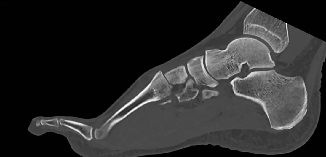
Ön ayak sabitken orta ayağın dönmesi veya ezilmesi, medial kuneiform ile ikinci metatars tabanı arasındaki Lisfranc bağ kompleksini ve tarsometatarsal eklemleri bozabilir. Kontrastsız BT, grafide gizli kalan küçük avulsiyonları, metatars taban kırıklarını ve kemik hizasındaki ince basamaklanmayı gösterir. Yük vermeden çekilmiş normal BT, yalnız ligamentöz instabiliteyi dışlamaz; kuşku sürüyorsa yük altında görüntüleme veya MR korelasyonu gerekebilir. Tedavi planını etkileyen kolon kayması, ikinci metatars tabanındaki kemik flek ve eşlik eden orta ayak kırıkları atlanmamalıdır.
Faz ve pencere
- Kontrastsız tanısal
- Tarsometatarsal eklem yüzleri, medial kuneiform-ikinci metatars tabanı ilişkisi, eklem aralıkları ve küçük avulsiyon parçaları kemik algoritmasında görülür. İyotlu kontrast kullanılmaz; standart yük vermeyen BT ligamentin kendisini ve yük altında ortaya çıkan instabiliteyi doğrudan göstermez. Aksiyel ve koronal kemik rekonstrüksiyonlarında C1-M2 ve M1-M2 aralıkları, M2-C2 hizası; sagittal planda dorsal basamak ve ark ilişkisi değerlendirilir.
Önerilen pencereler: Kemik (G 1800 / M 400).
BT bulguları
- M2-C2 hizası — ikinci metatars tabanının medial kenarı ara kuneiformun medial kortikal kenarıyla aynı doğrultuda olmalıdır; dışa kayma dizilimi bozar.
- C1-M2 ayrışması — medial kuneiform ile ikinci metatars tabanı arasındaki aralık asimetrik açılır veya eklem yüzleri paralelliğini kaybeder.
- Fleck fragmanı — Lisfranc bağının kemik tutunma noktasından kopan küçük parça C1-M2 aralığında ya da M2 tabanı yanında görülür.
- TMT eklem basamağı — bir veya daha fazla metatars tabanında dorsal, plantar ya da transvers yönde subluksasyon vardır.
- Tarsal veya metatarsal taban kırığı — M2 tabanı, kuneiformlar ve kuboidde eklem yüzüne ulaşan çatlaklar eşlik edebilir.
- Kolon uyumsuzluğu — medial, orta veya lateral metatars dizileri proksimal tarsal kemiklerle devamlı eksen oluşturmaz.
- Plantar kemik fragmanı — Lisfranc kompleksinin plantar tarafındaki avulsiyon parçaları ince aksiyel ve koronal rekonstrüksiyonlarda dikkatle aranır.
Ölçütler ve sınıflamalar
- Myerson-modifiye Hardcastle
- Tip A tüm metatarsların aynı yönde yer değiştirdiği tam uyumsuzluktur; tip B kısmi uyumsuzluktur ve medial tek kolon B1, lateral kolon B2 olarak ayrılır; tip C divergent paterninde birinci metatars mediale, diğerleri laterale yönelir.
- Myerson D alt grubu
- Eklenen D1 alt tipi yer değiştirmesiz, D2 alt tipi yer değiştirmiş yaralanmayı belirtir; bağ kaynaklı ve kemik kaynaklı ayrım D2 alt başlıklarında yapılabilir.
Normalde
Normal görünümde ikinci metatars tabanının medial kenarı ara kuneiformun medial kenarıyla aynı hatta seyretmeli; TMT eklem aralıkları karşılaştırıldığında simetrik olmalı ve ani genişleme ya da basamaklanma bulunmamalıdır. Aynı kesitlerde C1-M2 aralığını, M1-M2 ayrımını ve dorsal metatars taban yüksekliklerini karşı tarafla kıyaslayın; yük vermeyen BT'de görünür ayrışma bulunmaması bağ instabilitesini tek başına dışlamaz.
Normal BT ile kıyasla
Aynı bölgenin, kaynak veri setinde patoloji içermediği belirtilen bir BT incelemesini kesit kesit kaydırın; organ sınırlarını açarak anatomiyi hasta görüntüsüyle karşılaştırın. Küçük rastlantısal bulgular tümüyle dışlanmamıştır.
Kaynak: TotalSegmentator v2.01 veri seti (Wasserthal ve ark., Radiology: Artificial Intelligence 2023), CC BY 4.0, vaka s0685; organ sınırları veri setinin otomatik segmentasyonundan, birleştirilerek sadeleştirildi. Görüntüler 2,5 mm kesit aralığına yeniden örneklendi.
Ayırıcı tanı
- Lisfranc bağının ikinci metatars tabanından avulse olduğu olgularda izole ikinci metatars tabanı kırığı plantar-medial bölümde görülebilir.
- İzole kuneiform kırıkları nadirdir ve genellikle doğrudan travma bağlamında görülür; Lisfranc bağ yaralanmasıyla birlikte de bulunabilir.
- Orta ayak osteoartriti
- genellikle travma sonrası ve bağ/yumuşak doku yetersizliğine bağlı instabilite sonucu gelişir; ileri evrelere kadar bulgular hafif kalabilir.
- Nöropatik artropati
- nöropati varlığında kemik enflamasyonu, rezorpsiyon, mikrofraktürler ve yapısal değişikliklerle ilerleyen yıkıcı bir süreçtir; tek bir akut travmatik bağ zedelenmesine indirgenemez.
- Aksesuar ossiküller
- genellikle rastlantısal görüntülenen anatomik varyasyonlardır; ağrı sendromları, dejeneratif değişiklikler veya impingement gibi patolojilere yol açabilirler.
Tuzaklar
- Yük vermeyen BT'de eklem aralıkları normal görünebilir; ince ligament yırtığı için negatif kemik BT'sini instabilite yokluğu şeklinde yorumlamayın.
- Tek başına küçük flek fragmanı yönü göstermez; C1-M2 hizasını ve diğer TMT eklemlerini çok düzlemli rekonstrüksiyonlarda takip edin.
- M2 tabanının C2 ile ilişkisini yanlış referansla değerlendirme; M2'nin medial kenarı ara kuneiformun medial kenarıyla karşılaştırılır.
- Tarsometatarsal eklemlerde plantar yönlü avulsiyon parçalarını kaçırmayın; ince kesitli koronal ve aksiyel rekonstrüksiyonlarda eklem altı ve plantar korteksi dikkatlice tarayın.
Kendini dene
Orta ayak BT'sinde C1-M2 aralığında küçük kemik parçası ve M2 tabanında hizalanma bozukluğu görülüyor. En olası yaralanma hangisidir?
Cevabı göster
Lisfranc kompleksi yaralanması. Flek benzeri avulsiyon ile C1-M2 hizasının bozulması Lisfranc kompleksini lokalize eder. Chopart orta ayağı hindfoottan ayırır; Lisfranc bileşimi kuneiformlar, kuboid ve metatars bazlarından oluşur; naviküler kırık ön ayakta lokalize olur; kalkaneus kırığı topuğu etkiler.
Yük vermeden çekilmiş BT'de TMT hizaları korunuyor; klinikte plantar ekimoz ve belirgin orta ayak hassasiyeti sürüyor. Hangi yorum en doğrudur?
Cevabı göster
Ligament instabilitesi dışlanmaz. Standart BT kemik hizasını ve kırıkları gösterir, ancak yük altında açılan ligamentöz instabiliteyi dışlayamaz. Kırık veya Chopart çıkığı mevcut görüntüde kanıtlanmamıştır; osteoartrit için kronik eklem değişiklikleri aranır.
İlgili konular
Kaynaklar
Bu sayfadaki 42 cümle ve 2 quiz sorusu aşağıdaki kaynaklardan alıntılarla tek tek karşılaştırıldı; 11 cümle bu karşılaştırmada düzeltildi (2026-10-08).
- Lisfranc Injuries: Latest Updates on Diagnostics and Managementpmc.ncbi.nlm.nih.gov
- Inconsistent radiographic diagnostic criteria for lisfranc injuries: a systematic reviewpmc.ncbi.nlm.nih.gov
- Midfoot and Forefoot Injuries.pubmed.ncbi.nlm.nih.gov
- Comparison of Arthrodesis and Non-fusion to Treat Lisfranc Injuries.pubmed.ncbi.nlm.nih.gov
- Flexible Fixation Technique for Subtle Lisfranc Injuries.pubmed.ncbi.nlm.nih.gov
- Lisfranc injury: A review and simplified treatment algorithm.pubmed.ncbi.nlm.nih.gov
- Lisfranc complex injuries management and treatment: current knowledge.pubmed.ncbi.nlm.nih.gov
- [Lisfranc injuries].pubmed.ncbi.nlm.nih.gov
- Homolateral Lisfranc Dislocation 1-5 in a Collegiate Quarterback: A Case Study.pubmed.ncbi.nlm.nih.gov
- Lisfranc Injury Imaging and Surgical Management.pubmed.ncbi.nlm.nih.gov
- Anatomical Pathology of Subtle Lisfranc Injury.pubmed.ncbi.nlm.nih.gov
- Tarsometatarsal joint complex injuries: A study of injury pattern in complete homolateral lesions.pubmed.ncbi.nlm.nih.gov
- A transcuneiform fracture-dislocation of the midfoot.pubmed.ncbi.nlm.nih.gov
- ACR Appropriateness Criteria Acute Trauma to the Foot.pubmed.ncbi.nlm.nih.gov
- Outcome after nonoperative treatment of stable Lisfranc injuries. A prospective cohort study.pubmed.ncbi.nlm.nih.gov
- Evaluation of the tarsometatarsal joint using conventional radiography, CT, and MR imaging.pubmed.ncbi.nlm.nih.gov
- Lisfranc Dislocationpubmed.ncbi.nlm.nih.gov
- Passive and Dynamic Stabilizers of First Ray Alignment After TMT-I Fusion: The C1-C2 Intercuneiform Ligament and Peroneus Longus in a Sequential Cadaveric Destabilization Model.pubmed.ncbi.nlm.nih.gov
- Effectiveness of internal fixation versus arthrodesis on posttraumatic metatarsal-cuneiform joint dislocation: A systematic review and meta-analysis.pubmed.ncbi.nlm.nih.gov
- Glossary of terms for musculoskeletal radiology.pubmed.ncbi.nlm.nih.gov
- Weightbearing Computed Tomography vs Conventional Tomography for Examination of Varying Degrees of Lisfranc Injures: A Systematic Review of the Literature.pubmed.ncbi.nlm.nih.gov
- Lisfranc Injury Diagnosis: What Is the Diagnostic Reliability of New Radiographic Signs Using Three-dimensional CT?pubmed.ncbi.nlm.nih.gov
- A Narrative Review on Avulsion Fractures of the Upper and Lower Limbs.pubmed.ncbi.nlm.nih.gov
- Lisfranc Injuries: Latest Updates on Diagnostics and Management.pubmed.ncbi.nlm.nih.gov
- Anatomy of the Lisfranc joint complex: An illustrated review with surgical approach.pubmed.ncbi.nlm.nih.gov
- From Missed Diagnosis to Optimal Outcomes: A Comprehensive Review of Lisfranc Injuries.pubmed.ncbi.nlm.nih.gov
- A Systematic Approach to the Evaluation of Acute Musculoskeletal Injuries in the Emergency Department.pubmed.ncbi.nlm.nih.gov
- Imaging of osteoarthritis from the ankle through the midfoot.pubmed.ncbi.nlm.nih.gov
- Osteotomies: Indications, Imaging Appearance, Surgical Techniques, and Complications.pubmed.ncbi.nlm.nih.gov
- Evidence for deliberate burial of the dead by <i>Homo naledi</i>.pubmed.ncbi.nlm.nih.gov
- The oldest ceratosaurian (Dinosauria: Theropoda), from the Lower Jurassic of Italy, sheds light on the evolution of the three-fingered hand of birds.pubmed.ncbi.nlm.nih.gov
- Guidelines on multidisciplinary approaches for the prevention and management of diabetic foot disease (2020 edition).pubmed.ncbi.nlm.nih.gov
- Guide to lower extremity radiologic measurements: part 3 ankle and foot.pubmed.ncbi.nlm.nih.gov
- Pregnancy-related transverse arch deformation: a subject-specific finite element analysis of the contributions of body weight and tissue stiffness.pubmed.ncbi.nlm.nih.gov
- Bilateral congenital hallux varus with metatarsus adductus in a 6-year-old girl: A case report.pubmed.ncbi.nlm.nih.gov
- Patient-Reported Outcomes Following Lisfranc Fixation via Articular Bridge Plating Combined With the Suture Tape Ligament Reconstruction Technique.pubmed.ncbi.nlm.nih.gov
- Imaging Insights into Midfoot and Forefoot Disorders.pubmed.ncbi.nlm.nih.gov
- Hallux valgus: Advances in weightbearing CT imaging and three-dimensional assessment of first ray deformity.pubmed.ncbi.nlm.nih.gov
- Management of a Displaced, Osteoporotic Cuboid Fracture: A Case Report.pubmed.ncbi.nlm.nih.gov
- Evaluation of treatment planning discrepancies: CT versus plain radiographic findings in patients with foot and ankle trauma.pubmed.ncbi.nlm.nih.gov
- Fluoroscopic-guided procedures of the lower extremity.pubmed.ncbi.nlm.nih.gov
- Open dorsomedial fracture-dislocation of the medial cuneiform with concomitant tibialis anterior tendon rupture: a case report and review of the literature.pubmed.ncbi.nlm.nih.gov
- Magnetic resonance imaging of the ankle and foot.pubmed.ncbi.nlm.nih.gov
- High- and Low-Energy Lisfranc Injuries: A Prospective Study of Functional Outcomes and an Algorithm Based on Trauma Kinetics.pubmed.ncbi.nlm.nih.gov
- A rare Lisfranc-type injury involving dorsal dislocation of the intermediate cuneiform and second metatarsal.pubmed.ncbi.nlm.nih.gov
- Lisfranc Sports Injuries: What Do We Know So Far?pubmed.ncbi.nlm.nih.gov
- Classification in 157 patients with Lisfranc injuries using three-dimensional fracture lines and heat map.pubmed.ncbi.nlm.nih.gov
- Biomechanical analysis of elastic fixation for lisfranc injuries with finite elements method.pubmed.ncbi.nlm.nih.gov
- A modified three-incision approach to treating three-column Lisfranc injuries.pubmed.ncbi.nlm.nih.gov
- Fraturas do esqueleto apendicular e critérios de manejo cirúrgico: ensaio iconográfico.pubmed.ncbi.nlm.nih.gov
- Comparison Between Standard Screw and Internal Brace in Treatment of Subtle Lisfranc Injury.pubmed.ncbi.nlm.nih.gov
- Operative treatment of Lisfranc injuries in elite athletes: 2024 International Foot and Ankle Sports Consensus and systematic review.pubmed.ncbi.nlm.nih.gov
- Diagnostic evaluation and nonoperative management of Lisfranc injuries in athletes.pubmed.ncbi.nlm.nih.gov
- Surgical Techniques for Lapidus Arthrodesis: Approaches, Indications, and Outcomes.pubmed.ncbi.nlm.nih.gov
- Practical guidelines for the prevention and management of diabetic foot disease in China.pubmed.ncbi.nlm.nih.gov
- Charcot foot disease: A new approach.pubmed.ncbi.nlm.nih.gov
- Osseous morphology differences in the foot and ankle associated with Charcot-Marie-Tooth disease.pubmed.ncbi.nlm.nih.gov
- Outcome of Surgical Fixation for Midfoot Charcot Neuroarthropathy - A Systematic Review.pubmed.ncbi.nlm.nih.gov
- Anatomical variation in the ankle and foot: from incidental finding to inductor of pathology. Part II: midfooot and forefoot.pubmed.ncbi.nlm.nih.gov
- Imaging of pediatric foot disorders.pubmed.ncbi.nlm.nih.gov
- The state of trial registrations in the field of Orthopaedics in years 2015-2020. A meta-epidemiological study.pubmed.ncbi.nlm.nih.gov
- Beyond the Image Frame: An Art-Based Pedagogical Framework for Teaching Diagnostic Reasoning in Breast Ultrasound to Medical Students.pubmed.ncbi.nlm.nih.gov
- Prospective Validation of a Weightbearing CT Stability Threshold for Subtle Lisfranc Injuries.pubmed.ncbi.nlm.nih.gov
- Evaluation of Mild Cognitive Impairment through Perientorhinal/Hippocampal Imaging and Comprehensive Neuropsychological and Psychophysical Assessment.pubmed.ncbi.nlm.nih.gov
- The Role of Distal Medial Cuneiform Angle in Hallux Valgus: A Systematic Review.pubmed.ncbi.nlm.nih.gov
- The fibularis longus muscle revisited: comparative anatomy, developmental perspectives, and clinical relevance.pubmed.ncbi.nlm.nih.gov
- The missed chapter on midfoot: Chopart injuries.pubmed.ncbi.nlm.nih.gov
- Assessing Lisfranc Instability With Triplanar Weightbearing CT Alignment: Standardized Second Metatarsal Dorsal Offset.pubmed.ncbi.nlm.nih.gov
- Introduction of a modified Degan classification to specify treatment algorithms in fractures of the anterior process of the calcaneus.pubmed.ncbi.nlm.nih.gov
- Imaging of lower extremity infections: predisposing conditions, atypical infections, mimics, and differentiating features.pubmed.ncbi.nlm.nih.gov
- Three-Plane Alignment of the Second Metatarsal Improves Reliability of Weightbearing CT Measurements in Lisfranc Injury Assessment.pubmed.ncbi.nlm.nih.gov
- Increased Intercuneiform and Naviculocuneiform Joint Spacing in Hallux Valgus: A 3-Dimensional Distance Mapping Pilot Study.pubmed.ncbi.nlm.nih.gov
- Assessment of sensorimotor and functional differences between patients with surgically treated partially incongruent Lisfranc injuries and healthy controls: a cross-sectional study.pubmed.ncbi.nlm.nih.gov
- Targeting AHNAK ameliorates osteonecrosis of the femoral head and affects adipogenic pathways involving Smad/PPARγ.pubmed.ncbi.nlm.nih.gov
- Optimizing diagnosis and management of ONFH, FNSF, and SIF.pubmed.ncbi.nlm.nih.gov
- Surgical Management of Multi-Ligamentous Knee Injuries: Current Concepts and Case Report of a Complex KD-IV Case.pubmed.ncbi.nlm.nih.gov
- Diagnostic Accuracy of Artificial Intelligence Versus Musculoskeletal Radiologists for Foot and Ankle Fracture Detection on Radiographs.pubmed.ncbi.nlm.nih.gov
- Radiographic Outcomes of Open Reduction and Kirschner-Wire Fixation Versus Screw or Plate Fixation for Lisfranc Injuries: A Propensity Score-Weighted Comparative Study.pubmed.ncbi.nlm.nih.gov
- Three-Dimensional Morphometric Analysis of the Lisfranc Joint and Its Relationship to Injury.pubmed.ncbi.nlm.nih.gov
- Prevalence and Radiographic Morphology of Hallux Valgus in Adolescent Athletes.pubmed.ncbi.nlm.nih.gov
- Charcot-Marie-Tooth Cavovarus Foot: Current Concepts and Emerging Strategies for Deformity Correction.pubmed.ncbi.nlm.nih.gov
- Comparison of diagnostic performance of X‑ray, CT and MRI in patients with surgically confirmed subtle Lisfranc injuries.pubmed.ncbi.nlm.nih.gov
- Midfoot arthritis- current concepts review.pubmed.ncbi.nlm.nih.gov
- An old mismanaged Lisfranc injury treated by gradual deformity correction followed by the second-stage internal fixation.pubmed.ncbi.nlm.nih.gov
- Treatment of paediatric Lisfranc injuries: A systematic review and introduction of a novel treatment algorithm.pubmed.ncbi.nlm.nih.gov
- Acute foot and ankle injuries and time return to sport.pubmed.ncbi.nlm.nih.gov
- Lisfranc fracture-dislocation in a female soccer athlete.pubmed.ncbi.nlm.nih.gov
- Clinical efficacy analysis of elastic fixation for isolated ligamentous Lisfranc injuries.pubmed.ncbi.nlm.nih.gov
- Radiological Assessment of Charcot Neuro-Osteoarthropathy in Diabetic Foot: A Narrative Review.pubmed.ncbi.nlm.nih.gov
- Acute Charcot arthropathy as a first presentation of type 2 diabetes mellitus.pubmed.ncbi.nlm.nih.gov
- Clinical and Radiological Outcomes after Open Reduction and Internal Fixation of Lisfranc Injuries: A Single Centre Experience.pubmed.ncbi.nlm.nih.gov
- Cuboid Injuries.pubmed.ncbi.nlm.nih.gov
- Biomechanical comparison of mini-plate and screw fixation in minimally invasive distal metatarsal osteotomy for hallux valgus : a finite element and cadaveric study.pubmed.ncbi.nlm.nih.gov
- Comparative efficacy of double and triple arthrodesis in adult-acquired flatfoot deformity: a retrospective analysis.pubmed.ncbi.nlm.nih.gov
- Tenosynovial Giant Cell Tumors: A Musculoskeletal Case Series.pubmed.ncbi.nlm.nih.gov
- Lisfranc fleck sign: characteristics and clinical outcomes following fixation using a percutaneous position Lisfranc screw.pubmed.ncbi.nlm.nih.gov
- Demystifying the status of fracture healing using tomosynthesis: A case report.pubmed.ncbi.nlm.nih.gov
- Isolated medial cuneiform fracture: Rare avulsion of atypical tibialis posterior tendon insertion - A case report and review of literature.pubmed.ncbi.nlm.nih.gov
- Nonunion of Isolated Medial Cuneiform Fracture Fixed With a Compression Screw and Compression Staple: A Case Report.pubmed.ncbi.nlm.nih.gov
- Dental Emergencies.pubmed.ncbi.nlm.nih.gov
- Physeal fractures about the knee.pubmed.ncbi.nlm.nih.gov
- Hemorrhagic cholecystitis with auto-avulsion.pubmed.ncbi.nlm.nih.gov
Eğitim amaçlıdır; hasta başında tanı ve tedavi kararının yerine geçmez. İçerik yapay zekâ destekli bir süreçle güncel literatüre göre cümle cümle kaynaklarla karşılaştırıldı, ancak uzman radyolog incelemesinden geçmedi. Klinik kararlarda güncel kılavuzları, kurum protokollerini ve radyoloji raporunu esas alın.