Özet
Kemik enfarktüsü, medüller boşlukta gelişen iskemik kemik nekrozudur. Kemik enfarktında MR’da serpijinöz enfarkt morfolojisi görülebilir. Kemik lezyonlarında matriks mineralizasyonunun değerlendirilmesi ayırıcı tanıyı daraltmaya yardımcı olabilir. Kemik enfarktı alanında kortikal yıkım, agresif bir görüntüleme özelliğidir ve patolojik değerlendirmeyi gündeme getirir.
Okuma sırası
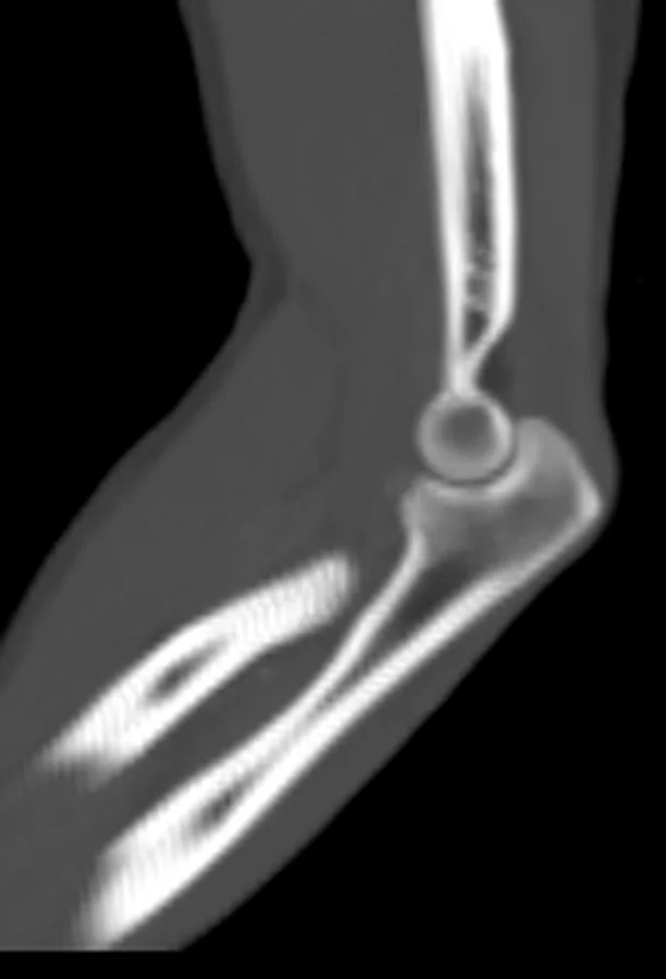
- Distal humerus medüllası. Koronal kesitte distal humerus medüllasını değerlendir; kemik lezyonlarında metafizer ve subartiküler yerleşimleri ayırt et.
- Medüller serpijinöz kenar. Sklerotik sınırı takip et; kemik osteonekrozunda serpijinöz enfarkt morfolojisini ara.
- Enfarkt merkezi. MR’da osteonekroz odağını değerlendir; serpijinöz enfarkt morfolojisi ve çift çizgi işareti görülebilir.
- Humerus korteksi ve periost. Son olarak korteksi değerlendir; kemik lezyonunda kortikal yıkım gibi agresif özellikler ileri patolojik değerlendirmeyi gerektirebilir.
Faz ve pencere
- Kontrastsız tanısal
- Olgun medüller enfarkt BT’de metafiz-diafiz boyunca uzanan yamalı veya serpijinöz sklerotik kenar ve görece radyolüsent merkezle seçilir; erken enfarkt BT’de silik olabilir.
- Kemik algoritması tanısal
- İnce kesit kemik rekonstrüksiyonunda medüller serpijinöz sklerozun devamlılığı, korteksin korunması, endosteal kontur ve eşlik eden kırık hattı değerlendirilir.
Önerilen pencereler: Kemik (G 1800 / M 400).
BT bulguları
- Kemik enfarktı medüller boşlukta gelişen bir kemik lezyonudur.
- BT’de lezyon merkezi serpijinöz sklerotik kenara göre görece düşük dansiteli olabilir; yağlı kemik iliği sinyalinin korunumu MR’da daha iyi değerlendirilir.
- Metafiz-diafiz yerleşimi, subkondral eklem yüzüne oturan osteonekrozdan ayrımda yardımcıdır.
- Tipik olgun enfarkt çevresinde kortikal bütünlük korunur; belirgin kortikal yıkım beklenen görünüm değildir.
- Agresif periostal reaksiyon ya da yumuşak doku kitlesi tipik enfarkt lehine değildir ve alternatif tanıyı gündeme getirir.
- Kırık varsa BT’de kırığın tipi ve yayılımı ayrıca değerlendirilir.
- Birden fazla benzer medüller odak varsa sistemik yatkınlıkla ilişkili çok odaklı enfarkt olasılığını belirt.
Normalde
Aynı düzeyde normal distal humerus ve proksimal radius/ulna medüllasında trabeküler yapı homojen görünür; odak çevresinde serpijinöz sklerotik sınır veya merkez-kenar atenüasyon farkı bulunmaz. Kıyaslamada lezyonun medüller yerleşimini, sklerotik sınırın konturunu, korteksin kesintisizliğini ve eklem altı subkondral kemiğe uzanıp uzanmadığını kontrol et.
Ayırıcı tanı
- Enkondrom
- medüller kıkırdak matriksinde noktasal veya halka-yay kalsifikasyonu ve bazen endosteal çentiklenme görülür.
- Kronik osteomiyelit
- sekestr, düzensiz kortikal yıkım, periostal yeni kemik veya sinüs yolu eşlik edebilir.
- Kemik adacığı ve diğer sklerotik lezyonlar, enfarktdan farklı olarak homojen skleroz veya düzensiz kortikal değişikliklerle ayrılabilir.
- Kemik metastazı
- bilinen primer kanser ve çoklu litik/sklerotik odaklar olasılığı artırır; tipik serpijinöz kenar ve görece korunmuş merkez kemik enfarktını destekler.
- Subkondral osteonekroz
- lezyon metafiz-diafiz medüllasında değil, eklem yüzünün hemen altındaki epifizde merkezlenir ve çökme gelişebilir.
Tuzaklar
- Erken evre iskemik kemik lezyonlarında görüntüleme bulguları belirgin olmayabilir ve lezyonun ilk aşamada normal yapıya benzer görünmesi mümkündür; bu durumda tanısal kesinlik için ileri görüntüleme yöntemleri değerlendirilmelidir.
- Kıkırdak kökenli lezyonların radyolojik özellikleri diğer medüller sklerotik süreçlerle örtüşebilir; lezyon içindeki mineralizasyon dağılımının analizi ayırıcı tanıda kritik önem taşır.
- Lezyon eklem yüzü altındaki epifizdeyse medüller kemik enfarktüsü etiketi yerine subkondral osteonekroz olasılığını değerlendir.
- Kırık eşlik ediyorsa, enfarktla ilişkisini ve varsa kortikal/trabeküler kesintiyi çok düzlemli kemik penceresinde ayrıca değerlendir.
Kendini dene
MR’da distal femur kondilleri ve proksimal tibialarda serpijinöz kenarlı medüller odaklar görülüyor. Bu görünüm en çok hangi kemik lezyonuyla ilişkilidir?
Cevabı göster
Kemik enfarktüsü. MR’da distal femur kondilleri ve proksimal tibialardaki osteonekroz odaklarında serpijinöz enfarkt morfolojisi ve çift çizgi işareti görülebilir.
Takipte çoğu olguda stabil kalan benign kıkırdak tümörü hangisidir?
Cevabı göster
Enkondrom. Enkondromlar, çoğunlukla uzun kemiklerde görülen benign kıkırdak tümörleridir. Enkondromların düşük dereceli kondrosarkomlardan radyolojik ve histopatolojik ayrımı zor olabilir.
Kaynaklar
Bu sayfadaki 35 cümle ve 2 quiz sorusu aşağıdaki kaynaklardan alıntılarla otomatik olarak karşılaştırıldı; 17 cümle bu karşılaştırmada düzeltildi (2026-10-09).
- What is the validity of self-reported fractures?pubmed.ncbi.nlm.nih.gov
- Application of 3-Dimensional Printing Technology in Rotational Osteotomy of Congenital Radioulnar Synostosis.pubmed.ncbi.nlm.nih.gov
- Prenatal and neonatal phenotype of Larsen of La Réunion Island syndrome (B4GALT7-linkeropathy).pubmed.ncbi.nlm.nih.gov
- Three-dimensional printing in paediatric orthopaedic surgery.pubmed.ncbi.nlm.nih.gov
- Screw length in volar locking plate fixation for distal radial fractures.pubmed.ncbi.nlm.nih.gov
- Ulnar collateral ligament dysfunction increases stress on the humeral capitellum: a finite element analysis.pubmed.ncbi.nlm.nih.gov
- Paget-Schroetter syndrome accompanied by pulmonary thromboembolism: A case report.pubmed.ncbi.nlm.nih.gov
- Rotational Anatomy of the Radius and Ulna: Surgical Implications.pubmed.ncbi.nlm.nih.gov
- Trabecular and cortical microstructure and fragility of the distal radius in women.pubmed.ncbi.nlm.nih.gov
- Multiple aneurysms of the radial artery in a woman with neurofibromatosis type 1 presenting as aneurysm rupture.pubmed.ncbi.nlm.nih.gov
- Bilateral Solitary Osteochondroma of the Bicipital Tuberosity Accompanied with Painful Clicking: A Case Report and Literature Review.pubmed.ncbi.nlm.nih.gov
- [Arthroscopic reconstruction of scaphoid nonunions].pubmed.ncbi.nlm.nih.gov
- Malignant Transformation of Musculoskeletal Lesions with Imaging-Pathology Correlation-Part 1: Bone Lesions.pubmed.ncbi.nlm.nih.gov
- Revisiting Fat Content in Bone Lesions: Paradigms in Bone Lesion Detection.pubmed.ncbi.nlm.nih.gov
- Conventional radiography for the assessment of focal bone lesions of the appendicular skeleton: fundamental concepts in the modern imaging era.pubmed.ncbi.nlm.nih.gov
- Benign incidental do-not-touch bone lesions.pubmed.ncbi.nlm.nih.gov
- Fifty Shades of PSMA-Avid Rib Lesions: A Comprehensive Review.pubmed.ncbi.nlm.nih.gov
- The roles of the nerve-immune axis in modulating bone regeneration.pubmed.ncbi.nlm.nih.gov
- Tissue regeneration strategies based on mesenchymal stem cell-derived extracellular vesicles: from bench to bedside.pubmed.ncbi.nlm.nih.gov
- Bone lesions of the tibia: Multimodal iconographic review and diagnostic algorithms, Part 2: Metaphyseal and epiphyseal lesions.pubmed.ncbi.nlm.nih.gov
- Electroacupuncture Modulates Multiple Pathways for Neuroprotection and Neurorepair in Ischemic Stroke.pubmed.ncbi.nlm.nih.gov
- Intraosseous lipomas: A report of six cases and a review of literature.pubmed.ncbi.nlm.nih.gov
- Intraosseous tumours of the knee.pubmed.ncbi.nlm.nih.gov
- Radiological manifestation of avascular necrosis (AVN) in sickle cell disease (SCD): a review of diagnostic imaging.pubmed.ncbi.nlm.nih.gov
- Canine Idiopathic Arteriopathy, Appendicular Bone Infarcts, and Neoplastic Transformation of Bone Infarcts in 108 Dogs ( Canis lupus familiaris ).pubmed.ncbi.nlm.nih.gov
- Universal catalytic endothelial lineage nanovesicles for targeting and rescuing ischemic endothelium.pubmed.ncbi.nlm.nih.gov
- Anatomy-Guided Microsurgical Resection of a Dominant Frontal Lobe Tumor Without Intraoperative Adjuncts: A Case Report from a Resource-Limited Context.pubmed.ncbi.nlm.nih.gov
- Effect of <i>Bouvardia ternifolia</i> Root Extract on Brain Structures, Oxidative Stress, and p53 Expression in a Rat Model of Cerebral Ischemia/Reperfusion.pubmed.ncbi.nlm.nih.gov
- SMARCB1-deficient sinonasal carcinoma in a pediatric patient: Imaging findings and differential diagnosis of a rare entity.pubmed.ncbi.nlm.nih.gov
- Investigating the role of complement 5a in systemic bone loss after myocardial infarction.pubmed.ncbi.nlm.nih.gov
- Insulin-like growth factor receptor signaling in physiology and disease.pubmed.ncbi.nlm.nih.gov
- Spectrum of Neuroimaging Findings in CNS Lymphoproliferative Disorders.pubmed.ncbi.nlm.nih.gov
- Characterization of spontaneous gross and histologic findings in free-ranging Egyptian rousettes (Rousettus aegyptiacus) from Uganda with a review of reported pathogens.pubmed.ncbi.nlm.nih.gov
- Paediatric cerebrovascular syndromes: imaging clues to a diagnosis.pubmed.ncbi.nlm.nih.gov
- The scientific basis of vascularized bone grafts: Models and perfusion assessments, a systematic review.pubmed.ncbi.nlm.nih.gov
- Bone in Parathyroid Diseases Revisited: Evidence From Epidemiological, Surgical and New Drug Outcomes.pubmed.ncbi.nlm.nih.gov
- Revision total elbow replacement.pubmed.ncbi.nlm.nih.gov
- Classification of Chondrosarcoma: From Characteristic to Challenging Imaging Findings.pubmed.ncbi.nlm.nih.gov
- Imaging Aspects of Enchondromas in Pediatric Patients.pubmed.ncbi.nlm.nih.gov
- Candida Femoral Osteomyelitis in a Young Adult Without Known Risk Factors for Immunosuppression: A Case Report.pubmed.ncbi.nlm.nih.gov
- A multiscale pathoanatomical atlas guides the design of functionally graded, anatomically intelligent implants for chronic osteomyelitis.pubmed.ncbi.nlm.nih.gov
- The quiet flame: whole-body MRI and the underestimated burden of pediatric chronic non-bacterial osteomyelitis (CNO).pubmed.ncbi.nlm.nih.gov
- Imaging of bone marrow pitfalls with emphasis on MRI.pubmed.ncbi.nlm.nih.gov
- Edema-like marrow signal intensity: a narrative review with a pictorial essay.pubmed.ncbi.nlm.nih.gov
- A review of the oral health of individuals with disabilities in Puerto Rico and among U.S. Hispanics.pubmed.ncbi.nlm.nih.gov
- Anatomy of the intraosseous arterial microvasculature of the adult lumbar vertebral body and its role in fracture healing: a literature review and historical perspective.pubmed.ncbi.nlm.nih.gov
- Multimodal imaging features to diagnose abdominal complications of sickle cell disease.pubmed.ncbi.nlm.nih.gov
- An Atlas of Neonatal Neurovascular Imaging Anatomy as Depicted with Microvascular Imaging: The Intracranial Veins.pubmed.ncbi.nlm.nih.gov
- Senescence-driven osteonecrosis of the femoral head in the elderly-a distinct pathophysiological entity.pubmed.ncbi.nlm.nih.gov
- CT of Sacral Fractures: Classification Systems and Management.pubmed.ncbi.nlm.nih.gov
- Long term surgical results of cases with aneurysmal bone cysts-a comprehensive clinical, functional and oncologic analysis.pubmed.ncbi.nlm.nih.gov
- Bone marrow edema in children: chronic nonbacterial osteomyelitis and its mimickers.pubmed.ncbi.nlm.nih.gov
- Cystic bone Lesions: Diagnostic pitfalls and therapeutic considerations.pubmed.ncbi.nlm.nih.gov
- Imaging of lower extremity infections: predisposing conditions, atypical infections, mimics, and differentiating features.pubmed.ncbi.nlm.nih.gov
- Central and peripheral areal BMD, bone microarchitecture, and strength after initiation of androgen deprivation therapy in men with prostate cancer: a two-year follow-up study.pubmed.ncbi.nlm.nih.gov
- Radiological and Histopathological Correlative Analysis of Bone Tumors and Tumor-Like Lesions at a Tertiary Care Center.pubmed.ncbi.nlm.nih.gov
- Bilateral Knee Osteonecrosis in an Adolescent With Leukemia on Prolonged Corticosteroids: Discordant Femoral-Tibial Findings on Bone Scan SPECT-CT.pubmed.ncbi.nlm.nih.gov
- Radial extracorporeal shock wave therapy improves upper limb function in chronic stroke patients: a randomized sham-controlled trial.pubmed.ncbi.nlm.nih.gov
- Application of Novel Biomaterials to Enhance Bone Regeneration in a Canine Non-Union Olecranon Fracture.pubmed.ncbi.nlm.nih.gov
- HR-pQCT measures of bone in autosomal dominant osteopetrosis highly correlate with fractures and inversely correlate with bone resorption markers.pubmed.ncbi.nlm.nih.gov
- Management of Tibial Adamantinoma: Balancing Oncologic Cure and Reconstructive Longevity.pubmed.ncbi.nlm.nih.gov
- Evolutionary course of the femoral head osteonecrosis: Histopathological - radiologic characteristics and clinical staging systems.pubmed.ncbi.nlm.nih.gov
- Pharmacological Targeting of Type H Endothelial Cells in Knee Osteoarthritis: From Molecular Signaling to Cellular Homeostasis.pubmed.ncbi.nlm.nih.gov
- Assessment, diagnostic criteria, and outcome measures for non-femoral head lower limb avascular necrosis in children and adolescents: a scoping review.pubmed.ncbi.nlm.nih.gov
- Benign bone and soft tissue tumors of the foot.pubmed.ncbi.nlm.nih.gov
- How Clinical and Radiological Findings in Chronic Mandibular Osteomyelitis Do Not Always Correlate: Diagnostic Dilemmas in Dental-Related Bone Inflammations.pubmed.ncbi.nlm.nih.gov
- Moth-Eaten Osteolysis of Multiple Foot Bones After a Conservatively Treated "Stable" Ankle Fracture: A Case Report and Literature Review.pubmed.ncbi.nlm.nih.gov
- Acromial Stress Fractures in Reverse Total Shoulder Arthroplasty: Diagnosis, Prevention, and Treatment.pubmed.ncbi.nlm.nih.gov
- The pathogenesis of cerebral small vessel disease and vascular cognitive impairment.pubmed.ncbi.nlm.nih.gov
- International Harmonization of Nomenclature and Diagnostic Criteria (INHAND): Nonproliferative and Proliferative Lesions of the Rabbit.pubmed.ncbi.nlm.nih.gov
- A 50-year perspective on the use and potential of artiodactyl calcanei in bone adaptation studies.pubmed.ncbi.nlm.nih.gov
- Fibrous dysplasia: Clinical features, deformities, management, and outcomes.pubmed.ncbi.nlm.nih.gov
- Analysis and Historical Evolution of Paediatric Bone Tumours: The Importance of Early Diagnosis in the Detection of Childhood Skeletal Malignancies.pubmed.ncbi.nlm.nih.gov
- Optimizing diagnosis and management of ONFH, FNSF, and SIF.pubmed.ncbi.nlm.nih.gov
- Spinal cord imaging in multiple sclerosis: from diagnosis to disease progression.pubmed.ncbi.nlm.nih.gov
- Prevalence and characteristics of benign cartilaginous tumours of the knee joint as identified on MRI scans.pubmed.ncbi.nlm.nih.gov
- Calcific Tendinitis of the Linea Aspera with Bilateral Deposits and Inflammatory Asymmetry Presenting as Unilateral Pain: A Case Report and Review of Literature.pubmed.ncbi.nlm.nih.gov
- Imaging of ewing sarcoma: an updated analysis including presenting features, prognostic imaging biomarkers, and treatment response assessment.pubmed.ncbi.nlm.nih.gov
- A KMT2C loss-of-function mutation in a JAK2-negative polycythemia vera-like myeloproliferative neoplasm: a case report.pubmed.ncbi.nlm.nih.gov
- Phylogeography of Bone Metastasis: Clonal Evolution, Skeletal Niche Adaptation, and Clinical Implications.pubmed.ncbi.nlm.nih.gov
- Common features of rare disease patients in the emergency department: a systematised literature review.pubmed.ncbi.nlm.nih.gov
- A radiology-based differential diagnostic model for pelvic chondrosarcoma using random forest algorithms.pubmed.ncbi.nlm.nih.gov
- Cavernous venous malformation of the frontal sinus: CT and MRI imaging findings.pubmed.ncbi.nlm.nih.gov
- Beyond Chordoma: A Comprehensive Review of Sacral Lesions.pubmed.ncbi.nlm.nih.gov
- The Resolution of a Biopsy-Proven Enchondroma in the Proximal Humerus Over a 14-Year Interval.pubmed.ncbi.nlm.nih.gov
- International Harmonization of Nomenclature and Diagnostic Criteria (INHAND): Non-proliferative and Proliferative Lesions of the Non-human Primate ( M. fascicularis ).pubmed.ncbi.nlm.nih.gov
- Glossary of terms for musculoskeletal radiology.pubmed.ncbi.nlm.nih.gov
- Imaging modalities for non-acute pathologies of the foot and ankle.pubmed.ncbi.nlm.nih.gov
- Chondromyxoid Fibroma: An Updated Review.pubmed.ncbi.nlm.nih.gov
- Liposclerosing Myxofibrous Tumor: A Separated Clinical Entity?pubmed.ncbi.nlm.nih.gov
- Molecular mechanisms, therapeutic strategies, and inter-organ fibrosis crosstalk of Gla proteins in cardiovascular disease.pubmed.ncbi.nlm.nih.gov
- The bone-cerebrovascular axis: effects of bone aging on neurovascular dysfunction and neurodegeneration.pubmed.ncbi.nlm.nih.gov
- Harnessing Natural Extracellular Vesicles to Combat Age-Related Diseases: From Aging Drivers to Therapeutic Opportunities.pubmed.ncbi.nlm.nih.gov
- Manifestations of radiation toxicity in the head, neck, and spine: An image-based review.pubmed.ncbi.nlm.nih.gov
- Commentary: an eye on PET quantification.pubmed.ncbi.nlm.nih.gov
- Musculoskeletal "don't touch" lesions: pictorial essay.pubmed.ncbi.nlm.nih.gov
- Value of 99m Tc-MDP SPECT/CT and 18 F-FDG PET/CT scanning in the evaluation of malignantly transformed fibrous dysplasia.pubmed.ncbi.nlm.nih.gov
- Evaluation of pain related behaviors and disease related outcomes in an immunocompetent mouse model of prostate cancer induced bone pain.pubmed.ncbi.nlm.nih.gov
Eğitim amaçlıdır; hasta başında tanı ve tedavi kararının yerine geçmez, tıbbi tavsiye değildir. Metinler yapay zekâ desteğiyle yazıldı ve otomatik bir adımda kaynak alıntılarıyla karşılaştırıldı; bu bir tıbbi doğrulama değildir ve içerik uzman radyolog incelemesinden geçmedi. Klinik kararlarda güncel kılavuzları, kurum protokollerini ve radyoloji raporunu esas alın.