
Özet
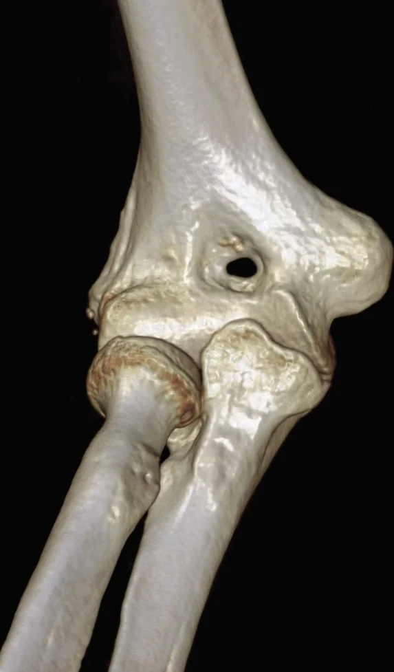
Distal humerus BT'sinde kemik içinde açıklık veya oval saydamlık görüldüğünde supratroklear foramen, iki fossayı ayıran ince septumun anatomik açıklığı olabilir. Bu anatomik varyant, distal humerusta olekranon ve koronoid fossalarını ayıran ince septumun perforasyonuyla bu iki fossayı birbirine bağlar. Direkt grafiler ve bilgisayarlı tomografi, canlı bireylerde koronoid-olekranon septumunun incelenmesi ve olası perforasyonun değerlendirilmesi amacıyla kullanılır. Septumun şeffaf veya radyolusens görünmesi gerçek perforasyondan ayırt edilmeli; iki fossayı ayıran kemikte tam bir geçişin varlığı bilgisayarlı tomografi ile doğrulanabilir.
Faz ve pencere
- Kontrastsız tanısal
- Bilgisayarlı tomografi, distal humerusda koronoid ve olekranon fossaları arasındaki septal perforasyonun varlığını değerlendirmek için kullanılabilir. Bilgisayarlı tomografi ve direkt röntgen, yaşayan bireylerde koronoid-olekranon septum perforasyonunun varlığı araştırılmak amacıyla kullanılmıştır.
Önerilen pencereler: Kemik (G 1800 / M 400).
BT bulguları
- Transseptal açıklık — olekranon ve koronoid fossalarını ayıran distal humerus septumunda gerçek kemik geçişi vardır.
- Fossa düzeyinde yerleşim — açıklık epikondil korteksinde değil, distal humerusun iki fossası arasında bulunur.
- Septumun saydam görünmesi gerçek perforasyonla karışabilir; sadece radyolusens görünmek perforasyon anlamına gelmez.
- Fossalarla iletişim — olekranon açıklığı iki fossa arasındaki kemik septumun perforasyonudur.
- Septum görünümü — perforasyon olmayan supratroklear septum ince, opak ya da radyolusent olabilir.
- Taraflar arası değişkenlik — karşı humerusta açıklık bulunması varyantı destekleyebilir, ancak tek taraflı olması anormal kılmaz.
Normalde
Normal distal humerusta olekranon fossası ile koronoid fossası arasında ince kemik septumu kalır; bu iki çukur arasında transkortikal geçiş yoktur. Bir saydamlığı değerlendirirken konumun septumda olmasına ve iki fossayla iletişim kurmasına bakın; saydam septum tek başına perforasyon göstermez.
Ayırıcı tanı
- İnce veya saydam septum
- kemik septum radyolusent görünebilir ancak gerçek perforasyon olmadan iki fossayı birbirine bağlayan tam bir geçiş söz konusu değildir.
- Distal humerus kırığı
- olekranon açıklığı normal anatomik varyanttır; bu varyantta düşük enerjili distal humerus kırıklarına yatkınlık bildirildiğinden travmatik kırık ayrıca değerlendirilmelidir.
- Supratroklear foramen sıklıkla dar medüller kanalla ilişkili olduğundan intramedüller tespit planlamasında dikkat edilmesi gereken bir varyanttır.
- Septumun incelmesi veya saydamlaşması
- kemik septum radyolusent görünebilir, ancak açıklık oluşmamışsa iki fossayı birleştiren gerçek perforasyon yoktur.
Tuzaklar
- Radyografide ince septumun saydam görünmesi gerçek perforasyonla karışabilir; kuşkulu olguda kesitsel BT septal açıklığın değerlendirilmesine yardımcı olur.
- Septumun saydam görünümü gerçek perforasyonla karışabilir; tanıda iki fossayı ayıran kemik septumda tam bir açıklığın varlığı doğrulanmalıdır.
- Supratroklear foramen, olekranon ve koronoid fossaları ayıran kemik septumunun perforasyonu olup nörovasküler kanal veya gerçek bir boşluk değildir.
Kendini dene
Distal humerus septumunda olekranon ve koronoid fossaları arasında gerçek kemik açıklığı görülüyor. En uygun adlandırma hangisidir?
Cevabı göster
Olekranon açıklığı (supratroklear foramen). Supratroklear foramen, humerus olekranon ve koronoid fossalarını ayıran septumun perforasyonudur [S5].
Saydam kemik odağı distal humerus metafizinde, olekranon-koronoid fossaları arasındaki septumun dışında ve iki fossayla bağlantısız. En uygun yorum hangisidir?
Cevabı göster
Bu alanı olekranon açıklığı olarak adlandırmak uygun değildir. Supratroklear foramen, iki fossa arasındaki septumun perforasyonudur [S5]; septum dışında ve fossalarla bağlantısız bir alan bu varyant olarak adlandırılamaz.
Kaynaklar
Bu sayfadaki 32 cümle ve 2 quiz sorusu aşağıdaki kaynaklardan alıntılarla tek tek karşılaştırıldı; 8 cümle bu karşılaştırmada düzeltildi (2026-10-09).
- Geometric morphometrics: does the appearance of the septal aperture depend on the shape of ulnar processes?pubmed.ncbi.nlm.nih.gov
- Cadaveric Feasibility Study Describing Transarticular Drilling of the Canine Anconeal Process through a Cranial Arthroscopic Portal.pubmed.ncbi.nlm.nih.gov
- Morphological study of the supratrochlear foramen in Canis lupus ssp.pubmed.ncbi.nlm.nih.gov
- Continuous decompression treatment of unicameral bone cyst in a small-breed dog: A case report.pubmed.ncbi.nlm.nih.gov
- Morphometric Characteristics of the Humerus and Ulna in Limbs Bearing the Supratrochlear Aperture (STA).pubmed.ncbi.nlm.nih.gov
- A radiographic investigation of the relationships between humeral cortical bone thickness, medullary canal width and the supratrochlear aperture (STA).pubmed.ncbi.nlm.nih.gov
- Bridging mouse and human anatomies; a knowledge-based approach to comparative anatomy for disease model phenotyping.pubmed.ncbi.nlm.nih.gov
- Forty new specimens of <i>Ichthyornis</i> provide unprecedented insight into the postcranial morphology of crownward stem group birds.pubmed.ncbi.nlm.nih.gov
- Unlocking Andean sigmodontine diversity: five new species of Chilomys (Rodentia: Cricetidae) from the montane forests of Ecuador.pubmed.ncbi.nlm.nih.gov
- A Systematic Revision of the Late Miocene Cercopithecid Monkeys From the Lemudong'o Formation, Kenya.pubmed.ncbi.nlm.nih.gov
- Osteology of the Hamadryas Baboon ( Papio hamadryas ).pubmed.ncbi.nlm.nih.gov
- A new stem-varanid lizard (Reptilia, Squamata) from the early Eocene of China.pubmed.ncbi.nlm.nih.gov
- Osteoid osteoma of the triquetrum: A rare cause of ulnar-sided wrist pain in an adolescent female.pubmed.ncbi.nlm.nih.gov
- Computed tomographic features of severe horn infection in a male Scimitar-horned oryx (Oryx dammah).pubmed.ncbi.nlm.nih.gov
- Magnetic Resonance Imaging-Based 3-Dimensional Models of the Pelvis and Hip Using Machine Learning for Automatic Bone Segmentation in a Dynamic Hip Impingement Simulation.pubmed.ncbi.nlm.nih.gov
- The Gross Anatomical and Histological Features of the Humerus in African Green Monkeys ( Chlorocebus sabaeus ) from Saint Kitts and Nevis, West Indies.pubmed.ncbi.nlm.nih.gov
- Feline Diaphyseal Fractures: Management and treatment options.pubmed.ncbi.nlm.nih.gov
- Traumatic joint luxations in cats: Reduce, repair, replace, remove.pubmed.ncbi.nlm.nih.gov
- Humeral Septal Aperture in Ancient Tombos Nubians.pubmed.ncbi.nlm.nih.gov
- Fractures of the coronoid process: state of the art.pubmed.ncbi.nlm.nih.gov
- Ultrasonographic differential diagnosis of medial elbow pain.pubmed.ncbi.nlm.nih.gov
- Imaging approach for jaw and maxillofacial bone tumors with updates from the 2022 World Health Organization classification.pubmed.ncbi.nlm.nih.gov
- The anatomical compartments and their connections as demonstrated by ectopic air.pubmed.ncbi.nlm.nih.gov
- Pediatric Supracondylar Humerus Fractures: Evaluation and Management Approach in Resource-limited Settings.pubmed.ncbi.nlm.nih.gov
- Dissection study of the innervation of the anterior elbow joint: laying the anatomical foundation for minimally invasive pain relief procedures to treat elbow joint pain.pubmed.ncbi.nlm.nih.gov
- Snapping triceps syndrome: a review of the literature and proposed operative treatment algorithm.pubmed.ncbi.nlm.nih.gov
- Osteomyelitis of the Jaw Bones and Its Mimics: Resolving the Diagnostic Enigma.pubmed.ncbi.nlm.nih.gov
- Cardiac multidetector computed tomography in infective endocarditis: a pictorial essay.pubmed.ncbi.nlm.nih.gov
- Multidetector computer tomography: evaluation of blunt chest trauma in adults.pubmed.ncbi.nlm.nih.gov
- Endoscopic endonasal technique: treatment of paranasal and anterior skull base malignancies.pubmed.ncbi.nlm.nih.gov
- Arthroscopic Excision of Osteoid Osteoma of the Elbow.pubmed.ncbi.nlm.nih.gov
- Proceedings of the 15th International Congress on Circumpolar Health.pubmed.ncbi.nlm.nih.gov
- Cranial anatomy, palaeoneurology, palaeobiology and stratigraphic age of the large-bodied ornithopod, <i>Muttaburrasaurus langdoni</i> Bartholomai and Molnar, 1981, from the mid-Cretaceous of Australia.pubmed.ncbi.nlm.nih.gov
- Special Considerations in Pediatric Endoscopic Skull Base Surgery.pubmed.ncbi.nlm.nih.gov
- Congenital heart disease: types, pathophysiology, diagnosis, and treatment options.pubmed.ncbi.nlm.nih.gov
- Lesions involving the outer surface of the bone in children: a pictorial review.pubmed.ncbi.nlm.nih.gov
- Olecranon flip osteotomy for distal humerus fractures.pubmed.ncbi.nlm.nih.gov
- A review of the surgical management of distal humerus fractures and nonunions: From fixation to arthroplasty.pubmed.ncbi.nlm.nih.gov
- Clinical Anatomy and Diagnostic Challenges in Peripheral Nerve Trauma for the Forensic Physician.pubmed.ncbi.nlm.nih.gov
- Skull Base Reconstruction by Subsite after Sinonasal Malignancy Resection.pubmed.ncbi.nlm.nih.gov
- Panorama of reconstruction of skull base defects: from traditional open to endonasal endoscopic approaches, from free grafts to microvascular flaps.pubmed.ncbi.nlm.nih.gov
- Comprehensive review on rhino-neurosurgery.pubmed.ncbi.nlm.nih.gov
- Anatomical and Radiological Aspects of the Supratrochlear Foramen in Brazilians.pubmed.ncbi.nlm.nih.gov
- Aperture in coronoid-olecranon septum: A radiological evaluation.pubmed.ncbi.nlm.nih.gov
- Flexing ChatGPT-4o's Diagnostic Muscle: Detection of Fractures in the Ossifying Pediatric Elbow on Radiographs.pubmed.ncbi.nlm.nih.gov
- Imaging of pediatric musculoskeletal trauma: Age-specific considerations and challenges.pubmed.ncbi.nlm.nih.gov
- Sonographic imaging of the radial nerve proper: Five potential entrapment sites.pubmed.ncbi.nlm.nih.gov
- Optic nerve sheath schwannoma: illustrative case.pubmed.ncbi.nlm.nih.gov
- Radiological Anatomy of the Shoulder, Elbow and Carpal Joints in Southern Giant Pouched Rats (<i>Cricetomys ansorgei</i>).pubmed.ncbi.nlm.nih.gov
- Missing Landmark Estimation Using Reverse Engineering: Challenges and Potential Solutions for the Study of Hominin Long Bones.pubmed.ncbi.nlm.nih.gov
- Modified Medial Para-Olecranon Pinning Versus Conventional Crossed Pinning for Displaced Pediatric Supracondylar Humerus Fractures: A Retrospective Comparative Cohort Study.pubmed.ncbi.nlm.nih.gov
- The Supratrochlear Foramen in Iron Age Humerus Remains from Iran: A Paleoepidemiological Case Report.pubmed.ncbi.nlm.nih.gov
- Gross Anatomy and Approach to the Humerus and Femur in the White-Eared Opossum (Didelphis albiventris).pubmed.ncbi.nlm.nih.gov
- Osteometric and Morphologic Study of Appendicular Skeletons of Female Simien Sheep in Wogera District, Northwest Ethiopia.pubmed.ncbi.nlm.nih.gov
- Computed Tomography Assessment of Healthy Elbow Joint Congruity in Dogs Being Affected by Pronation and Supination Angulation: A Cadaveric Study.pubmed.ncbi.nlm.nih.gov
- Humero-anconeal elbow incongruity in spaniel breed dogs with humeral intracondylar fissure: Arthroscopic findings.pubmed.ncbi.nlm.nih.gov
- The osteology of Triisodon crassicuspis (Cope, 1882): New insights into the enigmatic "archaic" placental mammal group "Triisodontidae".pubmed.ncbi.nlm.nih.gov
- Three-dimensional correction of cubitus varus deformity using patient-specific 3D-printed osteotomy guides.pubmed.ncbi.nlm.nih.gov
- Multimodality imaging review of ulnar nerve pathologies.pubmed.ncbi.nlm.nih.gov
- Diagnosis and evolution of the benign tumor osteochondroma.pubmed.ncbi.nlm.nih.gov
- How to Differentiate Pronator Syndrome from Carpal Tunnel Syndrome: A Comprehensive Clinical Comparison.pubmed.ncbi.nlm.nih.gov
Eğitim amaçlıdır; hasta başında tanı ve tedavi kararının yerine geçmez. İçerik yapay zekâ destekli bir süreçle güncel literatüre göre cümle cümle kaynaklarla karşılaştırıldı, ancak uzman radyolog incelemesinden geçmedi. Klinik kararlarda güncel kılavuzları, kurum protokollerini ve radyoloji raporunu esas alın.