Özet
Femur başı osteonekrozu genç ve orta yaşlı erişkinlerde steroid maruziyeti, alkol kullanımı, hemoglobinopati veya travma bağlamında görülebilir; erken evre hastalık asemptomatik de olabilir. BT, erken kemik iliği hastalığını dışlamak için uygun değildir; MR erken tanıda öne çıkar, ince kesit BT ise subkondral kırık ve baş çökmesini göstermede özellikle yararlıdır. Kırık hattı ya da kontur kaybının fark edilmemesi evreyi olduğundan düşük göstermeye ve eklem yüzü hasarının gözden kaçmasına yol açabilir. Subkondral kırığı saptamak ve lezyon yayılımını belirlemek amacıyla BT istenebilir; ancak tanı ve evrelemede MR daha sık kullanılır.
Okuma sırası
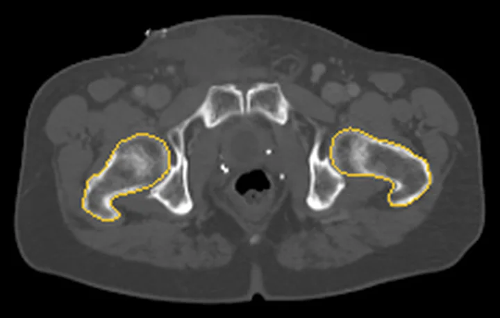
- Femur başları. Koronal kemik penceresinde iki femur başını aynı anda bul; süperior yük taşıyan yüzeylerin küreselliğini ve sağ-sol yoğunluk farkını gözle.
- Femur başı subkondral segmenti. Şüpheli başın içindeki kıvrımlı sklerotik sınırı kesitler boyunca izle; sınırın bir segmenti çevreleyip çevrelemediğini göster.
- Süperior subkondral kemik. Subkondral kortekste eklem yüzüne paralel ayrışma veya kresent çizgisi arayarak nekrozun kırık evresine geçip geçmediğini belirle.
- Femur başı konturu. Femur başı üst kontüründeki çökme ölçülür; ARCO evrelemesi için çökme derinliği ve yayılımı belirlenir.
- Kalça eklem aralığı ve karşı kalça. Son olarak eklem aralığı, asetabular çatı ve karşı kalçada ikincil kıkırdak-kemik hasarı ya da eş odak ara.
Faz ve pencere
- Kontrastsız tanısal
- Femur başında coğrafi sınırlı skleroz, serpijinöz subkondral kırık hattı (kresent) ve eklem yüzü çökmesi aranır.
Önerilen pencereler: Kemik (G 1800 / M 400), Yumuşak doku (G 400 / M 40).
BT bulguları
- Coğrafi serpijinöz skleroz — femur başının ağırlık taşıyan bölümünde nekrotik segmenti çevreleyen kıvrımlı yoğun kenar.
- Subkondral kresent — eklem yüzüne paralel ince yarık veya kortikal kesinti; BT subkondral kırığı göstermede duyarlıdır.
- Eklem yüzü çökmesi — femur başı süperior kontürünün düzleşmesi veya basamaklanması ile karakterizedir.
- Segmental femur başı deformasyonu — baş-boyun bileşkesine doğru uzanabilen fokal kontur kaybı.
- Nekrotik alan çevresinde sklerotik sınır — canlı ve etkilenmiş kemik arasındaki coğrafi geçişi belirginleştirebilir.
- Sekonder osteoartrit — ilerlemiş olguda eklem aralığı daralması, asetabular subkondral skleroz/kistler ve marjinal osteofitler.
- İki taraflı veya çok odaklı kemik tutulumu — karşı kalça ve görüntülenen diğer kemiklerde benzer odaklar sistemik risk bağlamını destekleyebilir.
Ölçütler ve sınıflamalar
- ARCO 2019
- Evre I: radyografi ve BT normal, MR veya sintigrafi anormal. Evre II: radyografide skleroz/osteoliz/kistler, subkondral kırık veya düzleşme yok. Evre III, radyografide kresent (subkondral kırık) bulgusuyla tanımlanır. Evre IV: eklem aralığı daralması, asetabular değişiklik veya eklem yıkımıyla osteoartrit.
Normalde
Normal femur başı kontürü küreseldir; subkondral kemikte serpiginöz sklerotik sınır veya kırık hattı yokluğu normaldir. Femur başı süperior eklem yüzü kontürü, subkondral çizgi devamlılığı ve serpijinöz sklerotik sınırın varlığı değerlendirilir.
Normal BT ile kıyasla
Aynı bölgenin, kaynak veri setinde patoloji içermediği belirtilen bir BT incelemesini kesit kesit kaydırın; organ sınırlarını açarak anatomiyi hasta görüntüsüyle karşılaştırın. Küçük rastlantısal bulgular tümüyle dışlanmamıştır.
Kaynak: TotalSegmentator v2.01 veri seti (Wasserthal ve ark., Radiology: Artificial Intelligence 2023), CC BY 4.0, vaka s1256; organ sınırları veri setinin otomatik segmentasyonundan, birleştirilerek sadeleştirildi. Görüntüler 2,5 mm kesit aralığına yeniden örneklendi.
Ayırıcı tanı
- Subkondral yetersizlik kırığı
- osteonekrotik segmenti çevreleyen düzgün sklerotik sınırdan çok lokal, düzensiz subkondral kırık ve çökme öne çıkar; erken ayrımda MR gerekir.
- Geçici kemik iliği ödemi sendromu
- BT'de belirgin serpijinöz nekrotik sınır veya kresent olmayabilir; yaygın kemik iliği ödemi MR bulgusudur.
- İleri kalça osteoartriti
- daralma ve osteofitler eklem genelinde dağılım gösterir; fokal femur başı nekroz sınırı beklenmez.
- Femur başı travmatik osteokondral kırığı
- travma öyküsüyle uyumlu keskin parça/eklem yüzü basamağı ve eşlik eden asetabular ya da femur boynu kırığı araştırılır.
Tuzaklar
- Normal veya belirsiz BT osteonekrozu dışlamaz; kemik iliği sinyal değişikliği ve erken nekrotik sınır için MR daha duyarlıdır.
- Kresenti otomatik olarak osteonekroz kabul etme: subkondral yetersizlik kırığı da çökme yapabilir; klinik bağlam ve MR bant morfolojisiyle ayır.
- Tek bir oblik aksiyel kesitte görünen kontur basamağını çökme sayma; koronal ve sagittal yeniden biçimlendirmelerde devamlılığını doğrula.
- Bilateral hastalıkta belirgin taraftaki çökme, karşı taraftaki daha erken lezyonu görünmez kılabilir; iki femur başını ayrı ayrı tara.
Kendini dene
Süperior femur başında serpijinöz sklerotik sınır ve ona komşu kresent görülüyor. En olası süreç hangisidir?
Cevabı göster
Avasküler femur başı nekrozu. Coğrafi sklerotik sınırla birlikte subkondral kresent, osteonekroz segmentinde kırık ve olası çökme düşündürür. Geçici ödem BT'de bu nekrotik sınır-kresent bileşimini oluşturmaz; artrit eklem içi/çevresi bulgularla, enkondrom ise medüller kıkırdak matriksiyle tanınır.
Femur başında odaksal serpijinöz sınır ve kresent yok; MR'da yaygın baş-boyun kemik iliği ödemi tarifleniyor. Hangi taklitçi düşünülür?
Cevabı göster
Geçici osteoporoz paterni. Yaygın kemik iliği ödemi ve belirgin nekrotik segment sınırının olmaması geçici kemik iliği ödemi sendromunu destekler. Yetersizlik kırığında subkondral bant; osteonekrozda coğrafi sınır; travmatik osteokondral kırıkta parça veya travma ilişkili yüzey hasarı aranır.
Kaynaklar
Bu sayfadaki 41 cümle ve 2 quiz sorusu aşağıdaki kaynaklardan alıntılarla otomatik olarak karşılaştırıldı; 7 cümle bu karşılaştırmada düzeltildi (2026-10-09).
- pmc.ncbi.nlm.nih.govpmc.ncbi.nlm.nih.gov
- Osteonecrosis of the Femoral Head.pubmed.ncbi.nlm.nih.gov
- Histopathological aspects and staging systems in non-traumatic femoral head osteonecrosis: an overview of the literature.pubmed.ncbi.nlm.nih.gov
- Advanced MRI Techniques for the Hip Joint: Focus on the Postoperative Hip.pubmed.ncbi.nlm.nih.gov
- Benefits of CT Scanning for the Management of Hip Arthritis and Arthroplasty.pubmed.ncbi.nlm.nih.gov
- Case report: Osteonecrosis of the femoral head after hip arthroscopy.pubmed.ncbi.nlm.nih.gov
- Current management options for osteonecrosis of the femoral head: part 1, diagnosis and nonoperative management.pubmed.ncbi.nlm.nih.gov
- MRI-detected bone marrow changes within 3 weeks after initiation of high-dose corticosteroid therapy: a possible change preceding the subsequent appearance of low-intensity band in femoral head osteonecrosis.pubmed.ncbi.nlm.nih.gov
- Low-intensity pulsed ultrasound enhances bone repair in a rabbit model of steroid-associated osteonecrosis.pubmed.ncbi.nlm.nih.gov
- Short-term bisphosphonate therapy could ameliorate osteonecrosis: a complication in childhood hematologic malignancies.pubmed.ncbi.nlm.nih.gov
- Anti-RNP Antibody: A Potential Novel Predictor for Osteonecrosis in Systemic Lupus Erythematosus.pubmed.ncbi.nlm.nih.gov
- From the radiologic pathology archives imaging of osteonecrosis: radiologic-pathologic correlation.pubmed.ncbi.nlm.nih.gov
- Perfusion image guided mechanical thrombectomy combined with tirofiban successfully revascularize systemic lupus erythematosus related acute large vessel occlusion: A case report.pubmed.ncbi.nlm.nih.gov
- A new preclinical femoral head osteonecrosis model in sheep.pubmed.ncbi.nlm.nih.gov
- Osteochondral mosaicplasty of the femoral head.pubmed.ncbi.nlm.nih.gov
- Shoulder Osteonecrosis: Pathogenesis, Causes, Clinical Evaluation, Imaging, and Classification.pubmed.ncbi.nlm.nih.gov
- Beyond the definitions of the phenotypic complications of sickle cell disease: an update on management.pubmed.ncbi.nlm.nih.gov
- Optimizing diagnosis and management of ONFH, FNSF, and SIF.pubmed.ncbi.nlm.nih.gov
- Beyond Cartilage Regeneration: Targeting the Subchondral Bone with Extracorporeal Shock Wave Therapy and Regenerative Medicine in Knee Osteoarthritis.pubmed.ncbi.nlm.nih.gov
- Fascia Lata and Tensor Fasciae Latae in Lower-Limb Reconstruction.pubmed.ncbi.nlm.nih.gov
- Diagnosis and Treatment of Nontraumatic Osteonecrosis of the Femoral Head: A Systematic Review and Meta-Analyses for the ARCO Clinical Practice Guideline Development Workgroup.pubmed.ncbi.nlm.nih.gov
- Bone marrow lesions related to bone marrow edema syndromes and osteonecrosis.pubmed.ncbi.nlm.nih.gov
- Osteoarthritis: Epidemiology, Diagnosis, and Treatment.pubmed.ncbi.nlm.nih.gov
- Kienböck Disease: Recent Advances in Understanding and Management.pubmed.ncbi.nlm.nih.gov
- Dynamic evolution of osseous structure in osteonecrosis of the femoral head and dynamic collapse risks: a preliminary CT image study.pubmed.ncbi.nlm.nih.gov
- Radiological manifestation of avascular necrosis (AVN) in sickle cell disease (SCD): a review of diagnostic imaging.pubmed.ncbi.nlm.nih.gov
- Atraumatic fractures of the femur.pubmed.ncbi.nlm.nih.gov
- Chinese Guideline for the Diagnosis and Treatment of Osteonecrosis of the Femoral Head in Adults.pubmed.ncbi.nlm.nih.gov
- Osteonecrosis in Youth with Sickle Cell Disease-A Narrative Review.pubmed.ncbi.nlm.nih.gov
- Artificial Intelligence Applications in MRI for the Diagnosis and Management of Osteonecrosis of the Femoral Head: A Comprehensive Review.pubmed.ncbi.nlm.nih.gov
- Advances in FAI Imaging: a Focused Review.pubmed.ncbi.nlm.nih.gov
- Emerging roles of growth factors in osteonecrosis of the femoral head.pubmed.ncbi.nlm.nih.gov
- Imaging in Hip Arthroplasty Management Part 2: Postoperative Diagnostic Imaging Strategy.pubmed.ncbi.nlm.nih.gov
- Three-Dimensional Printing of Medical Devices Used Directly to Treat Patients: A Systematic Review.pubmed.ncbi.nlm.nih.gov
- Macrophage immunometabolic reprogramming in inflammatory repair failure in osteonecrosis of the femoral head.pubmed.ncbi.nlm.nih.gov
- Senescence-driven osteonecrosis of the femoral head in the elderly-a distinct pathophysiological entity.pubmed.ncbi.nlm.nih.gov
- Hip Arthroscopy With Triangular Drilling and Suction Decompression for Osteonecrosis of the Femoral Head: A Retrospective Study.pubmed.ncbi.nlm.nih.gov
- Bilateral Knee Osteonecrosis in an Adolescent With Leukemia on Prolonged Corticosteroids: Discordant Femoral-Tibial Findings on Bone Scan SPECT-CT.pubmed.ncbi.nlm.nih.gov
- Systemic Sarcoidosis With Neurosarcoidosis Features as a Risk Factor for Multifocal Osteonecrosis.pubmed.ncbi.nlm.nih.gov
- Challenges in the management of bone metastases: focus on differentiated thyroid cancer and neuroendocrine tumors.pubmed.ncbi.nlm.nih.gov
- Osteonecrosis of the Femoral Head: Evolution of Contemporary Management Strategies.pubmed.ncbi.nlm.nih.gov
- Modified trapdoor procedures using autogenous tricortical iliac graft without preserving the broken cartilage for treatment of osteonecrosis of the femoral head: a prospective cohort study with historical controls.pubmed.ncbi.nlm.nih.gov
- <sup>18</sup>F-FDG PET/CT in the evaluation of femoral head osteonecrosis in patients with lymphoma.pubmed.ncbi.nlm.nih.gov
- Pharmacotherapy of Kienböck Disease and the Carpal Avascular Necrosis Family: A Narrative Review with a Four-Axis Translation Framework.pubmed.ncbi.nlm.nih.gov
- The Unique Features of Pediatric Sports Injuries: The Hip.pubmed.ncbi.nlm.nih.gov
- Cosmic Signs in Radiology: A Pictorial Review.pubmed.ncbi.nlm.nih.gov
- Post-COVID-19 Jaw Osteonecrosis: A Narrative Review.pubmed.ncbi.nlm.nih.gov
- Corticosteroid-associated osteonecrosis in paediatric compared with adult patients: are they the same disease?pubmed.ncbi.nlm.nih.gov
- Diagnostic biomarkers for non-traumatic osteonecrosis of the femoral head : a systematic review and pathophysiological cascade network.pubmed.ncbi.nlm.nih.gov
- Non-femoral head MRI radiomics as a potential imaging biomarker for identifying osteonecrosis of the femoral head in systemic lupus erythematosus patients.pubmed.ncbi.nlm.nih.gov
- Conservative and surgical treatment of wrist osteoarthritis: a systematic review.pubmed.ncbi.nlm.nih.gov
- Avascular Necrosis Secondary to Traumatic Posterior Hip Dislocation.pubmed.ncbi.nlm.nih.gov
- Hip Arthroscopic Management for Patients With Subchondral Stress Fractures of the Femoral Head: A Case-Control Study.pubmed.ncbi.nlm.nih.gov
- Evolutionary course of the femoral head osteonecrosis: Histopathological - radiologic characteristics and clinical staging systems.pubmed.ncbi.nlm.nih.gov
- Guidelines on multidisciplinary approaches for the prevention and management of diabetic foot disease (2020 edition).pubmed.ncbi.nlm.nih.gov
- Long-term safety and effectiveness of the "OptEase" vena cava filter.pubmed.ncbi.nlm.nih.gov
- Japan Endocrine Society Clinical Practice Guideline for the Diagnosis and Management of Pheochromocytoma and Paraganglioma 2025.pubmed.ncbi.nlm.nih.gov
- Bone marrow magnetic resonance imaging (MRI): morphological and functional features from reconversion to infiltration.pubmed.ncbi.nlm.nih.gov
- Imaging modalities for non-acute pathologies of the foot and ankle.pubmed.ncbi.nlm.nih.gov
- Chinese herbal Huo-Gu formula for the treatment of steroid-associated osteonecrosis of femoral head: A 14-year follow-up of convalescent SARS patients.pubmed.ncbi.nlm.nih.gov
- Super-acute massive osteolysis of the femoral head after acetabular fracture fixation : a comparative study with conventional post-traumatic osteonecrosis requiring total hip arthroplasty.pubmed.ncbi.nlm.nih.gov
- The Impact of Radiomics Image Analysis on Adult Hip Pathologies: A Scoping Review.pubmed.ncbi.nlm.nih.gov
- Quantitative contrast enhanced dual energy CT to predict avascular necrosis: a feasibility study of proximal humerus fractures.pubmed.ncbi.nlm.nih.gov
- Developmental Dysplasia of the Hip - Part 1.pubmed.ncbi.nlm.nih.gov
- Digitally Designed Bone; A 3D-patient-specific Allograft Shelf for Severe Adolescent Hip Dysplasia: From Digital Design to Clinical Reality-A Conceptual Case Report.pubmed.ncbi.nlm.nih.gov
- Radiographic evaluation of the painful adolescent and young adult hip.pubmed.ncbi.nlm.nih.gov
- Techniques of Deformity Correction in Adolescent Idiopathic Scoliosis-A Narrative Review of the Existing Literature.pubmed.ncbi.nlm.nih.gov
- Treatment of Steroid-Induced Osteonecrosis of the Distal Femoral Condyle Using Structural Bone Grafting.pubmed.ncbi.nlm.nih.gov
- Automatic diagnosis of osteonecrosis of the femoral head and segmentation of necrotic regions using deep learning at computed tomography: a real-world multicenter study.pubmed.ncbi.nlm.nih.gov
- Association of anterior necrotic boundary location with collapse progression in symptomatic osteonecrosis of the femoral head: analysis of ARCO 2021 type 2 lesions.pubmed.ncbi.nlm.nih.gov
- Characteristics of the Extent and Onset of Osteonecrosis of the Femoral Head After Femoral Neck System (FNS) Fixation: A Minimum Two-Year Follow-Up Comparative Study with Cannulated Screws.pubmed.ncbi.nlm.nih.gov
- Case Report: Combined posterolateral and direct anterior approaches for total hip arthroplasty in osteonecrosis of the femoral head with a rare massive osteophyte with literature review.pubmed.ncbi.nlm.nih.gov
- Anatomy-driven automated subsegmentation of pelvic and proximal femoral CT into clinically relevant subregions and landmarks.pubmed.ncbi.nlm.nih.gov
- Normative Values of Femoral Inclination Angle and Medial Proximal Femoral Angle: An MRI-Based Study.pubmed.ncbi.nlm.nih.gov
- Posterior Acetabular Wall Fracture Mimicking a Subcapital Femoral Neck Fracture on Plain Radiographs: A Diagnostic Pitfall and the Role of Intraoperative Fluoroscopy: A Case Report.pubmed.ncbi.nlm.nih.gov
- Initiation of Collapse in Osteonecrosis of the Femoral Head: A Patient-Specific Finite Element Simulation Study.pubmed.ncbi.nlm.nih.gov
- Femoral stem alignment and post-operative patient satisfaction: a retrospective analysis of total hip arthroplasty using computer-assisted equipment for placing cups.pubmed.ncbi.nlm.nih.gov
- A clinically applicable energy-based approach for evaluating bone strength.pubmed.ncbi.nlm.nih.gov
- Operative Procedures for Ultrasound Assessment of Extracranial Artery Disease: A Narrative Review by the Italian Society for Vascular Investigation (SIDV).pubmed.ncbi.nlm.nih.gov
- Engineered Xenogeneic Bone Scaffold with IL-10 Nanodelivery System: Immunomodulation and BMSC Fate Programming for Skull Defect Repair.pubmed.ncbi.nlm.nih.gov
- Systemic lupus erythematosus complicated by refractory <i>Mycobacterium avium complex</i> infection involving bilateral lower extremities: a case report and literature review.pubmed.ncbi.nlm.nih.gov
- Three-dimensional mapping of necrotic lesions for early-stage osteonecrosis of the femoral head.pubmed.ncbi.nlm.nih.gov
- Analysis of the efficacy of drilling decompression autologous bone marrow and allogeneic bone grafting in the treatment of HIV-positive patients with early osteonecrosis of the femoral head.pubmed.ncbi.nlm.nih.gov
- A review of the oral health of individuals with disabilities in Puerto Rico and among U.S. Hispanics.pubmed.ncbi.nlm.nih.gov
- Osteochondritis Dissecans of the Medial Femoral Condyle With Intra-Articular Meniscal Calcification: A Rare Case and Literature Review.pubmed.ncbi.nlm.nih.gov
- Glossary of terms for musculoskeletal radiology.pubmed.ncbi.nlm.nih.gov
- Osteochondral Lesions of the Talus: A Review on Talus Osteochondral Injuries, Including Osteochondritis Dissecans.pubmed.ncbi.nlm.nih.gov
- Microdamage accumulation in the femoral head and its association with subchondral insufficiency fractures: A retrospective study.pubmed.ncbi.nlm.nih.gov
- Subchondral Insufficiency Fracture of the Humeral Head: A Rare Case in a Young Person.pubmed.ncbi.nlm.nih.gov
- International Harmonization of Nomenclature and Diagnostic Criteria (INHAND): Non-proliferative and Proliferative Lesions of the Non-human Primate ( M. fascicularis ).pubmed.ncbi.nlm.nih.gov
- International Society of Paediatric Surgical Oncology (IPSO) Surgical Practice Guidelines.pubmed.ncbi.nlm.nih.gov
- Hip Arthroscopy-Assisted Management of Pipkin Types I and II Femoral Head Fracture-Dislocations: Mid-Term Clinical and Radiographic Outcomes.pubmed.ncbi.nlm.nih.gov
- A rare traumatic triad: Open bicondylar non-conjoint Hoffa fracture with partial tibial tubercle and medial epicondyle avulsions - A case report and biomechanical analysis.pubmed.ncbi.nlm.nih.gov
- Morphology-Guided Surgical Management of Complex Traumatic Hip Fracture-Dislocations and Acetabular Fractures: A Six-Case Series Illustrating Morphology-Based Surgical Approaches.pubmed.ncbi.nlm.nih.gov
- Bilateral Avascular Necrosis of the Femoral Head in a Patient With Sickle Cell Trait: A Case Report.pubmed.ncbi.nlm.nih.gov
- Avascular necrosis of the femoral head following hysteroscopic myomectomy: Evaluation of bone perfusion using dynamic contrast-enhanced MRI and intravoxel incoherent motion.pubmed.ncbi.nlm.nih.gov
- Real-World Outcomes of Autologous Osteoblast Implantation in Patients With Osteonecrosis of the Femoral Head: A Retrospective, Multicenter, Single-Arm Cohort Study in India.pubmed.ncbi.nlm.nih.gov
- Outcomes of the conservative hip preservation treatment in patients with osteonecrosis of the femoral head: With 4-year follow-up.pubmed.ncbi.nlm.nih.gov
- Safety, Feasibility, and Preliminary Efficacy of Allogeneic MSCs to Treat Advanced Femoral Head Osteonecrosis (ALOFEM): A Pilot Study in Young Onco-Hematological Patients.pubmed.ncbi.nlm.nih.gov
- A Descriptive Study on the Role of Magnetic Resonance Imaging in Staging Avascular Necrosis of the Hip Joint: Current Trends and Insights.pubmed.ncbi.nlm.nih.gov
- Advancements in 3D printing technologies for personalized treatment of osteonecrosis of the femoral head.pubmed.ncbi.nlm.nih.gov
- Femoral Head Osteonecrosis: Computed Tomography Not Needed to Identify Collapse When Using the Association Research Circulation Osseous Staging System.pubmed.ncbi.nlm.nih.gov
- A single sequence MRI-based deep learning radiomics model in the diagnosis of early osteonecrosis of femoral head.pubmed.ncbi.nlm.nih.gov
- Comparison of two joint-preserving treatments for osteonecrosis of the femoral head: core decompression and core decompression with additional cancellous bone grafting.pubmed.ncbi.nlm.nih.gov
- Updating Osteonecrosis of the Femoral Head.pubmed.ncbi.nlm.nih.gov
Eğitim amaçlıdır; hasta başında tanı ve tedavi kararının yerine geçmez, tıbbi tavsiye değildir. Metinler yapay zekâ desteğiyle yazıldı ve otomatik bir adımda kaynak alıntılarıyla karşılaştırıldı; bu bir tıbbi doğrulama değildir ve içerik uzman radyolog incelemesinden geçmedi. Klinik kararlarda güncel kılavuzları, kurum protokollerini ve radyoloji raporunu esas alın.