Özet
Primer kemik lenfoması (PBL), kemikte bir veya daha fazla lezyonla ortaya çıkan ve lenf nodu ya da başka ekstranodal tutulum bulunmayan malign lenfoid neoplazi olarak tanımlanır; femur sık tutulan kemikler arasındadır. BT, kemik iliği tutulumunu, kortikal destrüksiyonu ve yumuşak doku uzanımını gösterebilir; kemik iliği tutulumu ve yumuşak doku uzanımının kapsamı MRG ile daha iyi değerlendirilir. PBL’nin görüntüleme görünümü özgül değildir ve Ewing sarkomu ile osteomiyelit gibi durumları taklit edebilir; kortikal yıkıma göre büyük yumuşak doku kitlesi PBL lehine bir ipucudur.
Faz ve pencere
- Kontrastsız tanısal
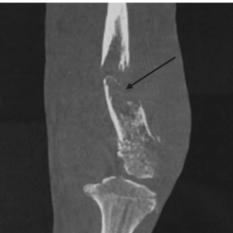
- BT, PBL’de kemik iliği tutulumunu, kortikal destrüksiyonu ve eşlik eden yumuşak doku uzanımını gösterebilir; kemik yıkımı permeatif veya güve yeniği tarzında olabilir. Kortikal yıkıma göre büyük kitle-korteks uyumsuzluğu PBL kuşkusunu artırır.
Önerilen pencereler: Kemik (G 1800 / M 400), Yumuşak doku (G 400 / M 40).
BT bulguları
- Metadiyafizer medüller lezyon — PBL uzun kemiklerin metafiz-diyafiz bileşkesinde sık görülür ve kemik iliğinden kaynaklanır.
- Permeatif veya güve yeniği yıkım — medüller kemikte litik-infiltratif yıkım ve geniş geçiş zonu görülebilir.
- Medüller yayılım — lezyon kemik iliği boyunca uzanabilir; MRG kemik iliği tutulumunun kapsamını BT’den daha iyi gösterir.
- Korteks-yumuşak doku uyumsuzluğu — büyük ekstraosseöz kitleye karşın dış korteksin geniş bölümü görece korunmuş kalabilir.
- Ekstraosseöz yumuşak doku kitlesi — kemik iliğinden başlayan lezyon femur dışına uzanıp çevre kompartımanları tutabilir.
- Katmanlı periostal reaksiyon — lameller veya çok katmanlı (“onion-skin”) periostal yeni kemik görülebilir.
- Patolojik kırık — PBL’ye patolojik kırık eşlik edebilir.
Normalde
BT, kemik iliği tutulumunu ve kortikal destrüksiyonu göstererek lezyonun yayılımının değerlendirilmesine katkı sağlar. PBL’de kemik iliği tutulumunun, yumuşak doku uzanımının ve kortikal destrüksiyonun kapsamını birlikte değerlendirin.
Normal BT ile kıyasla
Aynı bölgenin, kaynak veri setinde patoloji içermediği belirtilen bir BT incelemesini kesit kesit kaydırın; organ sınırlarını açarak anatomiyi hasta görüntüsüyle karşılaştırın. Küçük rastlantısal bulgular tümüyle dışlanmamıştır.
Kaynak: TotalSegmentator v2.01 veri seti (Wasserthal ve ark., Radiology: Artificial Intelligence 2023), CC BY 4.0, vaka s0685; organ sınırları veri setinin otomatik segmentasyonundan, birleştirilerek sadeleştirildi. Görüntüler 2,5 mm kesit aralığına yeniden örneklendi.
Ayırıcı tanı
- Ewing sarkomu
- diyafizer permeatif yıkım, lameller periostal reaksiyon ve yumuşak doku kitlesiyle PBL’yi taklit edebilir; bu görüntüleme bulguları tek başına tanıyı ayırt ettirmez.
- Osteosarkom
- bulutsu osteoid matriks ve agresif periostal reaksiyon (ör. sunburst veya Codman üçgeni) osteosarkom lehinedir.
- Osteomiyelit, PBL’yi görüntülemede taklit edebilir; sekestr görülmesi ayırıcı tanıda osteomiyeliti düşündürmelidir.
- Kemik metastazı
- çok odaklı kemik lezyonları veya bilinen primer malignite öyküsü ikincil kemik tutulumunu destekler.
- Kondrosarkom
- BT’de kortikal yıkım ve noktasal ya da amorf kalsifikasyonlar içeren yumuşak doku kitlesi görülebilir; bu agresif bulgular malignite değerlendirmesini gerektirir.
Tuzaklar
- Yumuşak doku kitlesinin boyutundan tek başına kemik korteksinin tamamen yıkıldığını varsaymayın; korteks-yumuşak doku oranı bu tümör için ayırt edici ipucudur.
- BT’de korteksin görece korunması PBL’yi dışlamaz; MRG kemik iliği tutulumunun ve kompartıman uzanımının kapsamını daha iyi gösterir.
- Primer kemik lenfoması tanımı yalnız femur görüntüsünden kurulamaz; uzak hastalık odağı tüm-vücut görüntülemeyle değerlendirilir ve kesin tanı histopatolojik incelemeyle konur.
Kendini dene
Femur medullasında permeatif yıkım ve geniş çevresel kitle var; korteks kitlenin boyutuna göre beklenenden iyi korunmuş. En olası tanı hangisidir?
Cevabı göster
Primer kemik lenfoması. Korteksin görece korunmasına karşın büyük yumuşak doku kitlesi bulunması PBL’yi düşündüren bir örüntüdür, ancak tek başına tanı koydurmaz. Osteosarkomda bulutsu osteoid matriks, kemik enfarktında serpijinöz sklerotik sınır, enkondromda ise halka-yay kondroid matriks görülebilir.
PBL’de konvansiyonel radyografi ve BT’de en sık görülen kemik yıkımı paterni hangisidir?
Cevabı göster
Permeatif veya güve yeniği tarzı litik yıkım. PBL, konvansiyonel radyografi ve BT’de en sık medüller kaviteden kaynaklanan permeatif veya güve yeniği tarzı litik yıkım paterni oluşturur.
İlgili konular
Kaynaklar
Bu sayfadaki 33 cümle ve 2 quiz sorusu aşağıdaki kaynaklardan alıntılarla otomatik olarak karşılaştırıldı; 21 cümle bu karşılaştırmada düzeltildi (2026-10-09).
- Primary bone lymphoma: pictorial essaypubmed.ncbi.nlm.nih.gov
- Primary Bone Lymphoma: A Review of the Literature with Emphasis on Histopathology and Histogenesispmc.ncbi.nlm.nih.gov
- Musculoskeletal Lymphoma: Imaging Features, Diagnosis, and Assessment of Treatment Responsepubs.rsna.org
- Radiographic Evaluation of Bone Tumorspubs.rsna.org
- The imaging of osteomyelitisqims.amegroups.org
- State-of-the-art imaging for diagnosis of metastatic bone diseasepmc.ncbi.nlm.nih.gov
- Subchondral insufficiency fracture of the knee: review of current concepts and radiological differential diagnoseslink.springer.com
- Atraumatic fractures of the femurpmc.ncbi.nlm.nih.gov
- Askin Tumor Presents with Ewing Sarcoma of the Femur in Young Female Patient: Case Report and Literature Reviewpmc.ncbi.nlm.nih.gov
- Primary Bone Diffuse Large B-cell Lymphoma (DLBCL) in a Young Adult: A Case Reportpmc.ncbi.nlm.nih.gov
- Analysis of imaging characteristics of primary malignant bone tumors in children.pubmed.ncbi.nlm.nih.gov
- Evaluation of primary bone lymphoma and the importance of positron emission tomography.pubmed.ncbi.nlm.nih.gov
- Case report: Primary bone lymphoma presenting as a painful supraclavicular lump.pubmed.ncbi.nlm.nih.gov
- Is True Whole-Body 18 F-FDG PET/CT Required in Pediatric Lymphoma? An IAEA Multicenter Prospective Study.pubmed.ncbi.nlm.nih.gov
- Primary lymphoma of bone in the femur complicated by a fracture and listeria osteomyelitis.pubmed.ncbi.nlm.nih.gov
- Primary Bone Anaplastic Large Cell Lymphoma Masquerading as Ewing Sarcoma: Diagnosis by Anchored Multiplex PCR.pubmed.ncbi.nlm.nih.gov
- Primary alveolar soft part sarcoma of the right femur and primary lymphoma of the left femur: A case report and literature review.pubmed.ncbi.nlm.nih.gov
- [Diagnostic usefulness of FDG-PET/CT in multiple primary osseous Hodgkin lymphoma].pubmed.ncbi.nlm.nih.gov
- Case of polyostotic primary bone lymphoma successfully treated with immunochemotherapy and consolidation radiotherapy.pubmed.ncbi.nlm.nih.gov
- Primary Bone Marrow B-Cell Lymphoma Undetected by Multiple Imaging Modalities That Initially Presented with Hypercalcemia.pubmed.ncbi.nlm.nih.gov
- Epiphyseal presentation of non-Hodgkin's lymphoma of bone in two pediatric patients--one with primary lymphoma of bone.pubmed.ncbi.nlm.nih.gov
- Complementary roles of bone scintigraphy and MR imaging in the detection and long-term follow-up of primary non-Hodgkin's bone lymphoma in a child-case report.pubmed.ncbi.nlm.nih.gov
- [Sjögren disease complicated by primary breast lymphoma: A case report].pubmed.ncbi.nlm.nih.gov
- Primary Bone Diffuse Large B-cell Lymphoma (DLBCL) in a Young Adult: A Case Report.pubmed.ncbi.nlm.nih.gov
- Pathological fracture of the femur without surgical fixation in primary bone diffuse large B-cell lymphoma (DLBCL): A rare case with full recovery.pubmed.ncbi.nlm.nih.gov
- Primary Synovial Sarcoma of the Prostate: A Case Report and Review of the Literature.pubmed.ncbi.nlm.nih.gov
- Primary Bone Lymphoma in Adults: A Report of Two Cases and Review of the Literature Highlighting the Role of Radiotherapy.pubmed.ncbi.nlm.nih.gov
- Multifocal osseous and cutaneous Rosai-Dorfman-Destombes disease presenting as an aggressive intramedullary femoral lesion.pubmed.ncbi.nlm.nih.gov
- Musculoskeletal Manifestations of T-Cell Lymphomas: Clinical Presentation, Imaging Features, Pathobiology, and Orthopedic Considerations-A Narrative Review.pubmed.ncbi.nlm.nih.gov
- Using WB-LDCT to differentiate between benign and malignant medullary lesions in the appendicular skeleton of patients with plasma cell diseases.pubmed.ncbi.nlm.nih.gov
- Imaging of ewing sarcoma: an updated analysis including presenting features, prognostic imaging biomarkers, and treatment response assessment.pubmed.ncbi.nlm.nih.gov
- Apparent diffusion coefficient can assist in differentiating between benign and malignant primary bone tumors in pediatric patients.pubmed.ncbi.nlm.nih.gov
- Bone lesions of the tibia: Multimodal iconographic review and diagnostic algorithms, Part 2: Metaphyseal and epiphyseal lesions.pubmed.ncbi.nlm.nih.gov
- Primary bone lymphoma with extensive imaging-defined necrosis on magnetic resonance imaging and <sup>18</sup>F-fluorodeoxyglucose positron emission tomography: A diagnostic pitfall.pubmed.ncbi.nlm.nih.gov
- Unraveling Translational Insights into Systemic Multi-Organ Toxicity of Cytosine Arabinoside (Ara-C): A Systematic Review of Preclinical Animal Evidence.pubmed.ncbi.nlm.nih.gov
- Non-Conventional Treatments for Conventional Chondrosarcoma.pubmed.ncbi.nlm.nih.gov
- 2025 American Thyroid Association Management Guidelines for Adult Patients with Differentiated Thyroid Cancer.pubmed.ncbi.nlm.nih.gov
- Malignant Transformation of Musculoskeletal Lesions with Imaging-Pathology Correlation-Part 1: Bone Lesions.pubmed.ncbi.nlm.nih.gov
- Analysis and Historical Evolution of Paediatric Bone Tumours: The Importance of Early Diagnosis in the Detection of Childhood Skeletal Malignancies.pubmed.ncbi.nlm.nih.gov
- Musculoskeletal manifestations of lymphoma: a case series.pubmed.ncbi.nlm.nih.gov
- Atlas of gross, histologic, and immunohistologic anatomy of the lymphoid system in Egyptian rousettes (Rousettus aegyptiacus).pubmed.ncbi.nlm.nih.gov
- Function and Regulation of Bone Marrow Adipose Tissue in Health and Disease: State of the Field and Clinical Considerations.pubmed.ncbi.nlm.nih.gov
- Paget's Disease of the Spine as a Catalyst for Osteosarcoma: A Narrative Review.pubmed.ncbi.nlm.nih.gov
- Molecular Pathogenesis, Tumor Microenvironment and Health Disparities in Select Pediatric Solid Tumors: An Integrative Narrative Review.pubmed.ncbi.nlm.nih.gov
- Practical approach to the diagnosis, management, and treatment of pediatric patients with bone fragility: an expert opinion.pubmed.ncbi.nlm.nih.gov
- Emphysematous osteomyelitis in hematological malignancies: tales of two cases.pubmed.ncbi.nlm.nih.gov
- Pictorial Review of Paediatric Limp.pubmed.ncbi.nlm.nih.gov
- Osteomyelitis of the Jaw Bones and Its Mimics: Resolving the Diagnostic Enigma.pubmed.ncbi.nlm.nih.gov
- Classification of Chondrosarcoma: From Characteristic to Challenging Imaging Findings.pubmed.ncbi.nlm.nih.gov
- Multimodality imaging of greater trochanter lesions.pubmed.ncbi.nlm.nih.gov
- Musculoskeletal tumors and tumor-like lesions with "dark" signal intensity on T2-weighted MR images: A pictorial review.pubmed.ncbi.nlm.nih.gov
- Clinical practice guidelines for the treatment of Ewing sarcoma (Spanish Sarcoma Research Group-GEIS).pubmed.ncbi.nlm.nih.gov
- Conventional radiography for the assessment of focal bone lesions of the appendicular skeleton: fundamental concepts in the modern imaging era.pubmed.ncbi.nlm.nih.gov
- Glossary of terms for musculoskeletal radiology.pubmed.ncbi.nlm.nih.gov
- Imaging of non-neuronopathic Gaucher disease: recent advances in quantitative imaging and comprehensive assessment of disease involvement.pubmed.ncbi.nlm.nih.gov
- Musculoskeletal Infection: The Great Mimickers on Imaging.pubmed.ncbi.nlm.nih.gov
- Carcinoma of Unknown Primary Origin Presenting as Rapidly Progressive Multifocal Skeletal Lesions and Fatal Systemic Thrombosis in a Dog: A Case Report.pubmed.ncbi.nlm.nih.gov
- Beyond Chordoma: A Comprehensive Review of Sacral Lesions.pubmed.ncbi.nlm.nih.gov
- Diagnostic imaging and emerging image-guided neurointervention in brain tumors: a narrative review.pubmed.ncbi.nlm.nih.gov
- A Rare Primary Adrenal Malignancy Manifesting as a Hemorrhagic Mass: Case Report and Literature Review.pubmed.ncbi.nlm.nih.gov
- Unexpected Massive Hemorrhage During Incision and Drainage of a Presumed Perianal Abscess: A Case of Pelvic Chondrosarcoma.pubmed.ncbi.nlm.nih.gov
- Calcified Chondroid Mesenchymal Neoplasm of the Temporomandibular Joint: A Rare and Recently Described Benign Tumor.pubmed.ncbi.nlm.nih.gov
- An analysis of the imaging features and diagnostic value of osteoid osteoma on DR, CT, and MRI.pubmed.ncbi.nlm.nih.gov
- Atypical presentation of intraosseous plasmacytoma in a patient with POEMS syndrome.pubmed.ncbi.nlm.nih.gov
- Chronic Osteomyelitis or Squamous Cell Carcinoma of the Mandible: A Case Report With Diagnostic Challenges.pubmed.ncbi.nlm.nih.gov
- Diffusion-Weighted Imaging in the Musculoskeletal System: Evolving Role in Modern Imaging Practice.pubmed.ncbi.nlm.nih.gov
- Integrated diagnostic evaluation of multiple malignant neoplasms via <sup>99m</sup>Tc-MDP three-phase bone imaging: A case report.pubmed.ncbi.nlm.nih.gov
Eğitim amaçlıdır; hasta başında tanı ve tedavi kararının yerine geçmez, tıbbi tavsiye değildir. Metinler yapay zekâ desteğiyle yazıldı ve otomatik bir adımda kaynak alıntılarıyla karşılaştırıldı; bu bir tıbbi doğrulama değildir ve içerik uzman radyolog incelemesinden geçmedi. Klinik kararlarda güncel kılavuzları, kurum protokollerini ve radyoloji raporunu esas alın.