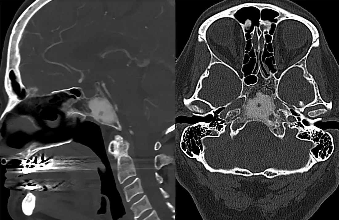

Özet
Rastlantısal veya ağrı nedeniyle incelenen kemik odağında BT, büyüme biçimini, kortikal bütünlüğü ve matriks mineralizasyonunu tanımlayarak risk değerlendirmesine katkı verir. Keskin sınır ve düzgün sklerotik kenar çoğu kez yavaş seyirli lezyonlarda görülür; kortikal destrüksiyon, geniş geçiş zonu ve ekstraosseöz kitle daha agresif biyolojiye işaret eder. BT özellikle mineralize matriksi ve kortikal kemik değişikliğini gösterir, ancak kemik iliği ve yumuşak doku yayılımının tam değerlendirmesinde MR tamamlayıcı olabilir. Agresif bulguların atlanması lezyon yayılımının olduğundan küçük raporlanmasına, mineralizasyon deseninin yanlış okunması ise gereksiz alarm veya gecikmeye yol açabilir.
Faz ve pencere
- Kontrastsız tanısal
- Medüller lezyonun sınırı, mineralizasyonu ve kortikal kemik değişiklikleri BT ile değerlendirilebilir.
- Kemik algoritması tanısal
- İnce kesit ve multiplanar kemik rekonstrüksiyonları, endosteal scalloping, korteks penetrasyonu, periostal kemik oluşumu ve lezyonun eklem yüzüyle ilişkisini ortaya koyar.
Önerilen pencereler: Kemik (G 1800 / M 400), Yumuşak doku (G 400 / M 40).
BT bulguları
- Dar geçiş zonu — lezyon sınırı normal medüller trabeküllerden keskin biçimde ayrılır; yavaş büyümeyi destekler.
- İnce sklerotik kenar bazı litik lezyonlarda görülebilir; tek başına benignliği kanıtlamaz.
- Silik, düzensiz sınır (poorly-defined margin), agresif büyüme ve yüksek Lodwick derecesi ile uyumludur.
- Kortikal destrüksiyon — korteksin fokal kesintisi veya medüller odaktan dışarı doğru uzanım agresiflik işaretidir.
- Kortikal penetrasyon (kısmi veya tam), lezyonun kemik dışına uzanımı ve agresif büyüme potansiyeli açısından Lodwick derecelendirmesinde belirleyicidir.
- Osteoid matriks — bulutumsu veya amorf yoğun kemik üretimi osteojenik tümörlerde görülebilir.
- Kondroid matriks — halka-yay biçimli kalsifikasyonlar lobüle kıkırdak matriksini düşündürür; tek başına benignlik kanıtı değildir.
- Fibröz matriks — buzlu cam dansitesi kemik ekspansiyonu içinde homojen ya da yamalı görünebilir.
Ölçütler ve sınıflamalar
- Lodwick ve değiştirilmiş Lodwick-Madewell büyüme derecelendirmesi
- Özgün Lodwick sistemi litik lezyonları IA (coğrafi, iyi sınırlı, sklerotik kenarlı), IB (coğrafi ve iyi sınırlı, sklerotik kenarsız), IC (coğrafi ama silik sınırlı), II (güve yeniği) ve III (permeatif) olarak derecelendirir. Değiştirilmiş Lodwick-Madewell sisteminde özgün IC, II; değişen/karma sınırlar IIIA; güve yeniği veya permeatif yıkım IIIB; başka görüntülemede saptanan radyografik olarak gizli lezyon IIIC olarak tanımlanır. Lodwick sistemi, litik kemik lezyonlarının radyografik büyüme hızını ve malignite olasılığını değerlendirmek için tasarlanmış bir algoritmadır; doğrudan histolojik tanı veya cross-sectional skorlamayı temsil etmez. Değiştirilmiş sürümün özgün Lodwick sisteminden üstün olduğu gösterilmemiştir.
Normalde
Litik lezyon değerlendirilirken kortekste penetrasyon olup olmadığı ve bunun kısmi mi tam mı olduğu incelenir. Lezyonlu düzeyle komşu normal segmenti kıyaslarken sınırın ne kadar geniş bir alanda silindiğini, kortikal iç yüzün oyulmasını, dış konturda kırılmayı ve mineralize desenin kemik iliği yerine lezyon matriksi içinde yer alıp almadığını inceleyin.
Normal BT ile kıyasla
Aynı bölgenin, kaynak veri setinde patoloji içermediği belirtilen bir BT incelemesini kesit kesit kaydırın; organ sınırlarını açarak anatomiyi hasta görüntüsüyle karşılaştırın. Küçük rastlantısal bulgular tümüyle dışlanmamıştır.
Kaynak: TotalSegmentator v2.01 veri seti (Wasserthal ve ark., Radiology: Artificial Intelligence 2023), CC BY 4.0, vaka s0685; organ sınırları veri setinin otomatik segmentasyonundan, birleştirilerek sadeleştirildi. Görüntüler 2,5 mm kesit aralığına yeniden örneklendi.
Ayırıcı tanı
- Enkondrom ile düşük dereceli kondrosarkomun görüntüleme bulgularıyla ayırımı zor olabilir.
- Osteoid osteom
- kortikal nidus çevresinde yoğun reaktif skleroz ve kortikal kalınlaşma vardır; küçük nidus ince kesit BT'de seçilebilir.
- Fibröz displazi
- genişleyen kemik segmenti içinde buzlu cam matriks, kortikal remodeling ve kemiğe karışan sınır oluşturur.
- Osteomiyelit
- litik yıkım ve periostal reaksiyona sekestr, kloaka veya yumuşak doku inflamasyonu eşlik edebilir.
- Osteoblastik metastaz
- medüller sklerotik odaklar özellikle çoklu dağılımda veya bilinen primer tümör bağlamında düşünülür.
Tuzaklar
- Keskin sklerotik kenar benignlik lehine olsa da her zaman histolojik benign tümör anlamına gelmez; lezyonun yeri, boyutu ve kortikal davranışı da kaydedilmelidir.
- Halka-yay mineralizasyonunu tek başına enkondrom tanısı saymayın; kondroid matriks benign ve malign tümörlerde görülebilir.
- Kortikal incelme ekspansil benign lezyonlarda remodeling olabilir; korteksin tam kesintisi ve dışarı uzanımını ayrı arayın.
- BT'de kitle görünmemesi kemik iliği yayılımını dışlamaz; yumuşak doku veya medüller kapsam klinik soruya göre MR ile tamamlanabilir.
Kendini dene
Tesadüfi metafiz odağı keskin sınırlı ve halka-yay kalsifikasyonlu; korteks korunmuş. Matriks hangisi?
Cevabı göster
Kondroid. Halka-yay biçimli kalsifikasyon kondroid matriksin tipik biçimidir. Osteosarkom osteoid matriks üretir; fibröz displazide buzlu cam görünümü görülebilir.
Litik lezyonda geniş belirsiz sınır, kortikal kesinti ve kemik dışı solid kitle saptandı. Bu kombinasyon neyi destekler?
Cevabı göster
Agresif biyoloji. Geniş geçiş zonu ile kortikal yıkım ve ekstraosseöz kitle agresif lezyon davranışını destekler. Yine de görüntüleme bulguları tek başına histolojik tanı koymaz; diğer seçenekler bu kombinasyonu açıklamaz.
İlgili konular
Kaynaklar
Bu sayfadaki 43 cümle ve 2 quiz sorusu aşağıdaki kaynaklardan alıntılarla tek tek karşılaştırıldı; 8 cümle bu karşılaştırmada düzeltildi (2026-10-09).
- The Lodwick classification for grading growth rate of lytic bone tumors: a decision tree approachpmc.ncbi.nlm.nih.gov
- Intraosseous schwannoma of the mobile spine: a report of twenty cases.pubmed.ncbi.nlm.nih.gov
- Primary Orbital Chondromyxoid Fibroma: A Rare Case.pubmed.ncbi.nlm.nih.gov
- Osteolytic-variant POEMS syndrome: an uncommon presentation of "osteosclerotic" myeloma.pubmed.ncbi.nlm.nih.gov
- Epithelioid hemangioma of the spine: Two cases.pubmed.ncbi.nlm.nih.gov
- Bone Scanning Findings in a Patient with Heat Stroke-Induced Rhabdomyolysis.pubmed.ncbi.nlm.nih.gov
- Chondroblastoma Of The Lumbar Vertebra.pubmed.ncbi.nlm.nih.gov
- Diagnostic imaging of fetal and pediatric orbital abnormalities.pubmed.ncbi.nlm.nih.gov
- 18 F-FDG PET/CT in the evaluation of cartilaginous bone neoplasms: the added value of tumor grading.pubmed.ncbi.nlm.nih.gov
- Tumors and pseudotumors of foot and ankle: Bone lesions.pubmed.ncbi.nlm.nih.gov
- Atypical Radiographic Features of Aggressive Vertebral Hemangiomas.pubmed.ncbi.nlm.nih.gov
- Giant frontal sinus osteoma and its potential consequences: illustrative case.pubmed.ncbi.nlm.nih.gov
- MRI enhancement patterns in 28 cases of clival chordomas.pubmed.ncbi.nlm.nih.gov
- Primary Benign Neoplasms of the Spine.pubmed.ncbi.nlm.nih.gov
- Chondromyxoid Fibroma of Distal Phalanx of the Great Toe: A Rare Clinical Entity.pubmed.ncbi.nlm.nih.gov
- When plasmacytoma defies expectations: An unusual maxillary presentation with mixed imaging features in a young patient.pubmed.ncbi.nlm.nih.gov
- Imaging features and differential diagnosis of benign osseous cystic lesions: Aneurysmal bone cyst versus femoral neck herniation pit.pubmed.ncbi.nlm.nih.gov
- Beyond Chordoma: A Comprehensive Review of Sacral Lesions.pubmed.ncbi.nlm.nih.gov
- Multifocal osseous and cutaneous Rosai-Dorfman-Destombes disease presenting as an aggressive intramedullary femoral lesion.pubmed.ncbi.nlm.nih.gov
- Transphyseal Proximal Humeral Aneurysmal Bone Cyst with Pathologic Fracture in a Child: A Case Report.pubmed.ncbi.nlm.nih.gov
- Giant cell tumor of the pubic bone with concomitant enchondroma: a case report.pubmed.ncbi.nlm.nih.gov
- Multimodal AI fusion: integrating MRI with PET/CT, histopathology, and liquid biopsy for bone tumor diagnosis.pubmed.ncbi.nlm.nih.gov
- Beyond Cartilage Regeneration: Targeting the Subchondral Bone with Extracorporeal Shock Wave Therapy and Regenerative Medicine in Knee Osteoarthritis.pubmed.ncbi.nlm.nih.gov
- Odontogenic Abscesses in Pet Rabbits: A Comprehensive Review of Pathogenesis, Diagnosis, and Treatment Advances.pubmed.ncbi.nlm.nih.gov
- A Pattern-based Imaging Approach to Pediatric Jaw Lesions.pubmed.ncbi.nlm.nih.gov
- Imaging features of primary tumors of the spine: A pictorial essay.pubmed.ncbi.nlm.nih.gov
- Computed Tomographic Lesion Descriptors in Foals With Hematogenous Osteomyelitis: Interobserver Agreement and Association With Outcome.pubmed.ncbi.nlm.nih.gov
- Extraosseous osteochondroma of the hypothenar region: a case report and literature review.pubmed.ncbi.nlm.nih.gov
- Review of imaging modalities and radiological findings of calvarial lesions.pubmed.ncbi.nlm.nih.gov
- Intraosseous tumours of the knee.pubmed.ncbi.nlm.nih.gov
- Surgically Treated Benign Bone Tumors and Tumor-like Conditions in the Pediatric Population-A 10-Year Institutional Experience.pubmed.ncbi.nlm.nih.gov
- A methodological framework for fractal analysis in monitoring periapical lesions after endodontic retreatment: a comprehensive parameter optimization study.pubmed.ncbi.nlm.nih.gov
- Conventional radiography for the assessment of focal bone lesions of the appendicular skeleton: fundamental concepts in the modern imaging era.pubmed.ncbi.nlm.nih.gov
- Cone-Beam Computed Tomography Imaging of Primary Non-Hodgkin Lymphoma of the Jaws: A 6-Case Series.pubmed.ncbi.nlm.nih.gov
- Anterior rib giant cell tumor of bone mimicking a malignant chest-wall tumor: a case report.pubmed.ncbi.nlm.nih.gov
- Giant expansile humeral lesion with soap-bubble appearance mimicking a primary bone tumor: Multiple myeloma as the final diagnosis.pubmed.ncbi.nlm.nih.gov
- Distinguishing Benign Notochordal Cell Tumors From Chordoma in a Patient With Multicentric Spinal Lesions.pubmed.ncbi.nlm.nih.gov
- Multimodal Imaging of Osteosarcoma: From First Diagnosis to Radiomics.pubmed.ncbi.nlm.nih.gov
- Global research trends and emerging themes in osteosarcoma metabolomics: a bibliometric and visualization analysis.pubmed.ncbi.nlm.nih.gov
- Unexpected Massive Hemorrhage During Incision and Drainage of a Presumed Perianal Abscess: A Case of Pelvic Chondrosarcoma.pubmed.ncbi.nlm.nih.gov
- Robot-assisted thoracoscopic resection of a costal cartilage chondrosarcoma mimicking anterior mediastinal teratoma: a case report.pubmed.ncbi.nlm.nih.gov
- Calcified Chondroid Mesenchymal Neoplasm of the Temporomandibular Joint: A Rare and Recently Described Benign Tumor.pubmed.ncbi.nlm.nih.gov
- Cavernous venous malformation of the frontal sinus: CT and MRI imaging findings.pubmed.ncbi.nlm.nih.gov
- Cyst-Resembling Fibrous Dysplasia of the Mandible: A Case Report.pubmed.ncbi.nlm.nih.gov
- Combining cryopreserved autografts and vascularized bone flap for craniofacial defect reconstruction in fibrous dysplasia: a case report.pubmed.ncbi.nlm.nih.gov
- Diagnostic Imaging Analysis of Humerus Bone Structure in Long-Legged Buzzard (Buteo rufinus): Radiology and CT-Scan Insights.pubmed.ncbi.nlm.nih.gov
- Fibrous dysplasia of the jaws: four cases with conservative and resective surgical management.pubmed.ncbi.nlm.nih.gov
- Routine MRI Signal Intensity as a Surrogate of Subchondral Bone Healing: Validation Against Micro-CT and Histology in an Ovine Model.pubmed.ncbi.nlm.nih.gov
- Malignant Transformation of Musculoskeletal Lesions with Imaging-Pathology Correlation-Part 1: Bone Lesions.pubmed.ncbi.nlm.nih.gov
- Classification of Chondrosarcoma: From Characteristic to Challenging Imaging Findings.pubmed.ncbi.nlm.nih.gov
- High-Grade Undifferentiated Pleomorphic Sarcoma of the Distal Tibia Presenting as a Radiographically Benign-Appearing Lesion: A Case Report and Review of the Literature.pubmed.ncbi.nlm.nih.gov
- Imaging of ewing sarcoma: an updated analysis including presenting features, prognostic imaging biomarkers, and treatment response assessment.pubmed.ncbi.nlm.nih.gov
- Radiomics in spinal research: a narrative review.pubmed.ncbi.nlm.nih.gov
- Primary osseous kaposiform hemangioendothelioma confined to long bones: a retrospective imaging analysis of 12 pediatric cases.pubmed.ncbi.nlm.nih.gov
- Classifications for radiographic evaluation of radiolucent bone lesions have poor inter- and intra-observer agreement.pubmed.ncbi.nlm.nih.gov
- Decoding calvarial lesions: Contemporary neurosurgical management integrating radiological and histopathological features.pubmed.ncbi.nlm.nih.gov
- Ewing Sarcoma-Diagnosis, Treatment, Clinical Challenges and Future Perspectives.pubmed.ncbi.nlm.nih.gov
- A radiology-based differential diagnostic model for pelvic chondrosarcoma using random forest algorithms.pubmed.ncbi.nlm.nih.gov
- Chondromyxoid Fibroma: An Updated Review.pubmed.ncbi.nlm.nih.gov
- Intra-articular Osteoid Osteoma of the Femoral Neck Mimicking Inflammatory Hip Disease: A Case Report and Review of the Literature.pubmed.ncbi.nlm.nih.gov
- Eight months of misdiagnosis: Intra-articular osteoid osteoma in a 48-year-old woman.pubmed.ncbi.nlm.nih.gov
- Osteoid osteoma of the triquetrum: A rare cause of ulnar-sided wrist pain in an adolescent female.pubmed.ncbi.nlm.nih.gov
- Giant Fibrous Dysplasia of the Sixth Rib Managed in a Rural, Resource-Limited Setting: A Case Report.pubmed.ncbi.nlm.nih.gov
- A rare calvarial aneurysmal bone cyst in an adult: Case report and literature review.pubmed.ncbi.nlm.nih.gov
- Intracranial aneurysmal bone cysts in association with fibrous dysplasia: Case report and scoping review of case reports.pubmed.ncbi.nlm.nih.gov
- Candida Femoral Osteomyelitis in a Young Adult Without Known Risk Factors for Immunosuppression: A Case Report.pubmed.ncbi.nlm.nih.gov
- A case of pelvic Ewing sarcoma misinterpreted as osteomyelitis: diagnostic and clinical implications.pubmed.ncbi.nlm.nih.gov
- Osteoblastic skeletal metastases mimicking high-bone-turnover metabolic bone disease in gastric adenocarcinoma.pubmed.ncbi.nlm.nih.gov
- Immune checkpoint inhibition in bone metastases: lesion response heterogeneity, the osteoimmune niche, and microenvironment-directed therapy.pubmed.ncbi.nlm.nih.gov
- Metastatic Leydig cell tumor: A case report and review of the literature.pubmed.ncbi.nlm.nih.gov
- A pictorial essay of thoracic wall diseases: multiple pathologies in the same anatomical site.pubmed.ncbi.nlm.nih.gov
- Benign incidental do-not-touch bone lesions.pubmed.ncbi.nlm.nih.gov
- Imaging modalities for non-acute pathologies of the foot and ankle.pubmed.ncbi.nlm.nih.gov
- Aggressive thoracic vertebral hemangioma managed with preoperative embolization and surgical decompression: A case report.pubmed.ncbi.nlm.nih.gov
- Case Report: Adult proximal humeral aneurysmal bone cyst: radical resection and reconstruction with osteoconductive allograft & reverse arthroplasty-Ecuador's first reported case and functional outcomes.pubmed.ncbi.nlm.nih.gov
- Acute sacral osteomyelitis mimicking hip pathology in a child: A diagnostic challenge.pubmed.ncbi.nlm.nih.gov
- Primary splenic angiosarcoma presenting with multiple bone destruction as the initial manifestation: a case report and narrative review.pubmed.ncbi.nlm.nih.gov
- Spatiotemporal mapping of osteomyelitis pathogenesis guides the design of functionally graded bone regeneration scaffolds.pubmed.ncbi.nlm.nih.gov
- Liposomal multimodal theranostics for bone disorders: from rational responsive delivery and biomimetic strategies to clinical translation.pubmed.ncbi.nlm.nih.gov
- Drug-Loaded Bioscaffolds for Osteochondral Regeneration.pubmed.ncbi.nlm.nih.gov
- Unveiling Primary Bone Tumors of the Spine: A Review of Essential Imaging Clues.pubmed.ncbi.nlm.nih.gov
- Diagnostic challenges and therapeutic approaches in pediatric osteosarcoma: A case report.pubmed.ncbi.nlm.nih.gov
- Osteosarcoma: A Comprehensive Morphological and Molecular Review with Prognostic Implications.pubmed.ncbi.nlm.nih.gov
- Multimodality imaging of greater trochanter lesions.pubmed.ncbi.nlm.nih.gov
- Cervical spine chordoma: Imaging features and diagnostic challenges of a rare C6 localization.pubmed.ncbi.nlm.nih.gov
- Tumour-to-tumour metastasis: head and neck squamous cell carcinoma to enchondroma.pubmed.ncbi.nlm.nih.gov
- Understanding Diagnostic Challenges in Ewing Sarcoma - A Report of Two Cases.pubmed.ncbi.nlm.nih.gov
- Secondary Malignant Transformation of Giant Cell Tumor of Bone Nine Years After Initial Curettage: A Case Report and Literature Review.pubmed.ncbi.nlm.nih.gov
- Giant Cell Tumor of the Distal Ulna With Contiguous Involvement of the Distal Radius: A Report of a Rare Case.pubmed.ncbi.nlm.nih.gov
- Purely osteolytic bone lesions in the early osteoarticular phase of synovitis, acne, pustulosis, hyperostosis and osteitis syndrome.pubmed.ncbi.nlm.nih.gov
- Imaging findings of basaloid squamous cell carcinoma: a case report.pubmed.ncbi.nlm.nih.gov
- Sacral benign notochordal cell tumor with atypical imaging features and 16-year spontaneous partial regression: A case report.pubmed.ncbi.nlm.nih.gov
- The solitary skull mass: An atypical presentation of anaplastic large cell lymphoma.pubmed.ncbi.nlm.nih.gov
- A rare case of thyroid cartilage chondroma: diagnostic challenges and the role of multimodal imaging and histopathology.pubmed.ncbi.nlm.nih.gov
- Case Report: Primary giant chondrosarcoma of the manubrium sterni.pubmed.ncbi.nlm.nih.gov
- Diagnostic Dilemma in a Distal Femoral Intramedullary Lesion: Enchondroma Mimicking Low-Grade Chondrosarcoma with Discordant Biopsy Findings: A Case Report.pubmed.ncbi.nlm.nih.gov
- Glossary of terms for musculoskeletal radiology.pubmed.ncbi.nlm.nih.gov
- Imaging of skull vault tumors in adults.pubmed.ncbi.nlm.nih.gov
- A Huge Intraosseous Hemangioma in the Maxillary Sinus Misdiagnosed as Osteosarcoma: A Case Report.pubmed.ncbi.nlm.nih.gov
- Proximal Tibial Giant Cell Tumor in a Skeletally Immature Child: A Diagnostic Dilemma and Review of the Literature.pubmed.ncbi.nlm.nih.gov
- Diagnostic and Treatment Strategies for Adult Non-Ossifying Fibromas: A Case Report and Literature Review.pubmed.ncbi.nlm.nih.gov
- Multi-modality imaging approach to bone tumors - State-of-the art.pubmed.ncbi.nlm.nih.gov
- Case Report: Giant cell reparative granuloma of the humerus and femur-two rare cases with atypical skeletal involvement.pubmed.ncbi.nlm.nih.gov
Eğitim amaçlıdır; hasta başında tanı ve tedavi kararının yerine geçmez. İçerik yapay zekâ destekli bir süreçle güncel literatüre göre cümle cümle kaynaklarla karşılaştırıldı, ancak uzman radyolog incelemesinden geçmedi. Klinik kararlarda güncel kılavuzları, kurum protokollerini ve radyoloji raporunu esas alın.