Özet
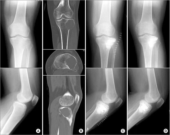
Dev hücreli kemik tümörü çoğunlukla iskelet olgunluğuna erişmiş genç erişkinlerde uzun kemik epifiz-metafizinde gelişen, lokal olarak agresif primer kemik tümörüdür. Kontrastsız BT, lezyonun litik yapısını, kortikal incelme veya yıkımı, subartiküler kemik desteğini ve patolojik kırığı gösterir; yumuşak doku ve tümör içi kanama için MR daha ayrıntılıdır. Distal femur, proksimal tibia ve distal radius sık bölgeler olduğundan eklem yüzüne uzanım özellikle aranmalıdır. Kortikal perforasyonun veya lezyon içindeki kırığın atlanması tümör yayılımını ve eklem çevresi kemik bütünlüğünü yanlış değerlendirmeye yol açabilir.
Faz ve pencere
- Kontrastsız tanısal
- Kemik algoritmalı kontrastsız BT'de epifizden metafize uzanan, eksantrik ve litik kemik kaybı; iç mineralize matriks olmaksızın kortikal incelme, ekspansiyon veya perforasyon görülebilir. Subartiküler kortikal desteği, kırık hattını ve yumuşak doku konturunu belirlemek için ince kesitler ile multiplanar rekonstrüksiyon kullanılır; kemik destrüksiyonunun gösterilmesi için intravenöz kontrast gerekli değildir.
Önerilen pencereler: Kemik (G 1800 / M 400), Yumuşak doku (G 400 / M 40).
BT bulguları
- Eksantrik epifiz-metafiz osteolizi — lezyon uzun kemiğin bir kenarından gelişir ve eklem altı epifize ulaşabilir.
- Keskin fakat sklerotik olmayan sınır — geçiş zonu çoğu kez belirgindir, ancak lezyon çevresinde kalın sklerotik halka tipik değildir.
- Kortikal incelme ve ekspansiyon — tümör korteksi dışa doğru şişirip ince kabuk haline getirebilir.
- Kortikal perforasyon — dış kemik sınırında kesinti ve periost dışına taşan tümör komponenti yerel agresifliği gösterir.
- Subartiküler uzanım — lezyon subkondral kemiğe dayanabilir; BT'de eklem yüzü altındaki ince kemik desteği izlenir.
- Patolojik kırık — incelmiş korteksten geçen kesinti, eklem çevresinde çökme veya deplasman eşlik edebilir.
- Sekonder anevrizmal kemik kisti değişikliği — kanama/kistik alanlar ve sıvı-sıvı seviyeleri eşlik edebilir; sıvı seviyeleri en iyi MR'da değerlendirilir.
- Mineralize tümör matriksinin yokluğu — belirgin osteoid ya da kondroid kalsifikasyon başka tanıları düşündürür.
Ölçütler ve sınıflamalar
- Campanacci radyografik sınıflaması
- Campanacci radyografik derecelendirmesi (histolojik derece veya TNM evresi değildir): Derece I — iyi sınırlı lezyon, ince olgun sklerotik kenar ve korteks tutulumu olmaması. Derece II — sınırları belirgin olmakla birlikte çevresel skleroz yoktur; korteks incelmış ve genişlemiştir ancak perforasyona uğramamıştır. Derece III — sınırlar belirsiz/agresiftir; kortikal yıkım ve kemik dışına taşan yumuşak doku kitlesi görülebilir.
Normalde
Normal distal femur epifiz-metafizinde trabeküler yapı subkondral plak boyunca sürer, kemik dış konturu düzgün ve korteks kesintisizdir; fokal eksantrik lusensi, ekspansiyon ya da patolojik kırık görülmez. Lezyonun eklem yüzüne olan mesafesi ve lezyonu saran etkilenmemiş kortikal kemik kalınlığı, yerel tekrar riski açısından BT'de değerlendirilmelidir.
Normal BT ile kıyasla
Aynı bölgenin, kaynak veri setinde patoloji içermediği belirtilen bir BT incelemesini kesit kesit kaydırın; organ sınırlarını açarak anatomiyi hasta görüntüsüyle karşılaştırın. Küçük rastlantısal bulgular tümüyle dışlanmamıştır.
Kaynak: TotalSegmentator v2.01 veri seti (Wasserthal ve ark., Radiology: Artificial Intelligence 2023), CC BY 4.0, vaka s0685; organ sınırları veri setinin otomatik segmentasyonundan, birleştirilerek sadeleştirildi. Görüntüler 2,5 mm kesit aralığına yeniden örneklendi.
Ayırıcı tanı
- Anevrizmal kemik kisti
- çoğunlukla daha genç hastalarda ilk iki dekatta görülen, metafiz bölgesinde yerleşimli, ekspansif ve septalı lezyonlardır; MR'da belirgin sıvı-sıvı seviyeleri karakteristik olmakla birlikte tanıyı tek başına kesinleştirmez.
- Telanjiektatik osteosarkom
- kanamalı kistik alanlar ve sıvı seviyeleriyle taklit edebilir; kalın nodüler solid komponent, agresif kortikal yıkım ve yumuşak doku kitle görünümü aranır.
- Kondroblastom
- genellikle açık büyüme plağı bulunan genç hastada epifizde sınırlı lezyondur; kenar sklerozu ve yaş/iskelet olgunluğu GCT'den ayrımda kullanılır.
- Hiperparatiroidiye bağlı kahverengi tümör
- litik kemik lezyonu olarak görünebilir; çoklu odaklar ve metabolik kemik hastalığı bağlamı ile ayrılır.
- Subartiküler dejeneratif kist
- eklem yüzüne komşu küçük subkondral boşluktur; eklem dejenerasyonu eşlik eder ve epifizden metafize uzanan ekspansil kitle oluşturmaz.
- Osteolitik metastaz veya plazmasitom
- litik odak yapabilir; yaş, bilinen malignite, çoklu odaklar ve medüller yayılım dağılımı ayırıcıdır.
Tuzaklar
- Sıvı-sıvı seviyeleri dev hücreli tümörün içindeki sekonder kistik değişiklikten kaynaklanabilir ve primer anevrizmal kist anlamına gelmez; altta yatan solid litik lezyonu BT'de yeniden arayın.
- İnce subkondral korteks aksiyel BT'de parsiyel hacim etkisiyle kaybolabilir; koronal/sagittal kemik rekonstrüksiyonlarda eklem yüzü altındaki kemik desteğini izleyin.
- Periostal reaksiyon veya Codman üçgeni genellikle patolojik kırıkla ilişkilendirilir; bu nedenle kortikal bütünlüğün devam ettiğini doğrulamak için ince kesitleri gözden geçirin.
- Açık fizisli çocukta saf epifiz yerleşimli lezyon klasik dev hücreli tümör dağılımına uymaz; yaş ve büyüme plağı durumunu anatomik örüntüyle birlikte yorumlayın.
Kendini dene
İskelet olgunluğunu tamamlamış kişide distal femurda eksantrik litik lezyon subkondral kemiğe uzanıyor; belirgin sklerotik halka yok. En olası tanı nedir?
Cevabı göster
Dev hücreli kemik tümörü. İskelet olgunluğu sonrası epifiz-metafize uzanan, eksantrik ve sklerotik kenarı olmayan litik lezyon dev hücreli tümör için tipiktir. Kondroblastom açık fizisli gençte daha sınırlı epifiz odağıdır; anevrizmal kist çoğunlukla metafizer ekspansil lezyon, dejeneratif kist ise eklem artropatisine eşlik eden küçük subkondral boşluktur.
Epifiz-metafiz litik tümörü içinde BT'de sıvı-sıvı seviyeleri görülüyor. Bu bulgu hangi ek değişiklikle açıklanabilir?
Cevabı göster
Sekonder anevrizmal kist. Dev hücreli tümör içinde sekonder anevrizmal kist değişikliği veya kanama sıvı-sıvı seviyelerine yol açabilir. Seviyeler tek başına primer kisti kanıtlamaz; enfarkt, osteoid osteom ve enkondrom bu ekspansil kanamalı kistik görünümü açıklamaz.
Kaynaklar
Bu sayfadaki 43 cümle ve 2 quiz sorusu aşağıdaki kaynaklardan alıntılarla tek tek karşılaştırıldı; 4 cümle bu karşılaştırmada düzeltildi (2026-10-08).
- State of the Art and New Concepts in Giant Cell Tumor of Bone: Imaging Features and Tumor Characteristicspmc.ncbi.nlm.nih.gov
- Giant Cell Tumour of Bone: A Comprehensive Review of Pathogenesis, Diagnosis, and Treatmentpmc.ncbi.nlm.nih.gov
- Locally Aggressive Connective Tissue Tumors.pubmed.ncbi.nlm.nih.gov
- Giant cell tumor of bone.pubmed.ncbi.nlm.nih.gov
- Chemotherapy for bone sarcomas in adults: the MD anderson experience.pubmed.ncbi.nlm.nih.gov
- The clinical approach toward giant cell tumor of bone.pubmed.ncbi.nlm.nih.gov
- Role of (Neo)adjuvant Denosumab for Giant Cell Tumor of Bone.pubmed.ncbi.nlm.nih.gov
- Multiple primary tumors: a case report and review of the literature.pubmed.ncbi.nlm.nih.gov
- Giant Cell Tumour of Bone: A Comprehensive Review of Pathogenesis, Diagnosis, and Treatment.pubmed.ncbi.nlm.nih.gov
- Giant Cell Tumor of the Acromion: Case Report and Literature Review.pubmed.ncbi.nlm.nih.gov
- State of the Art and New Concepts in Giant Cell Tumor of Bone: Imaging Features and Tumor Characteristics.pubmed.ncbi.nlm.nih.gov
- A High-grade Sarcoma Arising in a Patient With Recurrent Benign Giant Cell Tumor of the Proximal Tibia While Receiving Treatment With Denosumab.pubmed.ncbi.nlm.nih.gov
- Morphologic Changes After Denosumab Therapy in Patients with Giant Cell Tumor of the Spine: Report of Four Cases and a Review of the Literature.pubmed.ncbi.nlm.nih.gov
- Giant Cell Tumor of the Proximal Fibula in a 15-year-old Female: A Review and Case Report.pubmed.ncbi.nlm.nih.gov
- Benign Osseous Tumors and Tumor-Like Conditions.pubmed.ncbi.nlm.nih.gov
- Low-grade Central Osteosarcoma with High-grade Transformation in the Distal Tibia: A Rare Case Report.pubmed.ncbi.nlm.nih.gov
- Surgically Treated Benign Bone Tumors and Tumor-like Conditions in the Pediatric Population-A 10-Year Institutional Experience.pubmed.ncbi.nlm.nih.gov
- A Novel, Joint Sparing Surgical Approach to the Management of Giant Cell Tumor of the Distal Radius in a Pediatric Patient: A Case Report and Comprehensive Literature Review.pubmed.ncbi.nlm.nih.gov
- Sacral benign notochordal cell tumor with atypical imaging features and 16-year spontaneous partial regression: A case report.pubmed.ncbi.nlm.nih.gov
- [Primary bone tumors of the spine].pubmed.ncbi.nlm.nih.gov
- Conventional osteosarcoma of the ilium masquerading as a brown tumor in primary hyperparathyroidism: a rare case report and literature review.pubmed.ncbi.nlm.nih.gov
- Giant Cell Tumor of the Distal Ulna With Contiguous Involvement of the Distal Radius: A Report of a Rare Case.pubmed.ncbi.nlm.nih.gov
- Unnoticed Giant Cell Tumor of the Left Distal Tibia in a 52-Year-Old Male: A Rare Case Report.pubmed.ncbi.nlm.nih.gov
- A Rare Case of Giant Cell tumor of Bone in Distal Tibia.pubmed.ncbi.nlm.nih.gov
- Recurrent Giant Cell Tumor of the Lateral Condyle of the Distal Femur Managed by Repeated Extended Curettage and Bone Cementing: A Case Report.pubmed.ncbi.nlm.nih.gov
- Posttraumatic intradiploic cyst-like lesion of the frontal bone without cortical disruption: A case report.pubmed.ncbi.nlm.nih.gov
- Mandibular Brown Tumor as a Result of Secondary Hyperparathyroidism-Radiological and Clinical Pitfalls and Dilemmas.pubmed.ncbi.nlm.nih.gov
- Proximal Tibial Giant Cell Tumor in a Skeletally Immature Child: A Diagnostic Dilemma and Review of the Literature.pubmed.ncbi.nlm.nih.gov
- Secondary Malignant Transformation of Giant Cell Tumor of Bone Nine Years After Initial Curettage: A Case Report and Literature Review.pubmed.ncbi.nlm.nih.gov
- Images of giant cell tumor and chondroblastoma around the knee: retrospective analysis of 99 cases.pubmed.ncbi.nlm.nih.gov
- Imaging modalities for non-acute pathologies of the foot and ankle.pubmed.ncbi.nlm.nih.gov
- Updates in postoperative imaging modalities following musculoskeletal surgery.pubmed.ncbi.nlm.nih.gov
- Asymptomatic nonossifying fibroma-like lesions in neurofibromatosis type 1 patients.pubmed.ncbi.nlm.nih.gov
- Diagnosis and treatment of juvenile yellow granuloma in a child: A case report.pubmed.ncbi.nlm.nih.gov
- Intraosseous tumours of the knee.pubmed.ncbi.nlm.nih.gov
- Diagnostic and Treatment Strategies for Adult Non-Ossifying Fibromas: A Case Report and Literature Review.pubmed.ncbi.nlm.nih.gov
- Retrospective Study of Multimodality Imaging Features of Chondroblastoma.pubmed.ncbi.nlm.nih.gov
- Diagnosis and Current Treatment of Aneurysmal Bone Cysts.pubmed.ncbi.nlm.nih.gov
- Transphyseal Proximal Humeral Aneurysmal Bone Cyst with Pathologic Fracture in a Child: A Case Report.pubmed.ncbi.nlm.nih.gov
- Proximal fibulectomy in the management of proximal fibular aneurysmal bone cyst: technical considerations and outcome; a retrospective case series.pubmed.ncbi.nlm.nih.gov
- Weed out the Unwanted: Endosurgical Management of a Foreign Body with Cystic Lesion.pubmed.ncbi.nlm.nih.gov
- Peripheral Giant Cell Granuloma Mimicking Central Giant Cell Granuloma in a Pediatric Patient: A Diagnostic Challenge.pubmed.ncbi.nlm.nih.gov
- Myxoma Unveiled: Insights from Central and Peripheral Variants through Two Rare Cases.pubmed.ncbi.nlm.nih.gov
- Beyond the Tooth-Bearing Zone: Unusual Presentation of an Odontogenic Fibromyxoma in the Mandibular Condyle.pubmed.ncbi.nlm.nih.gov
- Clinical results of knee juxta-articular giant-cell tumors treated with bone cement filling and internal fixation after extensive curettage.pubmed.ncbi.nlm.nih.gov
- Malignancy-mimicking osteitis fibrosa cystica secondary to primary hyperparathyroidism.pubmed.ncbi.nlm.nih.gov
- Brown's Tumor Presenting as a Pathological Humerus Fracture: A Rare Diagnostic Pitfall.pubmed.ncbi.nlm.nih.gov
- Acetabular Giant Cell Tumor Presenting with Protrusio Acetabuli: A Rare Case Report.pubmed.ncbi.nlm.nih.gov
- Clival Aneurysmal Bone Cyst With Cavernous Sinus Extension: A Diagnostic Challenge.pubmed.ncbi.nlm.nih.gov
- Use of Fibrin Sealant Combined With Demineralized Freeze-Dried Bone Allograft in the Management of a Recurrent Mandibular Aneurysmal Bone Cyst: A Case Report.pubmed.ncbi.nlm.nih.gov
- Imaging features and differential diagnosis of benign osseous cystic lesions: Aneurysmal bone cyst versus femoral neck herniation pit.pubmed.ncbi.nlm.nih.gov
- Protuberant fibro-osseous lesion ("Bullough bump") presenting as a large occipital mass in a patient with cervical spinal stenosis.pubmed.ncbi.nlm.nih.gov
- Beyond Chordoma: A Comprehensive Review of Sacral Lesions.pubmed.ncbi.nlm.nih.gov
- Biomaterials at the interface of bone-derived factors and inter-organ communication: Current evidence and future perspectives.pubmed.ncbi.nlm.nih.gov
- Post-infection physeal bar resection of the proximal tibia: a case report and narrative review of the literature.pubmed.ncbi.nlm.nih.gov
- Combining polydioxanone-based scaffold and allogeneic adipose-derived mesenchymal stem cells enhances articular cartilage repair - a pre-clinical study.pubmed.ncbi.nlm.nih.gov
- Three-dimensional-printed porous implant combined with autograft reconstruction for giant cell tumor in proximal tibia.pubmed.ncbi.nlm.nih.gov
- Safety and Efficacy of Unicondylar Knee Prosthesis Treatment for Unicompartmental Osteoarthritis of the Knee Joint.pubmed.ncbi.nlm.nih.gov
- Recurrence of Giant Cell Tumor of Bone and Related Risk Factors: A Retrospective Analysis of 340 Cases From a Single Center.pubmed.ncbi.nlm.nih.gov
- Dual-target neoadjuvant therapy with denosumab and sunitinib for giant cell tumor of bone: a sarcoma center experience.pubmed.ncbi.nlm.nih.gov
- Giant Cell Tumor of the Sacrum: A Narrative Review of Management Challenges and Modalities.pubmed.ncbi.nlm.nih.gov
- Free Fibula Flap Arthrodesis After En Bloc Resection of Distal Radius Giant Cell Tumors: Functional and Oncologic Outcomes From a Single-institution Experience.pubmed.ncbi.nlm.nih.gov
- Surgical and radiological outcomes of giant cell tumor of the bone: prognostic value of Campanacci grading and selective use of denosumab.pubmed.ncbi.nlm.nih.gov
- Functional Outcome and Complications of Wide Excision of the Distal End of Radius and Reconstruction with Ulnar Transposition in Campanacci Grade III Giant Cell Tumor: A Longitudinal Study of 14 Cases in South India.pubmed.ncbi.nlm.nih.gov
- The best solution is the simplest: advances in surgical and minimally invasive management of aneurysmal and simple bone cysts.pubmed.ncbi.nlm.nih.gov
- Exploratory quantitative magnetic resonance imaging characterization of histopathologic subtype heterogeneity in osteosarcoma.pubmed.ncbi.nlm.nih.gov
- Aneurysmal Bone Cyst of Calcaneum in a Young Adult: A Rare Case Report.pubmed.ncbi.nlm.nih.gov
- Rare giant cell tumour of the left parieto-occipital skull in an 8-year-old female patient: a case report.pubmed.ncbi.nlm.nih.gov
- Current Position of Nuclear Medicine Imaging in Primary Bone Tumors.pubmed.ncbi.nlm.nih.gov
- High-resolution ultrasound and MRI in the evaluation of the forefoot and midfoot.pubmed.ncbi.nlm.nih.gov
- Cyst formation in the subchondral bone following cartilage repair.pubmed.ncbi.nlm.nih.gov
- Staged Surgical Resection of Giant Multicentric Cranial Plasma Cell Neoplasms: A Case Report.pubmed.ncbi.nlm.nih.gov
- Decoding calvarial lesions: Contemporary neurosurgical management integrating radiological and histopathological features.pubmed.ncbi.nlm.nih.gov
- Axial Spondyloarthritis: Mimics and Pitfalls of Imaging Assessment.pubmed.ncbi.nlm.nih.gov
- Glossary of terms for musculoskeletal radiology.pubmed.ncbi.nlm.nih.gov
- International Harmonization of Nomenclature and Diagnostic Criteria (INHAND): Non-proliferative and Proliferative Lesions of the Non-human Primate ( M. fascicularis ).pubmed.ncbi.nlm.nih.gov
- Multimodal imaging in the diagnosis of bone giant cell tumors: A retrospective study.pubmed.ncbi.nlm.nih.gov
- A Pattern-based Imaging Approach to Pediatric Jaw Lesions.pubmed.ncbi.nlm.nih.gov
- Radiological features of central giant cell granuloma: comparative study of 7 cases and literature review.pubmed.ncbi.nlm.nih.gov
- Chondroblastoma of the femoral head: curettage without dislocation.pubmed.ncbi.nlm.nih.gov
- Imaging Findings of Pediatric Rheumatic Disorders: JIA in the PRINTO Era and Autoimmune Interferonopathies.pubmed.ncbi.nlm.nih.gov
- Long term outcome of surgical treatment of chondroblastoma: analysis of local control and growth plate/articular cartilage related complications.pubmed.ncbi.nlm.nih.gov
- Bone lesions of the tibia: Multimodal iconographic review and diagnostic algorithms, Part 2: Metaphyseal and epiphyseal lesions.pubmed.ncbi.nlm.nih.gov
- Primary Limb Leiomyosarcoma With Multifocal Musculoskeletal Soft Tissue Metastasis: A Case Report and Literature Review.pubmed.ncbi.nlm.nih.gov
- Patient-specific osteoarticular reconstruction of the proximal tibia for giant cell tumor using a 3D-printed trabecular implant and hinged knee arthroplasty: a case report.pubmed.ncbi.nlm.nih.gov
- Phosphaturic Mesenchymal Tumor: From Invisibility to Bedridden Morbidity.pubmed.ncbi.nlm.nih.gov
- Histopathological and radiographic characteristics of central giant cell granulomas: a retrospective study of 45 cases.pubmed.ncbi.nlm.nih.gov
- Case Report: Arthroscopic management of a giant Baker cyst containing numerous rice body-like loose bodies in a patient with rheumatoid arthritis.pubmed.ncbi.nlm.nih.gov
- Pseudomyogenic hemangioendothelioma of the thoracolumbar spine: A case-based review of a rare cause of vertebral collapse and neurological deficit.pubmed.ncbi.nlm.nih.gov
- Optimal management of giant cell tumor in the tibial plateau: A case report on curettage and cement reinforcement.pubmed.ncbi.nlm.nih.gov
- Chondrosarcoma arising from long-standing Dysplasia Epiphysealis Hemimelica of the proximal humerus: A case report.pubmed.ncbi.nlm.nih.gov
- Non-Ossifying Fibromas: A 2025 Review.pubmed.ncbi.nlm.nih.gov
- Review of imaging modalities and radiological findings of calvarial lesions.pubmed.ncbi.nlm.nih.gov
- Diffuse-Type Tenosynovial Giant Cell Tumor of the Hip With Acetabular Bone Involvement: A Case Report With Radiologic-Pathologic Correlation.pubmed.ncbi.nlm.nih.gov
- Autopsy-Confirmed Giant Pleural Hematoma Due to Spontaneous Hemothorax in ALK-Rearranged and TP53-Mutant Lung Adenocarcinoma: A Case Report.pubmed.ncbi.nlm.nih.gov
- Intralesional resection and autologous fibular graft reconstruction for a giant cell tumor of the fourth metacarpal with a secondary aneurysmal bone cyst: a case report.pubmed.ncbi.nlm.nih.gov
- Management of aneurysmal bone cysts of the spine and sacrum: A narrative review.pubmed.ncbi.nlm.nih.gov
- A rare calvarial aneurysmal bone cyst in an adult: Case report and literature review.pubmed.ncbi.nlm.nih.gov
- Benign bone and soft tissue tumors of the foot.pubmed.ncbi.nlm.nih.gov
- Benign incidental do-not-touch bone lesions.pubmed.ncbi.nlm.nih.gov
- Target-Specific Extracorporeal Shockwave Therapy Modulates TLR4/NF-κB/MMP13 Signaling Within the Joint Microenvironment to Improve Osteochondral Pathology in Patellofemoral Osteoarthritis.pubmed.ncbi.nlm.nih.gov
- Temporal progression of bone loss, trabecular microarchitectural deterioration, and mechanical impairment in a rat model of collagen-induced arthritis.pubmed.ncbi.nlm.nih.gov
- Spatiotemporal mapping of osteomyelitis pathogenesis guides the design of functionally graded bone regeneration scaffolds.pubmed.ncbi.nlm.nih.gov
- Giant Cell Tumor of the Distal Humerus: A Rare Location With a Good Response to Denosumab.pubmed.ncbi.nlm.nih.gov
- Clinical and Radiological Concordance of Bone Tumours Using Histopathology as the Gold Standard: A Pilot Study.pubmed.ncbi.nlm.nih.gov
- Risk Factors for Pathologic Fracture in Dogs With Confirmed or Suspected Appendicular Osteosarcoma Treated With Low- or Standard-Dose-Rate Palliative External Beam Radiation Therapy.pubmed.ncbi.nlm.nih.gov
- Giant expansile humeral lesion with soap-bubble appearance mimicking a primary bone tumor: Multiple myeloma as the final diagnosis.pubmed.ncbi.nlm.nih.gov
- Low-grade osteogenic tumor with secondary aneurysmal bone cyst: Distinguishing osteoblastoma from low-grade osteosarcoma in the setting of noncanonical molecular findings.pubmed.ncbi.nlm.nih.gov
- In Situ Profiling of Nanoscale Strains Uncovers Mechano-Architectural Predictors of Aging and Osteoarthritis Emergence.pubmed.ncbi.nlm.nih.gov
- Autologous chondrocyte implantation in osteochondral defects of the capitulum: clinical and radiological outcome in a case series.pubmed.ncbi.nlm.nih.gov
- The arrow and the target-description of the "Robin Hood" technique for fixation of tibial spine avulsion fractures: a technical note.pubmed.ncbi.nlm.nih.gov
- Using microwave ablation in combination with curettage and reconstruction with a vascularized iliac bone graft guided by a three-dimensional printed model for the treatment of a grade III talar giant cell tumor: a case report.pubmed.ncbi.nlm.nih.gov
Eğitim amaçlıdır; hasta başında tanı ve tedavi kararının yerine geçmez. İçerik yapay zekâ destekli bir süreçle güncel literatüre göre cümle cümle kaynaklarla karşılaştırıldı, ancak uzman radyolog incelemesinden geçmedi. Klinik kararlarda güncel kılavuzları, kurum protokollerini ve radyoloji raporunu esas alın.