
Özet
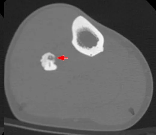
Osteoid osteom çoğunlukla çocuk ve genç erişkinlerde görülen, küçük niduslu benign osteojenik tümördür; tipik olarak geceleri artabilen fokal ağrıya yol açar. İnce kesit kontrastsız BT, özellikle kortikal lezyonda nidusu ve olası mineralize merkezini radyografiden veya MR'daki yaygın ödemden daha doğrudan gösterir. Femur ve tibia sık yerleşimlerdir; spinal tutulumda nidus genellikle posterior elemanlarda, pedikül veya faset komşuluğunda aranır. Nidusun atlanması reaktif ödemin yanlışlıkla tüm lezyon sanılmasına ve tanının gecikmesine neden olabilir.
Faz ve pencere
- Kontrastsız tanısal
- İnce kesit kemik rekonstrüksiyonlarında korteks içinde yuvarlak veya oval, düşük atenüasyonlu nidus; içinde noktasal, amorf ya da halkasal mineralizasyon ve çevresinde değişen derecede reaktif skleroz görülebilir. İnce kesit kontrastsız BT, özellikle yoğun reaktif skleroz içine gömülü olan küçük radyolüsent nidusu en belirgin şekilde gösterdiği için tanısal altın standarttır.
Önerilen pencereler: Kemik (G 1800 / M 400), Yumuşak doku (G 400 / M 40).
BT bulguları
- Kortikal nidus — düzgün sınırlı, yuvarlak veya oval küçük radyolüsent odak, kalınlaşmış korteks içinde seçilir.
- Nidus mineralizasyonu — noktasal, amorf ya da halkasal yoğunluk nidusun merkezinde veya içinde yer alır.
- Reaktif kortikal skleroz — nidusu çevreleyen kalınlaşmış, yoğun korteks özellikle uzun kemik diyafizinde belirgindir.
- Periostal yeni kemik — uzunlamasına kortikal kalınlaşma ile birlikte düzgün periostal kemik oluşumu görülebilir.
- Vasküler oluk — nidusa doğru uzanan ince lineer/serpijinöz kortikal kanal besleyici damar yolunu gösterebilir.
- Posterior eleman odağı — omurga tutulduğunda nidus pedikül, lamina veya faset komşuluğunda; çevre sklerozu aynı tarafta bulunabilir.
- Eklem yakınındaki nidus — eklem içi/juxtaartiküler yerleşimde belirgin kortikal skleroz az olabilir; çevresel reaktif değişiklik nidusu örtebilir.
Normalde
Aynı seviyedeki normal femur korteksi düzgün kalınlıkta, kesintisiz ve çevre medullayla düzenli geçişlidir; fokal lusensi, noktasal mineralize merkez veya çevresel sklerotik kalınlaşma görülmez. Tanısal ince kesit BT, yoğun reaktif skleroz içine gömülü olan küçük radyolüsent nidusu ve merkezdeki kalsifikasyonu diğer modalitelere göre çok daha belirgin şekilde gösterir.
Normal BT ile kıyasla
Aynı bölgenin, kaynak veri setinde patoloji içermediği belirtilen bir BT incelemesini kesit kesit kaydırın; organ sınırlarını açarak anatomiyi hasta görüntüsüyle karşılaştırın. Küçük rastlantısal bulgular tümüyle dışlanmamıştır.
Kaynak: TotalSegmentator v2.01 veri seti (Wasserthal ve ark., Radiology: Artificial Intelligence 2023), CC BY 4.0, vaka s0685; organ sınırları veri setinin otomatik segmentasyonundan, birleştirilerek sadeleştirildi. Görüntüler 2,5 mm kesit aralığına yeniden örneklendi.
Ayırıcı tanı
- Osteoblastom
- nidus benzeri osteoid yapım görülebilir; ancak lezyon daha hacimli ve sıklıkla eksenel kemiklerde ekspansildir, belirgin reaktif skleroz daha az tipiktir.
- Brodie apsesi
- alt akut enfeksiyon nedeniyle intraosseöz kavite ve çevresel skleroz yapabilir; tanıda klinik tablo ve MR'daki ödem/kavite görünümü BT bulgularından ayırt edici olabilir.
- Stres reaksiyonu veya stres kırığı da kortikal kalınlaşma yapabilir; bu durumda lezyonda nidus yerine kırık hattı ve periostal reaksiyon aranır.
- Osteoblastik metastaz
- yoğun sklerotik odak oluşturabilir; tek odaklı kortikal reaksiyon ve nidus yerine çoklu medüller odaklar ya da bilinen bir primer malignite öyküsü daha olasıdır.
Tuzaklar
- MR'da geniş kemik iliği ve yumuşak doku ödemi ile skleroz ön planda çıkabilir; bu durumda ince kesit BT nidusu ve çevre ilişkisini kesin olarak ortaya koyar.
- MR'daki geniş kemik iliği ve yumuşak doku ödemi küçük nidustan çok daha çarpıcı olabilir; BT'de odaklanmış kortikal inceleme tanıya götüren nidusu ortaya çıkarabilir.
- Spinal osteoid osteomda vertebra cismi yerine posterior elemanları ve faset çevresini inceleyin; komşu faset artropatisi tek başına nidus kabul edilmemelidir.
Kendini dene
Genç erişkinde gece ağrısı var. Femur korteksindeki yoğun kalınlaşmanın merkezinde mineralize küçük bir nidus görülüyor. En olası tanı hangisidir?
Cevabı göster
Osteoid osteom. Kortikal nidus ve çevresindeki belirgin reaktif kemik yanıtı osteoid osteomu destekler. Brodie apsesi kaviter enfeksiyon odağıdır; osteoblastom genellikle daha hacimli ekspansil lezyondur, stres kırığında ise nidus yerine kırık hattı beklenir.
Bir omurga BT'sinde tek taraflı faset komşuluğunda küçük radyolüsent nidus ve çevresel skleroz var. Öncelikle hangi lezyon düşünülür?
Cevabı göster
Osteoid osteom. Posterior eleman veya faset çevresindeki küçük radyolüsent nidus ve yerel skleroz osteoid osteom lehinedir. Faset osteoartriti eklem daralması ve osteofit ile karakterizedir; vertebra korpusunda hemanjiyom veya metastaz gibi odaklar ayırıcı tanıda değerlendirilir.
Kaynaklar
Bu sayfadaki 36 cümle ve 2 quiz sorusu aşağıdaki kaynaklardan alıntılarla tek tek karşılaştırıldı; 11 cümle bu karşılaştırmada düzeltildi (2026-10-08).
- Presentation of osteoid osteoma in the fifth metatarsal: a case report and review of the literaturepmc.ncbi.nlm.nih.gov
- Intraosseous osteoid osteoma of the temporomandibular joint: a case report and review of the literaturepmc.ncbi.nlm.nih.gov
- Interventional Radiology in the Treatment of Musculoskeletal Tumourspmc.ncbi.nlm.nih.gov
- Arthroscopic treatment of osteoid osteoma in the posterior proximal tibia: A case report and literature reviewpmc.ncbi.nlm.nih.gov
- Osteoid Osteoma, a Diagnostic Problem: A Series of Atypical and Mimicking Presentations and Review of the Recent Literaturepmc.ncbi.nlm.nih.gov
- Osteoid osteoma.pubmed.ncbi.nlm.nih.gov
- Bone and soft tissue tumors of hip and pelvis.pubmed.ncbi.nlm.nih.gov
- Pediatric Musculoskeletal Interventional MRI.pubmed.ncbi.nlm.nih.gov
- Bony sequestrum: a radiologic review.pubmed.ncbi.nlm.nih.gov
- Imaging Analyses of Bone Tumors.pubmed.ncbi.nlm.nih.gov
- Image-Guided Spinal Ablation: A Review.pubmed.ncbi.nlm.nih.gov
- Osteoid osteoma of the hip: imaging features.pubmed.ncbi.nlm.nih.gov
- Osteoid osteoma: a pictorial review.pubmed.ncbi.nlm.nih.gov
- [Osteoidosteoma. From diagnosis to treatment].pubmed.ncbi.nlm.nih.gov
- Temperature Monitoring in Hyperthermia Treatments of Bone Tumors: State-of-the-Art and Future Challenges.pubmed.ncbi.nlm.nih.gov
- Dactylitis: A pictorial review of key symptoms.pubmed.ncbi.nlm.nih.gov
- [Imaging of non-traumatic spinal diseases in children].pubmed.ncbi.nlm.nih.gov
- Osteoid osteoma and osteoblastoma.pubmed.ncbi.nlm.nih.gov
- Differential Diagnosis of Facet Joint Disorders.pubmed.ncbi.nlm.nih.gov
- What's new in pediatric bone sarcomas and benign bone tumors.pubmed.ncbi.nlm.nih.gov
- Surgically Treated Benign Bone Tumors and Tumor-like Conditions in the Pediatric Population-A 10-Year Institutional Experience.pubmed.ncbi.nlm.nih.gov
- Robot-assisted radiofrequency ablation versus open resection for osteoid osteoma: an analysis of perioperative outcomes and a rare case of malignant transformation.pubmed.ncbi.nlm.nih.gov
- An analysis of the imaging features and diagnostic value of osteoid osteoma on DR, CT, and MRI.pubmed.ncbi.nlm.nih.gov
- Risk factors for recurrence and complications of CT-guided radiofrequency ablation for the treatment of osteoid osteoma : a multi-institutional analysis.pubmed.ncbi.nlm.nih.gov
- Patterns and Timing of Local Recurrence in Benign Bone Tumors.pubmed.ncbi.nlm.nih.gov
- O-arm Navigation-assisted Excision of Cervical Osteoid Osteoma Adjacent to the Vertebral Artery: Technical Note and Case Report.pubmed.ncbi.nlm.nih.gov
- Full-endoscopic resection of a thoracic facet joint osteoblastoma: A case report.pubmed.ncbi.nlm.nih.gov
- Surgery of the tumors of the spine.pubmed.ncbi.nlm.nih.gov
- Osteoid osteoma of the femoral neck as a rare cause of hip pain in a 50-year-old male: A case report.pubmed.ncbi.nlm.nih.gov
- Interest of targeted computed tomography guided by scintigraphy in the diagnosis of an osteoid osteoma of the talus: A surgical case report.pubmed.ncbi.nlm.nih.gov
- Intra-articular Osteoid Osteoma of the Intermediate Cuneiform Bone Causing Articular Degeneration: A Rare Case and Literature Review.pubmed.ncbi.nlm.nih.gov
- Eight months of misdiagnosis: Intra-articular osteoid osteoma in a 48-year-old woman.pubmed.ncbi.nlm.nih.gov
- Osteoid osteoma of the triquetrum: A rare cause of ulnar-sided wrist pain in an adolescent female.pubmed.ncbi.nlm.nih.gov
- A case report of triquetral osteoid osteoma: from diagnostic challenge to complete recovery.pubmed.ncbi.nlm.nih.gov
- Imaging of Skull Base Tumors.pubmed.ncbi.nlm.nih.gov
- Imaging of pediatric calvarial and skull base tumors: A COG Diagnostic Imaging Committee/SPR Oncology Committee/ASPNR White Paper.pubmed.ncbi.nlm.nih.gov
- Unveiling Primary Bone Tumors of the Spine: A Review of Essential Imaging Clues.pubmed.ncbi.nlm.nih.gov
- CT-guided radiofrequency ablation of a tibial osteoid osteoma in pregnancy.pubmed.ncbi.nlm.nih.gov
- Intra-articular Osteoid Osteoma of the Femoral Neck Mimicking Inflammatory Hip Disease: A Case Report and Review of the Literature.pubmed.ncbi.nlm.nih.gov
- Low-grade osteogenic tumor with secondary aneurysmal bone cyst: Distinguishing osteoblastoma from low-grade osteosarcoma in the setting of noncanonical molecular findings.pubmed.ncbi.nlm.nih.gov
- Intramedullary osteosclerosis of the tibia with inflammatory features: Multimodality imaging findings.pubmed.ncbi.nlm.nih.gov
- Imaging Review of Pediatric Benign Osteocytic Tumors and Latest Updates on Management.pubmed.ncbi.nlm.nih.gov
- Arthroscopic Excision of Delayed Diagnosed Intra-articular Osteoid Osteoma of the Elbow: A Case Report.pubmed.ncbi.nlm.nih.gov
- Case report: Osteoid osteoma of the acetabulum treated with arthroscopy-assisted radiofrequency ablation.pubmed.ncbi.nlm.nih.gov
- Percutaneous calcium phosphate cement injection for unicameral bone cysts: mid- to long-term clinical and radiographic outcomes.pubmed.ncbi.nlm.nih.gov
- Diagnostic Imaging Analysis of Humerus Bone Structure in Long-Legged Buzzard (Buteo rufinus): Radiology and CT-Scan Insights.pubmed.ncbi.nlm.nih.gov
- Case Report: Total hip arthroplasty via the direct anterior approach using a cementless revision stem without corrective osteotomy for fibrous dysplasia.pubmed.ncbi.nlm.nih.gov
- Beyond Chordoma: A Comprehensive Review of Sacral Lesions.pubmed.ncbi.nlm.nih.gov
- Current Position of Nuclear Medicine Imaging in Primary Bone Tumors.pubmed.ncbi.nlm.nih.gov
- Brodie's Abscess of the Thumb: A Rare Complication of a Dog Bite.pubmed.ncbi.nlm.nih.gov
- <i>Arcanobacterium haemolyticum</i> Chronic Osteomyelitis and Bacteremia Complicated by Intraosseous Abscess: A Case Report.pubmed.ncbi.nlm.nih.gov
- Coccidioides Immitis and Mssa Brodie's Abscess of The Calcaneus in The Setting of Pulmonary Valley Fever: A Case Report.pubmed.ncbi.nlm.nih.gov
- Conventional radiography for the assessment of focal bone lesions of the appendicular skeleton: fundamental concepts in the modern imaging era.pubmed.ncbi.nlm.nih.gov
- The most common bone tumors of the upper extremity in childhood and adolescence and their treatment: a review of the current literature.pubmed.ncbi.nlm.nih.gov
- Long-term safety and effectiveness of the "OptEase" vena cava filter.pubmed.ncbi.nlm.nih.gov
- Enigmatic Cases of Finger Proximal Interphalangeal Joint Swelling: Case Series and Focused Review of the Literature.pubmed.ncbi.nlm.nih.gov
- Imaging Findings of Pediatric Rheumatic Disorders: JIA in the PRINTO Era and Autoimmune Interferonopathies.pubmed.ncbi.nlm.nih.gov
- Unmasking the great imitators-noninfectious conditions masquerading as spinal tuberculosis in a developing country: A single-center case series analysis.pubmed.ncbi.nlm.nih.gov
- Anatomy and pathology of facet joint.pubmed.ncbi.nlm.nih.gov
- Bone lesions of the tibia: Multimodal iconographic review and diagnostic algorithms, Part 1: Diagnostic algorithms, dysplasia and diaphyseal lesions.pubmed.ncbi.nlm.nih.gov
- Presentation of osteoid osteoma in the fifth metatarsal: a case report and review of the literature.pubmed.ncbi.nlm.nih.gov
- Giant osteoid osteoma of mandible in a 24-year-old female: a case report.pubmed.ncbi.nlm.nih.gov
- Interventional Radiology in the Treatment of Musculoskeletal Tumours.pubmed.ncbi.nlm.nih.gov
- An osteoid osteoma coexisting with a ganglion cyst in the fifth metatarsal head: A rare case report.pubmed.ncbi.nlm.nih.gov
- Full-endoscopic pedicle-based approach for resection of osteoid osteoma in the lumbar spine: a case report and review of current literature.pubmed.ncbi.nlm.nih.gov
- The Effect of More than 50% Resection of the Inferior Articular Process on Spinal Stability in Unilateral Single-Segment Interlaminar Endoscopic Lumbar Discectomy.pubmed.ncbi.nlm.nih.gov
- Consensus practice guidelines on sacroiliac joint complex pain from a multispecialty, international working group.pubmed.ncbi.nlm.nih.gov
- SPECT/CT in the Evaluation of Suspected Skeletal Pathology.pubmed.ncbi.nlm.nih.gov
- A Lumbar Spinal Osteoid Osteoma Associated with Kyphoscoliosis, Head Drop, and Pelvic Obliquity: A Case Report.pubmed.ncbi.nlm.nih.gov
- Epiphyseal enchondroma masking as osteoid osteoma: a case report.pubmed.ncbi.nlm.nih.gov
- Painless osteoid osteoma of the distal phalanx of a toe.pubmed.ncbi.nlm.nih.gov
- Cortical Lytic Lesion of the Proximal Tibia in an 8-Year-Old Child: A Pediatric Diagnostic Conundrum.pubmed.ncbi.nlm.nih.gov
- Osteoid Osteoma Mimicking Brachial Plexopathy in a Child.pubmed.ncbi.nlm.nih.gov
- Computed tomography-guided radiofrequency ablation in the treatment of intra-articular proximal femoral osteoid osteoma.pubmed.ncbi.nlm.nih.gov
- A comparative study assessing the efficacy and safety of radiofrequency ablation versus surgical treatment for osteoid osteoma: retrospective analysis in a single institution.pubmed.ncbi.nlm.nih.gov
- Endoscopic resection for cervical and thoracic spinal osteoid osteoma: A case series.pubmed.ncbi.nlm.nih.gov
- Osteoid Osteoma of the Femoral Neck Masquerading as Avascular Necrosis: A Diagnostic Dilemma and Management Success Story.pubmed.ncbi.nlm.nih.gov
- Osteoblastoma of the Middle Phalanx of the Hand Initially Diagnosed as Melorheostosis.pubmed.ncbi.nlm.nih.gov
- Brodie's Abscess in a Pediatric Patient: A Diagnostic Pitfall.pubmed.ncbi.nlm.nih.gov
- Intraosseous tumours of the knee.pubmed.ncbi.nlm.nih.gov
Eğitim amaçlıdır; hasta başında tanı ve tedavi kararının yerine geçmez. İçerik yapay zekâ destekli bir süreçle güncel literatüre göre cümle cümle kaynaklarla karşılaştırıldı, ancak uzman radyolog incelemesinden geçmedi. Klinik kararlarda güncel kılavuzları, kurum protokollerini ve radyoloji raporunu esas alın.