
Özet
Tipik vertebral hemanjiyom, vertebra korpusu içinde yağlı ve damarsal bileşenlerle yeniden düzenlenmiş trabeküllerden oluşan, çoğu kez tesadüfen saptanan benign bir lezyondur. Kontrastsız BT'de kalınlaşmış trabeküllerin aksiyel polka-dot ve sagital/koronal corduroy görünümü tanıyı destekler; tipik lezyon korpus sınırları içinde kalır. Bu trabeküler deseni tanımak, özellikle kanser öyküsü olan hastada lezyonun yanlışlıkla metastaz sayılmasını önler. Kortikal genişleme, kemik dışına uzanım veya epidural doku varsa tipik görünümden uzaklaşılır ve agresif özellikler ayrıca araştırılır.
Faz ve pencere
- Kontrastsız tanısal
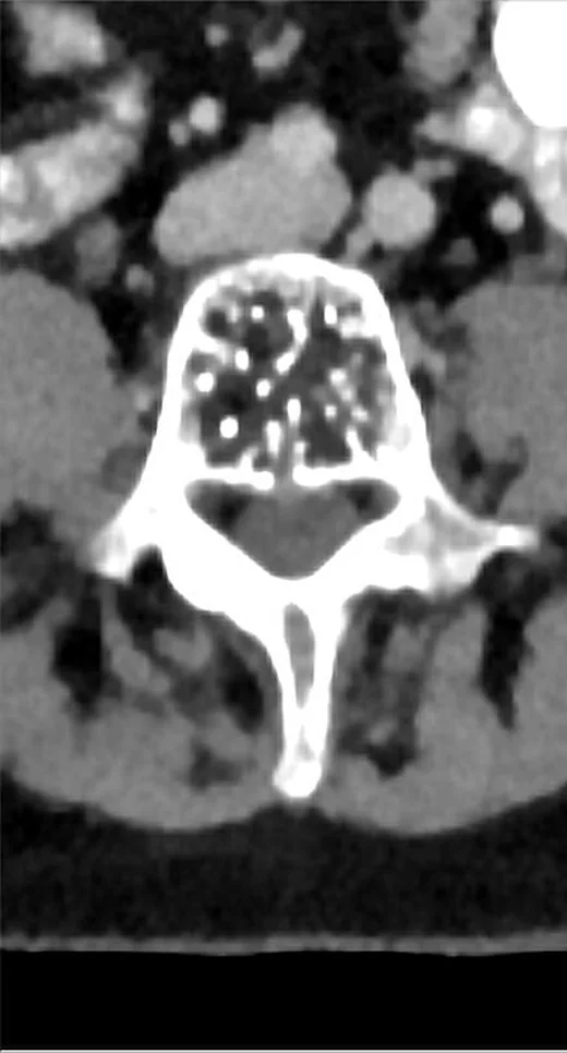
- Aksiyel BT'de düşük dansiteli stroma içinde noktasal yüksek dansiteli kalın trabeküller polka-dot görünümü verir; sagital/koronal planda dikey kalın trabeküller corduroy çizgilenmesi oluşturur. Tipik lezyonda vertebra konturu genişlemez ve ekstraosseöz kitle beklenmez; kontrast fazı tanı için gerekmez.
- Kemik algoritması tanısal
- BT’de kalınlaşmış trabeküller ve korteks değerlendirilebilir; sagital ve koronal BT görüntülerinde kalın trabeküllerin oluşturduğu corduroy görünümü de aranabilir.
Önerilen pencereler G genişlik, M merkez; değerler Hounsfield birimi (HU)
- KemikG 1800 / M +400
BT bulguları
- Polka-dot işareti — aksiyel kesitte hipodens stroma içindeki kalınlaşmış trabeküllerin kesit yüzleri dağınık yoğun noktalar gibi görünür.
- Corduroy işareti — sagital veya koronal planda kalın vertikal trabeküller paralel çizgilenme oluşturur.
- Yağlı stromaya bağlı görece düşük genel dansite — kalın trabeküller lezyonun daha düşük dansiteli zemini içinde seçilir.
- Korpus içi sınırlı yerleşim — tipik lezyon vertebra korpusu içinde kalır ve dış konturu genişletmez.
- Korunmuş korteks — agresif olmayan tipik hemanjiyomda kortikal destrüksiyon veya belirgin kortikal balonlaşma beklenmez.
- Ekstraosseöz komponent yokluğu — epidural veya paravertebral kitle tipik hemanjiyom için beklenen görünüm değildir.
- Önceki incelemelerde stabilite, hızlı büyüme olmadığını destekler; tanıda trabeküler yapı, yağ içeriği, korteks ve ekstraosseöz uzanım birlikte değerlendirilir.
- Tipik hemanjiyomda yağlı stroma nedeniyle MR’da T1 ve T2 sinyalleri genellikle yüksektir; atipik lezyonlarda yağ oranı azaldıkça T1 sinyali düşebilir.
Ölçütler ve sınıflamalar
- Görüntüleme görünümüne göre vertebral hemanjiyom
- Tipik lezyon yağ içeriği ve belirgin polka-dot/corduroy trabekül işaretleriyle; atipik lezyon daha az yağlı, daha vasküler ve işaretleri silik olabilir; agresif lezyonda kortikal genişleme/yıkım, posterior eleman veya kemik dışı yumuşak doku uzanımı görülebilir.
Normalde
Normal vertebra korpusunda trabeküller ince ve düzenli ağ oluşturur; kalın yoğun odaklar arasında polka-dot noktalanma veya sagital vertikal çizgilenme görülmez. Aynı korpusu komşu vertebralarla karşılaştırıp trabeküler kalınlaşmanın yerini, korteksin korunmasını ve lezyonun korpus dışına taşıp taşmadığını kontrol edin.
Normal BT ile kıyasla
Aynı bölgenin, kaynak veri setinde patoloji içermediği belirtilen bir BT incelemesini kesit kesit kaydırın; organ sınırlarını açarak anatomiyi hasta görüntüsüyle karşılaştırın. Küçük rastlantısal bulgular tümüyle dışlanmamıştır.
Kaynak: TotalSegmentator v2.01 veri seti (Wasserthal ve ark., Radiology: Artificial Intelligence 2023), CC BY 4.0, vaka s1338; organ sınırları veri setinin otomatik segmentasyonundan, birleştirilerek sadeleştirildi. Görüntüler 2,5 mm kesit aralığına yeniden örneklendi.
Ayırıcı tanı
- Metastaz vertebra korpusuyla sınırlı hemanjiyomun ayırıcı tanısında yer alır; tipik hemanjiyomda aksiyel polka-dot ve sagital/koronal corduroy görünümleri kalınlaşmış trabekülleri yansıtır.
- Kemik adacığı (enostoz)
- homojen yoğun kemik odağıdır; hipodens stroma içinde düzenli noktasal kalın trabeküller oluşturmaz.
- Atipik vertebral hemanjiyom
- yağ oranı daha düşük olduğu için klasik CT işaretleri belirgin olmayabilir; MR'da tipik yağlı sinyal örüntüsü de zayıflayabilir.
- Litik metastaz
- trabeküler kemik kaybı, kortikal erozyon veya yumuşak doku kitlesi tipik sınırlı hemanjiyom morfolojisine uymaz.
Tuzaklar
- Polka-dot işaretini tek bir yoğun nokta gibi yorumlamayın; çok sayıda nokta kalın trabeküllerin kesit görünümüdür ve aksiyel kemik penceresinde birlikte aranır.
- Klasik işaretlerin seçilmemesini hemanjiyomu dışlamak için kullanmayın; atipik veya agresif hemanjiyomlarda yağ azlığı ve trabekül kaybı tipik deseni silebilir.
- Kortikal dışa taşmayı tipik lezyonla uyumlu saymayın; genişleme, posterior eleman tutulumu veya kemik dışı kitle agresif özellikleri gündeme getirir.
- Kanser öyküsü bulunan hastada iyi huylu trabeküler deseni gözden kaçırıp odağı metastaz olarak etiketlemeyin; iç mimariyi ve eşlik eden destrüksiyonu beraber değerlendirin.
- Kanser öyküsü olan hastada vertebra korpusundaki lezyon metastaz ayırıcı tanısında yer alır; tipik hemanjiyom görünümü tanıyı desteklese de atipik veya agresif görünümler metastazı taklit edebilir.
Kendini dene
Aksiyel kontrastsız BT'de T11 korpusunda hipodens stroma içinde çok sayıda yoğun nokta, sagitalde kalın vertikal çizgiler ve korunmuş kontur var. En olası tanı nedir?
Cevabı göster
Tipik vertebral hemanjiyom. Polka-dot ve corduroy işaretleri kalınlaşmış trabeküllerin iki düzlemdeki görünümüdür ve tipik hemanjiyomu destekler. Aksiyel BT'de hipodens stroma içindeki noktasal yüksek dansiteli kalın trabeküller polka-dot görünümünü oluşturur ve tipik vertebral hemanjiyomu destekler.
Tipik vertebral hemanjiyom tanımlı lezyonda sonradan kortikal balonlaşma ve epidural yumuşak doku görülüyor. Kortikal balonlaşma ve epidural yumuşak doku uzanımı vertebral hemanjiyomda hangi davranış özelliğini düşündürür?
Cevabı göster
Agresif hemanjiyom. Kortikal genişleme ve epidural uzanım agresif hemanjiyom özellikleridir ve tipik, korpus içinde sınırlı görünümden farklıdır. Kemik adacığı ve osteoporoz epidural kitle yapmaz; bu görünüm normal varyant olarak geçiştirilemez.
Kaynaklar
Bu sayfadaki 42 cümle ve 2 quiz sorusu aşağıdaki kaynaklardan alıntılarla otomatik olarak karşılaştırıldı; 7 cümle bu karşılaştırmada düzeltildi (2026-10-09).
- Vertebral hemangiomas: a review on diagnosis and managementpmc.ncbi.nlm.nih.gov
- Bone up on spinal osseous lesions: a case review seriespmc.ncbi.nlm.nih.gov
- Imaging of Benign Tumors of the Osseous Spinepmc.ncbi.nlm.nih.gov
- [Skeletal hemangiomas].pubmed.ncbi.nlm.nih.gov
- [Spinal tumors].pubmed.ncbi.nlm.nih.gov
- A systematic approach to vertebral hemangioma.pubmed.ncbi.nlm.nih.gov
- The lipid-poor hemangioma: an investigation into the behavior of the "atypical" hemangioma.pubmed.ncbi.nlm.nih.gov
- Cranio-orbital primary intraosseous haemangioma.pubmed.ncbi.nlm.nih.gov
- Imaging Findings and Clinical Features of Primary Spinal Epithelioid Hemangioendothelioma.pubmed.ncbi.nlm.nih.gov
- Diagnostic imaging confusion in infectious spondylitis.pubmed.ncbi.nlm.nih.gov
- Imaging review of the atypical spinal epidural space pathologies.pubmed.ncbi.nlm.nih.gov
- Atypical Radiographic Features of Aggressive Vertebral Hemangiomas.pubmed.ncbi.nlm.nih.gov
- 18F-FDG PET/CT Findings in a Woman With Kaposiform Hemangioendothelioma in the Sacrum.pubmed.ncbi.nlm.nih.gov
- Case Report: 18 F-FDG PET/CT Demonstrating Malignant Spread of a Pulmonary Epithelioid Hemangioendothelioma.pubmed.ncbi.nlm.nih.gov
- Extradural lumbar nerve root and ganglion capillary hemangioma: case report.pubmed.ncbi.nlm.nih.gov
- Imaging features and atypical signs of symptomatic vertebral haemangioma: a retrospective single-centre analysis of 118 patients.pubmed.ncbi.nlm.nih.gov
- Beyond Chordoma: A Comprehensive Review of Sacral Lesions.pubmed.ncbi.nlm.nih.gov
- Unveiling Primary Bone Tumors of the Spine: A Review of Essential Imaging Clues.pubmed.ncbi.nlm.nih.gov
- Revisiting Fat Content in Bone Lesions: Paradigms in Bone Lesion Detection.pubmed.ncbi.nlm.nih.gov
- Aggressive thoracic vertebral hemangioma managed with preoperative embolization and surgical decompression: A case report.pubmed.ncbi.nlm.nih.gov
- Imaging intraosseous hemangiomas: Typical and atypical features across modalities.pubmed.ncbi.nlm.nih.gov
- Aggressive lumbar vertebral hemangioma presenting with foot drop mimicking spinal tuberculosis: A case report.pubmed.ncbi.nlm.nih.gov
- Unusual button sequestrum in multifocal epithelioid hemangioma of bone: A case report and literature review.pubmed.ncbi.nlm.nih.gov
- Pregnancy-related aggressive vertebral hemangioma presenting with radiculopathy: illustrative case.pubmed.ncbi.nlm.nih.gov
- Vertebral haemangiomas in palaeopathology: skeletal evidence from Etruscan Italy.pubmed.ncbi.nlm.nih.gov
- Vertebral hemangiomas: a review on diagnosis and management.pubmed.ncbi.nlm.nih.gov
- Primary splenic angiosarcoma presenting with multiple bone destruction as the initial manifestation: a case report and narrative review.pubmed.ncbi.nlm.nih.gov
- Case Report: Diagnosis of multiple juvenile angiomatosis in a newborn calf.pubmed.ncbi.nlm.nih.gov
- Rare anatomical localizations of hydatid cysts (2020-2025): Narrative review of presentation, imaging, and management.pubmed.ncbi.nlm.nih.gov
- Commentary: an eye on PET quantification.pubmed.ncbi.nlm.nih.gov
- Bone Metastasis from Renal Cancer Coinciding with the Same Anatomical Position as a Vertebral Hemangioma: A Collision Lesion Case Report.pubmed.ncbi.nlm.nih.gov
- Giant vertebral hemangioma masquerading as aggressive tumor: Tc-99m tagged RBC scan can help to solve the diagnostic conundrum!pubmed.ncbi.nlm.nih.gov
- Delayed Diagnosis of McCune-Albright Syndrome in Adulthood Revealed by Spinal Fibrous Dysplasia: Report of 2 Cases.pubmed.ncbi.nlm.nih.gov
- Occipital Bone Angiomatosis and Chronic Subdural Hematoma Causing Severe Cerebellar and Brainstem Compression and Obstructive Hydrocephalus in a Young Adult Cat.pubmed.ncbi.nlm.nih.gov
- Report-Documented Frequency and Spectrum of Incidental Findings on Emergency Abdominopelvic Computed Tomography: A Retrospective Clinical Data Study from Saudi Arabia.pubmed.ncbi.nlm.nih.gov
- Vertebral body osteonecrosis: Evolving imaging features over time.pubmed.ncbi.nlm.nih.gov
- Dumbbell-shaped thoracic epidural capillary hemangioma mimicking schwannoma: a case report.pubmed.ncbi.nlm.nih.gov
- Epidural cavernous hemangioma misdiagnosed as a schwannoma: A case report.pubmed.ncbi.nlm.nih.gov
- PHACE syndrome: a systematic literature review and illustrative case report of a patient with severe cerebrovascular and neurodevelopmental sequelae.pubmed.ncbi.nlm.nih.gov
- Application of unilateral biportal endoscopy in minimally invasive treatment of non-aggressive spinal benign tumors: technical note and preliminary clinical study.pubmed.ncbi.nlm.nih.gov
- Anatomical and biological reconstruction of traumatic complete burst fractures of the thoracolumbar vertebrae using pedicle fixation combined with stent-armed kyphoplasty and intracorporeal bone grafting: technique rationale, 7-year experience and clinical, imaging, and histological outcomes.pubmed.ncbi.nlm.nih.gov
- Clinical, Radiological, and Pathological Features of Intraosseous Hibernoma: A Systematic Review of Case Reports and Case Series.pubmed.ncbi.nlm.nih.gov
- Incidental Findings in Lung Cancer Screening.pubmed.ncbi.nlm.nih.gov
- Fifty Shades of PSMA-Avid Rib Lesions: A Comprehensive Review.pubmed.ncbi.nlm.nih.gov
- Association of lumbar vertebral hemangiomas with low back pain, morphological changes, and quality of life: a cross-sectional study.pubmed.ncbi.nlm.nih.gov
- An aggressive cervical hemangioma involving the vertebral artery: a case report.pubmed.ncbi.nlm.nih.gov
- The "Eagle Ray Sign" of the lumbar epidural space: A case of vascular-dominant spinal angiolipoma mimicking Aetobatus spp. morphology with favorable outcome and literature review.pubmed.ncbi.nlm.nih.gov
- Aggressive epithelioid vertebral haemangioma with atypical imaging features presenting as compressive myelopathy: a case series.pubmed.ncbi.nlm.nih.gov
- Challenging a Benign, Elusive Tumor: Atypical Spinal Osteoblastomas in the Thoracic Spine with Surgical Resection and Hemi-Vertebral Body Reconstruction via a Posterior Approach-A Two-Case Series.pubmed.ncbi.nlm.nih.gov
- Sacral dimple: clinical perspectives of lesions hidden beneath the skin.pubmed.ncbi.nlm.nih.gov
- Radiomics in spinal research: a narrative review.pubmed.ncbi.nlm.nih.gov
- ESR Essentials: bone marrow MRI in oncology-practice recommendations by the European Society of Musculoskeletal Radiology.pubmed.ncbi.nlm.nih.gov
- Spinal lesions: a comprehensive radiologic overview.pubmed.ncbi.nlm.nih.gov
- Primary intraosseous anastomosing hemangioma of the sacrum: illustrative case.pubmed.ncbi.nlm.nih.gov
- <sup>18</sup>F-FDG PET/CT findings of intermediate bone tumors of the spine.pubmed.ncbi.nlm.nih.gov
- Clinical retrospective analysis of 14 cases of superficial siderosis of the central nervous system.pubmed.ncbi.nlm.nih.gov
- Phenotypic Spectrum and Diagnostic Challenges in Klippel-Trenaunay Syndrome: A Case Series.pubmed.ncbi.nlm.nih.gov
- Pseudomyogenic hemangioendothelioma of the thoracolumbar spine: A case-based review of a rare cause of vertebral collapse and neurological deficit.pubmed.ncbi.nlm.nih.gov
- False Positive Findings of <sup>68</sup>Ga-DOTATOC PET/CT: A Systematic Review.pubmed.ncbi.nlm.nih.gov
- Stentoplasty-assisted Anterior Column Reconstruction with Navigation-guided Spinopelvic Fixation in an L5 Pathological Fracture Secondary to Vertebral Hemangioma - A Case Report.pubmed.ncbi.nlm.nih.gov
- Multilevel Lumbar CT Attenuation Beyond L1: Comparison with QCT-Derived Volumetric Bone Mineral Density and Prevalent Fragility Fracture Status in a Diagnostic Referral Cohort.pubmed.ncbi.nlm.nih.gov
- Management of a Rare Case of Cavernous Medullary Intraosseous Hemangioma in Proximal Tibia of a 38-year-old Female.pubmed.ncbi.nlm.nih.gov
- The value of magnetic resonance imaging and computed tomography in the study of spinal disorders.pubmed.ncbi.nlm.nih.gov
- Aggressive T1 vertebral haemangioma initially thought to be plasmacytoma: lessons from CT-MRI correlation.pubmed.ncbi.nlm.nih.gov
- Pathological Fracture of Thoracic Spine Aneurysmal Bone Cyst: A Case With Successful Outcome Following Debulking, Posterior Instrumentation, and Allogenic Bone Impaction Grafting.pubmed.ncbi.nlm.nih.gov
- Long-term safety and effectiveness of the "OptEase" vena cava filter.pubmed.ncbi.nlm.nih.gov
- Perspective/Short Review: Diagnosis and Treatment of Aggressive Vertebral Hemangiomas and Review of 2 Cases.pubmed.ncbi.nlm.nih.gov
- Simultaneous DOTATATE uptake in three non-neuroendocrine pathologies: Follicular lymphoma, Crohn's disease, and vertebral hemangioma.pubmed.ncbi.nlm.nih.gov
- [Primary bone tumors of the spine].pubmed.ncbi.nlm.nih.gov
- Genetic Bone Diseases: A Scoping Review of Pathology, Symptoms, Diagnosis, Treatment, and New Horizons.pubmed.ncbi.nlm.nih.gov
- Postmenopausal Osteoporosis: From Molecular Pathways to Therapeutic Targets-A Mechanism-to-Practice Framework Integrating Pharmacotherapy, Fall Prevention, and Adherence into Patient-Centered Care.pubmed.ncbi.nlm.nih.gov
- Multidisciplinary management of spinal metastases: what the radiologist needs to know.pubmed.ncbi.nlm.nih.gov
- Imaging of Benign Tumors of the Osseous Spine.pubmed.ncbi.nlm.nih.gov
- Magnetic Resonance Imaging correlates of benign and malignant alterations of the spinal bone marrow.pubmed.ncbi.nlm.nih.gov
- Sacroiliitis mimics: a case report and review of the literature.pubmed.ncbi.nlm.nih.gov
- Camurati-Engelmann disease: unique variant featuring a novel mutation in TGFβ1 encoding transforming growth factor beta 1 and a missense change in TNFSF11 encoding RANK ligand.pubmed.ncbi.nlm.nih.gov
- Cystic bone Lesions: Diagnostic pitfalls and therapeutic considerations.pubmed.ncbi.nlm.nih.gov
- [Imaging Findings of Spinal Metastases with Differential Diagnosis: Focusing on Solitary Spinal Lesion in Older Patients].pubmed.ncbi.nlm.nih.gov
- Musculoskeletal Pitfalls on Molecular Imaging Studies of Oncologic Patients: How to Stay Out of Trouble.pubmed.ncbi.nlm.nih.gov
- Extrapulmonary tuberculosıs: an old but resurgent problem.pubmed.ncbi.nlm.nih.gov
- Capillary hemangioma involving both intracranial and entire spinal dura: a case report.pubmed.ncbi.nlm.nih.gov
- Unresectable Primary Enteric-Type Thymic Adenocarcinoma Treated With FOLFOX Chemotherapy: A Case Report.pubmed.ncbi.nlm.nih.gov
Eğitim amaçlıdır; hasta başında tanı ve tedavi kararının yerine geçmez, tıbbi tavsiye değildir. Metinler yapay zekâ desteğiyle yazıldı ve otomatik bir adımda kaynak alıntılarıyla karşılaştırıldı; bu bir tıbbi doğrulama değildir ve içerik uzman radyolog incelemesinden geçmedi. Klinik kararlarda güncel kılavuzları, kurum protokollerini ve radyoloji raporunu esas alın.