Özet
Spondiloz, disk-son plak ünitesinde gelişen osteofitik ve sklerotik dejeneratif kemik değişikliklerini tanımlayan bir görüntüleme terimidir. Kontrastsız BT, omurga dejenerasyonunda kemik yapılarını, son plak sklerozunu ve osteofitleri değerlendirmede temel yöntemdir. Aynı hastada birden fazla düzey etkilenebilir; morfolojik dejenerasyonun belirtiyle eşleşmesi ayrıca gerekir. Posterior osteofit-disk kompleksi kanal veya foramen darlığını gizleyebileceğinden yalnız anterior kemik çıkıntılarına odaklanmak nöroforaminal etkilenmeyi atlatabilir.
Okuma sırası
- C5-C6 unkovertebral eklemler. Aksiyel C5-C6 kesitinde korpusun posterolateral kenarında ve nöral foramenin önünde uzanan iki unkovertebral eklemi bulun; osteofitin başladığı eklem konturunu izleyin.
- Daralmış ipsilateral C5-C6 nöral foramen. Unkovertebral osteofitin foramene uzanımını değerlendirin; foramene uzanan kemik çıkıntıları sinirlere bası yapabilir.
- Karşı taraf C5-C6 nöral foramen. Karşı taraftaki C5-C6 foramenini referans alın; açıklık farkının aynı aksiyel kesitte gerçekten görüldüğünü doğrulayın.
- C5-C6 son plaklar ve disk aralığı. Sagittal MPR’de disk aralığı ve son plak kenarlarına dönün; osteofitlerin tek düzeyli mi yoksa birden çok komşu seviyede mi bulunduğunu gösterin.
- Spinal kanal ve anterolateral vertebra kenarları. Son olarak posterior osteofitlerin kanal ön konturuna uzanımını ve uzun segmentte akıcı köprüleşme varsa disk aralıklarının korunup korunmadığını inceleyin.
Faz ve pencere
- Kontrastsız tanısal
- Kemik algoritmasında son plak kenarlarındaki marjinal osteofitler, endplate sklerozu ve disk aralığındaki dejeneratif daralma görülür; servikal aksiyel ve oblik MPR'lerde unkovertebral osteofitlerin foramene uzanımı aranır.
Önerilen pencereler G genişlik, M merkez; değerler Hounsfield birimi (HU)
- KemikG 1800 / M +400
- Yumuşak dokuG 400 / M +40
BT bulguları
- Vertebra korpusunun anterior veya lateral kenarından çıkan, son plak konturunu izleyen marjinal osteofit
- Son plaklarda karşılıklı skleroz, düzensizlik ve kronik dejeneratif yeniden şekillenme
- Osteofit eşliğinde intervertebral disk aralığı kaybı; vakum fenomeni görülebilir ancak zorunlu değildir
- Servikal unkovertebral eklemde kabalaşma ve posterolaterale uzanan osteofitin aynı taraf nöral forameni daraltması
- Posterior vertebral kenar osteofitinin spinal kanal ön konturuna çıkıntı yapması
- Birden fazla komşu korpusta akıcı anterolateral köprüleşme varsa sıradan fokal spondilozdan farklı ossifikasyon paterni
- Spondiloz değerlendirmesinde disk aralığı ve son plak bulgularına ek olarak faset eklem artrozunu, spinal kanal darlığını ve foramene uzanan osteofitlerin sinir basısına yol açıp açmadığını inceleyin.
Ölçütler ve sınıflamalar
- Resnick-Niwayama DISH örüntüsünde kesintisiz anterolateral ossifikasyon
- En az 4 komşu vertebra
- Resnick-Niwayama radyografik DISH ölçütleri
- Resnick-Niwayama ölçütleri; en az dört komşu vertebra boyunca akıcı osteofitleri, intervertebral disk aralığının korunmasını, faset ve kostovertebral eklemlerde ankiloz bulunmamasını ve sakroiliak eklem anormalliği olmamasını içerir.
Normalde
Yaşla ilişkili dejeneratif değişikliklerde servikal disk yüksekliği ve nöral foramen boyutları azalabilir.
Normal BT ile kıyasla
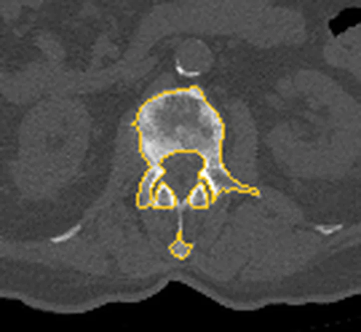
Aynı bölgenin, kaynak veri setinde patoloji içermediği belirtilen bir BT incelemesini kesit kesit kaydırın; organ sınırlarını açarak anatomiyi hasta görüntüsüyle karşılaştırın. Küçük rastlantısal bulgular tümüyle dışlanmamıştır.
Kaynak: TotalSegmentator v2.01 veri seti (Wasserthal ve ark., Radiology: Artificial Intelligence 2023), CC BY 4.0, vaka s1338; organ sınırları veri setinin otomatik segmentasyonundan, birleştirilerek sadeleştirildi. Görüntüler 2,5 mm kesit aralığına yeniden örneklendi.
Ayırıcı tanı
- DISH
- en az dört komşu vertebrayı tutan akıcı osteofitler ve disk aralıklarının korunmasıyla tanımlanır; bu uzun segmentli görünüm fokal dejeneratif osteofit örüntüsünden ayrımda yardımcı olur.
- Ankilozan spondilit
- ince marjinal sindesmofitler, vertebra köşesi erozyonları ve sakroiliit eşlik edebilir; kaba dejeneratif osteofit paterni beklenmez
- Disk hernisi
- disk materyalinin disk aralığı sınırlarının dışına fokal yer değiştirmesidir; posteriora uzanan herniasyon spinal kanal içindeki nöral yapıları etkileyebilir.
- Vertebral osteomiyelit
- son plak destrüksiyonu, disk aralığının inflamatuvar etkilenmesi ve paravertebral değişiklikler sıradan sklerotik spondilozdan farklıdır
- Osteokondrom veya başka kemik lezyonu
- fokal ekzofitik oluşumun medüller devamlılığı ve tipik son plak marjininden bağımsız konumu incelenir
Tuzaklar
- Akıcı anterolateral köprüleşme DISH ile karışabilir; köprülerin tek tek disk aralığından kaynaklanan kısa osteofitler mi yoksa uzun ligamentöz ossifikasyon mu olduğunu sagittal MPR'de izle.
- Servikal unkovertebral osteofit intervertebral foramene uzanarak sinirleri sıkıştırabilir ve radikülopatiye yol açabilir.
- Dejeneratif Modic tip 1 kemik iliği değişiklikleri, özellikle disk içi sıvı birikimi eşlik ettiğinde, erken spondilodiskiti taklit edebilir.
- BT, servikal spondilozda osteofit ve kemik foraminal darlığı değerlendirmede kullanılır; konvansiyonel MR kemik anormalliklerini göstermede yetersiz kalabilir.
Kendini dene
C5-C6'da unkovertebral kemik çıkıntısı aynı taraf nöral foramenin ön bölümünü daraltıyor. En olası değişiklik nedir?
Cevabı göster
Unkovertebral spondiloz. Unkovertebral eklemden çıkan osteofitin foramene devam etmesi servikal spondilozun tipik kemik paternidir. DISH uzun segment akıcı anterolateral ossifikasyon, ankilozan spondilit ince sindesmofit/sakroiliit, osteomiyelit ise destrüktif son plak-disk değişikliğiyle ayrılır.
Birden fazla komşu vertebrada uzun, akıcı anterolateral köprüleşme ve görece korunmuş disk aralıkları hangi paterni düşündürür?
Cevabı göster
DISH. Uzun ve akıcı anterolateral ligamentöz ossifikasyon ile disk aralıklarının korunması DISH örüntüsüdür. Fokal spondiloz genellikle tekil marjinal osteofit ve disk dejenerasyonu; disk hernisi yumuşak doku; spondilolizis ise pars interartikülaris defekti oluşturur.
İlgili konular
Kaynaklar
Bu sayfadaki 38 cümle ve 2 quiz sorusu aşağıdaki kaynaklardan alıntılarla otomatik olarak karşılaştırıldı; 11 cümle bu karşılaştırmada düzeltildi (2026-10-09).
- ABCs of the degenerative spinepmc.ncbi.nlm.nih.gov
- Lumbar spondylosis: clinical presentation and treatment approachespmc.ncbi.nlm.nih.gov
- Advances in the Pathogenesis, Diagnosis and Treatment of Bow Hunter's Syndrome: A Comprehensive Review of the Literature.pubmed.ncbi.nlm.nih.gov
- Cervical spondylosis: a rare and curable cause of vertebrobasilar insufficiency.pubmed.ncbi.nlm.nih.gov
- Radiological evidence of spontaneous spinal arthrodesis in patients with lower lumbar spondylolisthesis.pubmed.ncbi.nlm.nih.gov
- Microscopic anterior foraminal decompression combined with anterior lumbar interbody fusion.pubmed.ncbi.nlm.nih.gov
- Relationship between facet tropism and facet joint degeneration in the sub-axial cervical spine.pubmed.ncbi.nlm.nih.gov
- Dyspnea Associated with Cervical Spondylotic Radiculopathy: A Case Report and Review of Literatures.pubmed.ncbi.nlm.nih.gov
- What are the reliable radiological indicators of lumbar segmental instability?pubmed.ncbi.nlm.nih.gov
- A Surgical Case of Bow Hunter's Syndrome Diagnosed by Cervical Rotational MRA.pubmed.ncbi.nlm.nih.gov
- Subaxial Vertebral Artery Rotational Occlusion Syndrome: An Overview of Clinical Aspects, Diagnostic Work-Up, and Surgical Management.pubmed.ncbi.nlm.nih.gov
- CT and MRI characteristics of presumptive hypervitaminosis A in a cat.pubmed.ncbi.nlm.nih.gov
- Surgical management of cervical spondylotic myelopathy.pubmed.ncbi.nlm.nih.gov
- Cervical osteophyte resulting in compression of the jugular foramen: Case report.pubmed.ncbi.nlm.nih.gov
- Facet joint degeneration of the cervical spine: a computed tomographic analysis of 320 patients.pubmed.ncbi.nlm.nih.gov
- Complex cervical spondylotic myelopathy: a report of two cases and literature review.pubmed.ncbi.nlm.nih.gov
- Impact of Cervical Spine Immobility on Carotid Endarterectomy: A Report of Two Cases.pubmed.ncbi.nlm.nih.gov
- Clinical effects and prognostic factors of combined laminoplasty with keyhole decompression for multilevel cervical spondylotic myelopathy: analysis based on cervical sagittal parameters.pubmed.ncbi.nlm.nih.gov
- Added value of 3D fast-field-echo (FRACTURE) sequences for cervical spondylosis diagnosis: a prospective multi-reader non-inferiority study.pubmed.ncbi.nlm.nih.gov
- Retro-odontoid pseudotumor and subaxial spinal osteophytes: instability as the etiology and stabilization as the treatment. Illustrative cases.pubmed.ncbi.nlm.nih.gov
- Anterior Cervical Osteophyte Thinning for Forestier's Disease: A Case Series, Scoring System and Treatment Algorithm.pubmed.ncbi.nlm.nih.gov
- Comparative efficacy and safety of unilateral biportal endoscopic posterior cervical foraminotomy versus anterior cervical discectomy and fusion for single-level cervical radiculopathy: a retrospective cohort study.pubmed.ncbi.nlm.nih.gov
- Anterior cervical osteophyte-related dysphagia in a long-term growth hormone user: a case report.pubmed.ncbi.nlm.nih.gov
- Ossification of the Nuchal Ligament With Text Neck Syndrome.pubmed.ncbi.nlm.nih.gov
- Cervical epidural plasmacytoma causing recurrent radiculopathy and revealing plasma cell leukemia.pubmed.ncbi.nlm.nih.gov
- Dorsal C1-2 Spinal Cord Compression After Reduction of Atlantoaxial Posterior Dislocation Associated With Os Odontoideum: A Case Report and Literature Review.pubmed.ncbi.nlm.nih.gov
- Degenerative lumbosacral spondylolisthesis: Instability as the primary pathology and stabilization as definitive treatment. Experience with 28 cases treated by only fixation without any decompression.pubmed.ncbi.nlm.nih.gov
- Uni-portal non-coaxial spinal endoscopic surgery via crossing midline approach for cervical radiculopathy: a case series of four patients.pubmed.ncbi.nlm.nih.gov
- Conservative Management of a Symptomatic Anterior Limbus Vertebra in a Non-athletic Patient: A Case Report.pubmed.ncbi.nlm.nih.gov
- Seeding the Spine: Salmonella Java Discitis and Osteomyelitis After Traveller's Bacteraemia.pubmed.ncbi.nlm.nih.gov
- Subacute and Chronic Low-Back Pain: From MRI Phenotype to Imaging-Guided Interventions.pubmed.ncbi.nlm.nih.gov
- Self-stabilizing zero-profile 3D-printed vertebral body versus traditional titanium mesh-cage with plate in anterior cervical corpectomy and fusion: an open-label randomized controlled trial on efficacy and complications for cervical spondylotic myelopathy.pubmed.ncbi.nlm.nih.gov
- Patterns of spinal new bone formation in patients with axial spondyloarthritis, psoriatic arthritis, rheumatoid arthritis and healthy controls: a low-dose CT study.pubmed.ncbi.nlm.nih.gov
- Quantitative Disk and Metabolic Bone PET-MRI for Understanding Disk-Endplate Crosstalk in Degeneration of the Lumbar Spine.pubmed.ncbi.nlm.nih.gov
- Pathogenic mechanisms of adult degenerative scoliosis.pubmed.ncbi.nlm.nih.gov
- From Modic changes to the disc-endplate-bone marrow complex: imaging stratification, immune remodeling, and translational implications in intervertebral disc degeneration.pubmed.ncbi.nlm.nih.gov
- Case Report: Ochronotic arthropathy mimicking spondyloarthritis: a case-based review of diagnostic pitfalls and a novel likely pathogenic HGD variant.pubmed.ncbi.nlm.nih.gov
- Single-incision four-level oblique lateral interbody fusion and cement-augmented fixation in a severely osteoporotic lumbar spine: a case report.pubmed.ncbi.nlm.nih.gov
- Oblique lateral interbody fusion versus transforaminal lumbar interbody fusion for single-level lumbar adjacent segment disease: a retrospective comparative study of clinical and radiographic outcomes.pubmed.ncbi.nlm.nih.gov
- Ultrasonic Bone Scalpel in Anterior Cervical Discectomy and Fusion Enhances Outcomes and Foraminal Decompression in Cervical Radiculopathy: A Retrospective Cohort Study.pubmed.ncbi.nlm.nih.gov
- Is zero-profile fixation safe in consecutive two-level anterior cervical discectomy and fusion with partial uncinate resection? A finite element analysis.pubmed.ncbi.nlm.nih.gov
- Foraminal Decompression Technique During ACDF for Cervical Radiculopathy that Provides a Better Outcome: Total Uncinatectomy vs Partial Uncoforaminotomy.pubmed.ncbi.nlm.nih.gov
- Biomechanical assessment of endoscopic cross-over top decompression in cervical spinal stenosis: a finite element analysis.pubmed.ncbi.nlm.nih.gov
- Comparative efficacy of unilateral biportal endoscopy versus endoscopic foraminoplasty decompression for single-level lumbar spinal stenosis: a retrospective cohort study.pubmed.ncbi.nlm.nih.gov
- Case Report: Concurrent esophageal and spinal cord compression in cervical spondylosis: integrated anterior osteophytectomy and zero-profile ACDF for dual pathology decompression.pubmed.ncbi.nlm.nih.gov
- Genetics and pathophysiology of diffuse idiopathic skeletal hyperostosis.pubmed.ncbi.nlm.nih.gov
- Biomaterials for vertebral body repair: interior augmentation and total replacement-a literature review of bone fusion outcomes in clinical studies.pubmed.ncbi.nlm.nih.gov
- Beyond Resnick and Niwayama: a structured narrative literature review on the clinical and imaging phenotypes of diffuse idiopathic skeletal hyperostosis (DISH).pubmed.ncbi.nlm.nih.gov
- The Importance of Instrumentation Length in Ankylosing Spinal Disorders and Thoracolumbar Fractures.pubmed.ncbi.nlm.nih.gov
- Annular Repair Using an All-Suture Anchor Under Unilateral Biportal Endoscopy: Technical Note and Case Series.pubmed.ncbi.nlm.nih.gov
- Application of the pivot forward combined leveraging technique in the treatment of thoracolumbar fractures: a finite element analysis and clinical study.pubmed.ncbi.nlm.nih.gov
- Extrapedicular approach for basivertebral nerve radiofrequency ablation: a conceptual review.pubmed.ncbi.nlm.nih.gov
- International Pain and Spine Intervention Society Emergency Protocols: Intra-arterial Injection.pubmed.ncbi.nlm.nih.gov
- Risk of subsidence following ACDF with the coalition™ (Globus Medical) cage: a multivariable analysis identifying preoperative radiculopathy as the primary risk factor.pubmed.ncbi.nlm.nih.gov
- Acute thoracolumbar fractures in patients with a rigid spine: a computed tomography study.pubmed.ncbi.nlm.nih.gov
- Spinal mobility and functional limitations in patients with diffuse idiopathic skeletal hyperostosis compared to healthy controls and patients with axial spondyloarthritis.pubmed.ncbi.nlm.nih.gov
- Acute Respiratory Distress in a Hospitalized Patient Revealing Cervical Diffuse Idiopathic Skeletal Hyperostosis (DISH).pubmed.ncbi.nlm.nih.gov
- The Genetic Architecture of High Bone Mass.pubmed.ncbi.nlm.nih.gov
- Progression to Radiographic Axial Spondyloarthritis: A Narrative Review on Timeline and Novel Prediction Factors.pubmed.ncbi.nlm.nih.gov
- Radiographic sacroiliitis progression in psoriatic arthritis.pubmed.ncbi.nlm.nih.gov
- Development of structural damage in the spine in patients with PsA: findings from a longitudinal cohort study.pubmed.ncbi.nlm.nih.gov
- The Konakondla-Telfeian-Shen (KTS) Pyramid: a safe entry zone for ventral thoracic pathologies.pubmed.ncbi.nlm.nih.gov
- Posterior Lumbar Arthrodesis Using a Stand-Alone Hydrophilic Synthetic Bone Graft in the Setting of Postoperative Osteomyelitis/Discitis: A Case Report.pubmed.ncbi.nlm.nih.gov
- <i>Mycobacteroides abscessus</i> complex vertebral osteomyelitis and discitis of the thoracolumbar spine: a systematic review of management and outcomes with illustrative case.pubmed.ncbi.nlm.nih.gov
- Differentiating Tuberculous and Pyogenic Spondylodiscitis: Part II: MRI/CT Features, Prediction Models, and the Role of PET/CT-A Narrative Review.pubmed.ncbi.nlm.nih.gov
- [Primary bone tumors of the spine].pubmed.ncbi.nlm.nih.gov
- Subscapular Fossa Osteochondroma: A Case Report of an Anatomical Surgical Approach.pubmed.ncbi.nlm.nih.gov
- Atypical presentation of L3 vertebral body osteochondroma mimicking cauda equina syndrome: a case report.pubmed.ncbi.nlm.nih.gov
- Intervertebral Foramen Width Is an Important Factor in Deciding Additional Uncinate Process Resection in ACDF-a Retrospective Study.pubmed.ncbi.nlm.nih.gov
- Safety and efficacy of direct nerve root decompression via anterior cervical discectomy and fusion with uncinectomy for cervical radiculopathy.pubmed.ncbi.nlm.nih.gov
- The Impact of Single-Level ACDF on Neural Foramen and Disc Height of Surgical and Adjacent Cervical Segments: A Case-Series Radiological Analysis.pubmed.ncbi.nlm.nih.gov
- Recommendations of the International Consensus Meeting (ICM) 2025 on Spinal Infections: Part A Pyogenic Spondylodiscitis.pubmed.ncbi.nlm.nih.gov
- Spinal brucellosis - A mimicker of spinal tuberculosis: An analysis of 10 patients from a tertiary care center in India.pubmed.ncbi.nlm.nih.gov
- Anterior Cervical Foraminotomy for Radiculopathy After Cervical Artificial Disc Replacement: Technique Description and Case Report.pubmed.ncbi.nlm.nih.gov
- Alleviating cervical radiculopathy by manipulative correction of reversed cervical lordosis: 4 years follow-up.pubmed.ncbi.nlm.nih.gov
- Case Report: Combined posterolateral and direct anterior approaches for total hip arthroplasty in osteonecrosis of the femoral head with a rare massive osteophyte with literature review.pubmed.ncbi.nlm.nih.gov
- Bow hunter's syndrome secondary to lateral and posterior spondylosis: illustrative case.pubmed.ncbi.nlm.nih.gov
- Giant cervical facet joint osteophyte causing myelopathy: illustrative case.pubmed.ncbi.nlm.nih.gov
- Intervertebral Canals and Intracanal Ligaments as New Terms in Terminologia anatomica .pubmed.ncbi.nlm.nih.gov
- Transtubular Anterior Cervical Foraminotomy for the Treatment of Compressive Cervical Radiculopathy: Surgical Results and Complications in a Consecutive Series of Cases.pubmed.ncbi.nlm.nih.gov
- Study protocol for a randomised controlled multicentre study: the Foraminotomy ACDF Cost-Effectiveness Trial (FACET) in patients with cervical radiculopathy.pubmed.ncbi.nlm.nih.gov
- Diagnosing Dysphagia in Forestier Syndrome: A Dynamic Digital Radiology Application.pubmed.ncbi.nlm.nih.gov
- Artificial intelligence in spine care: A scoping review of diagnostic applications.pubmed.ncbi.nlm.nih.gov
- The Value of Dynamic MRI in Cervical Radiculopathy: A Report of Two Cases.pubmed.ncbi.nlm.nih.gov
- Determination of the Most Suitable Cut-Off Point of the Cervical Foraminal Cross-Sectional Area at the C5/6 Level to Predict Cervical Foraminal Bony Stenosis.pubmed.ncbi.nlm.nih.gov
- Preliminary Quantitative MRI Assessment After Combined Posterior Endoscopic Cervical Discectomy and Foraminotomy: An Exploratory Retrospective Cohort Study.pubmed.ncbi.nlm.nih.gov
- Diagnosis and surgery for cervical synovial cysts: A review.pubmed.ncbi.nlm.nih.gov
- Cervical DISH, is there any place for multilevel cervical arthroplasty? A case report metal-on-metal two-level cervical disc arthroplasty with 2-year follow-up and quality of life assessment.pubmed.ncbi.nlm.nih.gov
- When it is not sacroiliitis.pubmed.ncbi.nlm.nih.gov
- Spinal Involvement in Patients with Chronic Non-Bacterial Osteomyelitis (CNO): An Analysis of Distinctive Imaging Features.pubmed.ncbi.nlm.nih.gov
- Pivotal role of the synovioentheseal complex in the imaging of arthritis and rheumatic diseasespubmed.ncbi.nlm.nih.gov
- Magnetic resonance imaging (MRI) vs. computed tomography (CT) in the diagnosis and classification of spondylolysis and spondylolisthesis-a narrative review.pubmed.ncbi.nlm.nih.gov
- Contemporary clinical perspectives on chronic low back pain: The biology, mechanics, etc. underpinning clinical and radiological evaluation.pubmed.ncbi.nlm.nih.gov
- What a pain in the back: etiology, diagnosis and future treatment directions for discogenic low back pain.pubmed.ncbi.nlm.nih.gov
- Computed Tomographic Evaluation of the Morphometry of the Intervertebral Foramina and Intervertebral Discs of the Cervical Spine.pubmed.ncbi.nlm.nih.gov
- Behind the Procedure: Understanding Complications in Anterior Cervical Surgery.pubmed.ncbi.nlm.nih.gov
- Distinguishing bone marrow edema patterns on MRI between spondyloarthritis and diffuse idiopathic skeletal hyperostosis.pubmed.ncbi.nlm.nih.gov
- Congenital and Breed-Associated Vertebral Anomalies in Dogs and Cats: Developmental Anatomy, Genetic Determinants, and Biological Significance.pubmed.ncbi.nlm.nih.gov
- A Primer on Anatomy, Biophysics, Pathology, Imaging and Treatment of the Intervertebral Disc and the Anterior Spinal Column: The Discogenic, Intervertebral, Spinal Column (DISC) ASPN Workgroup.pubmed.ncbi.nlm.nih.gov
- Navigation-Assisted Bilateral Troughing Technique for Lumbar Laminectomy: A Stepwise Approach to Safe, Teachable, and Efficient Decompression.pubmed.ncbi.nlm.nih.gov
- Diffuse Idiopathic Skeletal Hyperostosis and Ankylosing Spondylitis: A Challenging Case and Review of the Literature.pubmed.ncbi.nlm.nih.gov
- Imaging Characteristics of Diffuse Idiopathic Skeletal Hyperostosis: More Than Just Spinal Bony Bridges.pubmed.ncbi.nlm.nih.gov
- Axial Spondyloarthritis: Mimics and Pitfalls of Imaging Assessment.pubmed.ncbi.nlm.nih.gov
- What Is New and What Is Next for SAPHO Syndrome Management: A Narrative Review.pubmed.ncbi.nlm.nih.gov
- Intact Posterior Tension Band in Spinal Fractures in Patients With Spinal Ankylosing Disorders - Is it a Myth or a Reality? A Systematic Review.pubmed.ncbi.nlm.nih.gov
- Navigating Low Back Pain: A Multidisciplinary Approach to Diagnosis, Management, and Education.pubmed.ncbi.nlm.nih.gov
- The value of magnetic resonance imaging and computed tomography in the study of spinal disorders.pubmed.ncbi.nlm.nih.gov
- Lower C2 slope and milder uncovertebral joint degeneration are risk factors for pseudarthrosis after single-level anterior cervical corpectomy and fusion (ACCF): retrospective study of 102 patients with minimum 2-year follow-up.pubmed.ncbi.nlm.nih.gov
- Resection of the posterior longitudinal ligament in anterior cervical decompression surgery: a retrospective study of the clinical and radiographic outcomes in Thailand.pubmed.ncbi.nlm.nih.gov
- Dysphagia Secondary to Cervical Hyperostosis in Diffuse Idiopathic Skeletal Hyperostosis: A Case Report.pubmed.ncbi.nlm.nih.gov
- The Infraneural Approach to Lumbar Transforaminal Epidural Steroid Injections: A Practical Review for Trainees and Interventionalists.pubmed.ncbi.nlm.nih.gov
- Dimensional changes of the neuroforamina in subaxial cervical spine during in vivo dynamic flexion-extension.pubmed.ncbi.nlm.nih.gov
Eğitim amaçlıdır; hasta başında tanı ve tedavi kararının yerine geçmez, tıbbi tavsiye değildir. Metinler yapay zekâ desteğiyle yazıldı ve otomatik bir adımda kaynak alıntılarıyla karşılaştırıldı; bu bir tıbbi doğrulama değildir ve içerik uzman radyolog incelemesinden geçmedi. Klinik kararlarda güncel kılavuzları, kurum protokollerini ve radyoloji raporunu esas alın.