Özet
Osteoblastik vertebra metastazı, tümörün tetiklediği yeni kemik yapımıyla korpus ve posterior elemanlarda fokal ya da yaygın skleroz oluşturur; prostat kanseriyle sık ilişkilidir, ancak tek başına primer tümörü kanıtlamaz. Kontrastsız BT kemik yoğunluğunu, dağılımı, trabeküler yapıyı ve vertebra yüksekliğini göstererek metastatik yükü ve yapısal değişikliği tarif eder. Pedikül tutulumu, korteks konturu ve eşlik eden litik alanlar paternin yorumunu değiştirir. Bulguların atlanması yaygın kemik hastalığının ve eşlik eden kırık ya da epidural yumuşak doku bileşeninin eksik değerlendirilmesine yol açabilir.
Faz ve pencere
- Kontrastsız tanısal
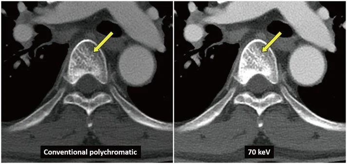
- Kemik penceresinde vertebra iliği içinde çevre kemiğe göre artmış dansiteli fokal, multifokal veya yaygın sklerotik odaklar izlenir; kortikal kontur, pediküller ve korpus yüksekliği aynı incelemede değerlendirilir. Kontrast fazı kemik skleroz paternini tanımak için gerekli değildir.
- Kemik algoritması tanısal
- Sagittal ve koronal rekonstrüksiyonlar sklerozun korpus boyunca dağılımını, posterior elemanlara geçişini ve varsa çökme/kompresyon deformitesini gösterir; aksiyel kesitlerde pedikül simetrisi karşılaştırılır.
Önerilen pencereler G genişlik, M merkez; değerler Hounsfield birimi (HU)
- KemikG 1800 / M +400
- Yumuşak dokuG 400 / M +40
BT bulguları
- Fokal osteoskleroz — korpus trabeküler alanında çevre iliğe göre belirgin artmış kemik yoğunluğu.
- Multifokal dağılım — aynı veya farklı spinal düzeylerde birbirinden ayrı sklerotik odaklar.
- Diffüz skleroz — vertebra korpusunun büyük bölümünde yaygın yoğunluk artışı; diğer omurga ve pelvis kemikleriyle dağılım karşılaştırılır.
- Pedikül sklerozu — pedikül içinde artmış yoğunluk veya kontur kalınlaşması metastatik posterior eleman tutulumunu düşündürür.
- Trabeküler mimari değişikliği — yoğunlaşmış ve düzensiz trabeküller sklerotik odağın iç yapısını gösterir.
- Korpus yüksekliği ve son plaklar — sklerotik vertebrada çökme, son plak deformitesi veya kırık eşlik edebilir.
- Mikst bileşen — yoğun sklerotik zemin içinde düşük dansiteli litik alanlar aynı lezyonun karma yapısını ortaya koyar.
- SINS (Spinal Instability Neoplastic Score): 0–6 stabil, 7–12 olası instabilite, 13–18 instabil; 7 ve üzeri cerrahi konsültasyon gerektirir.
- BT’de kemik yapısal değişikliği görülmese bile vertebra kemik iliğinde metastatik lezyonlar bulunabilir; MR kemik iliği anormalliklerini saptamada duyarlıdır.
Ölçütler ve sınıflamalar
- Radyolojik kemik metastazı paterni
- Osteolitik patern kemik/trabekül kaybı; osteoblastik (sklerotik) patern yeni yoğun kemik oluşumu; mikst patern aynı hastalık odağında hem litik hem sklerotik değişikliği ifade eder.
Normalde
Sağlıklı vertebralarda kemik yoğunluğu düzeyler arasında değişebilir; şüpheli odağı komşu vertebralarla karşılaştırın. Aynı düzeyde korpusun orta kısmını, pedikülleri ve komşu vertebraları karşılaştırarak odak dağılımını ve yükseklik korunmasını belirleyin.
Normal BT ile kıyasla
Aynı bölgenin, kaynak veri setinde patoloji içermediği belirtilen bir BT incelemesini kesit kesit kaydırın; organ sınırlarını açarak anatomiyi hasta görüntüsüyle karşılaştırın. Küçük rastlantısal bulgular tümüyle dışlanmamıştır.
Kaynak: TotalSegmentator v2.01 veri seti (Wasserthal ve ark., Radiology: Artificial Intelligence 2023), CC BY 4.0, vaka s1338; organ sınırları veri setinin otomatik segmentasyonundan, birleştirilerek sadeleştirildi. Görüntüler 2,5 mm kesit aralığına yeniden örneklendi.
Ayırıcı tanı
- Tipik vertebral hemanjiyom
- kalınlaşmış trabeküllerin aksiyel polka-dot veya sagittal corduroy düzeni bulunabilir; düzgün odak sınırı ve tipik iç mimari metastaz taklidini azaltır.
- Kemik adacığı (enostoz), genellikle ek inceleme veya girişim gerektirmeyen benign bir lezyondur; bilinen kanseri olan hastalarda benign kemik lezyonları metastaz sanılabileceğinden bunların tanınması gereksiz inceleme ve zararlı girişimleri önleyebilir.
- Paget hastalığı
- vertebra büyümesi, kortikal kalınlaşma ve kaba trabeküler yeniden yapılanma ile daha yaygın mimari değişiklik yapar.
- Osteopoikiloz
- çok sayıda benzer boyutlu, simetrik yerleşimli kemik adacığı görünümü metastatik multifokal odaklardan farklıdır.
- Tedavi sonrası kemik metastazlarında skleroz, kemik flare fenomeni ve mikst yanıt, lezyon yanıtının değerlendirilmesini güçleştirebilir.
Tuzaklar
- Her sklerotik odağı metastaz saymayın; hemanjiyom trabeküler işaretini, kemik adacığının sınırını ve dağılım paternini kontrol edin.
- Sklerozu primer tümörün kesin göstergesi gibi yorumlamayın; tümör tipleri litik, blastik veya mikst görünüm verebilir.
- Önceki inceleme olmadan tedaviye bağlı sklerotik yanıtı yeni metastazdan ayırmak mümkün olmayabilir; değişimi karşılaştırmalı tarif edin.
- Sklerotik yoğunluğun vertebra yüksekliğini koruduğunu varsaymayın; çökme, posterior duvar ve epidural yumuşak doku ayrıca aranmalıdır.
Kendini dene
Bir çalışmada prostat kaynaklı spinal metastazların en sık görülen kemik paterni hangisiydi?
Cevabı göster
Osteoblastik. Çalışmada prostat metastazlarının %62'si osteoblastik, %24'ü mikst ve %14'ü osteolitik olarak sınıflandırılmıştır; bu oranlar söz konusu çalışmanın bulgularıdır.
Spinal metastazlar BT'de hangi patern sınıflarıyla tanımlanabilir?
Cevabı göster
Osteolitik, osteoblastik veya mikst. BT görüntülemesinde spinal metastazlar osteolitik, osteoblastik veya mikst patern olarak değerlendirilebilir.
İlgili konular
Kaynaklar
Bu sayfadaki 39 cümle ve 2 quiz sorusu aşağıdaki kaynaklardan alıntılarla otomatik olarak karşılaştırıldı; 11 cümle bu karşılaştırmada düzeltildi (2026-10-09).
- Vertebral Sarcoidosis Mimicking Metastatic Disease in a Patient With Prior Melanoma: A Diagnostic Challengepmc.ncbi.nlm.nih.gov
- A Fully Automated Pipeline for Vertebral Structural Assessment From Medical Images. Application Under Metastatic Conditionspmc.ncbi.nlm.nih.gov
- Beyond SINS: A Critical Review of Biomechanical, Microstructural, and Radiomic Biomarkers for Predicting Fracture Risk in Spinal Metastasespmc.ncbi.nlm.nih.gov
- Multidisciplinary Exploration of Computed‐Tomographic and Ancient‐DNA Data of an Iron Age Skull From Latvia With Multiple Lytic Bone Lesions: Differential Diagnosis Between Metastatic Carcinoma, Multiple Myeloma and Skeletal Tuberculosispmc.ncbi.nlm.nih.gov
- Local Control and Vertebral Compression Fractures After Stereotactic Body Radiotherapy for Spinal Metastasespmc.ncbi.nlm.nih.gov
- Imaging characteristic analysis of metastatic spine lesions from breast, prostate, lung, and renal cell carcinomas for surgical planning: Osteolytic versus osteoblastic.pubmed.ncbi.nlm.nih.gov
- Neoplastic ascites in osteosarcoma: a case report.pubmed.ncbi.nlm.nih.gov
- Solitary upper thoracic vertebral metastasis as an initial presentation of pancreatic cancer: A case report.pubmed.ncbi.nlm.nih.gov
- Type, size, and position of metastatic lesions explain the deformation of the vertebrae under complex loading conditions.pubmed.ncbi.nlm.nih.gov
- Percutaneous Image-Guided Electrochemotherapy of Spine Metastases: Initial Experience.pubmed.ncbi.nlm.nih.gov
- 3D parametric finite element models to assess the mechanical role of the bone-lesion interface in human metastatic vertebrae.pubmed.ncbi.nlm.nih.gov
- Automatic detection of lytic and blastic thoracolumbar spine metastases on computed tomography.pubmed.ncbi.nlm.nih.gov
- Volume of Lytic Vertebral Body Metastatic Disease Quantified Using Computed Tomography-Based Image Segmentation Predicts Fracture Risk After Spine Stereotactic Body Radiation Therapy.pubmed.ncbi.nlm.nih.gov
- The role of bone metastases on the mechanical competence of human vertebrae.pubmed.ncbi.nlm.nih.gov
- Prediction of compressive strength of vertebral body with metastatic lesions based on quantitative computed tomography-based subject-specific finite element models.pubmed.ncbi.nlm.nih.gov
- Bone health in advanced prostate cancer: pathophysiology and management strategies.pubmed.ncbi.nlm.nih.gov
- Osteoblastic skeletal metastases mimicking high-bone-turnover metabolic bone disease in gastric adenocarcinoma.pubmed.ncbi.nlm.nih.gov
- Anabolics in Cancer Patients: To Use or Not to Use?pubmed.ncbi.nlm.nih.gov
- Osteomyelitis of the Jaw Bones and Its Mimics: Resolving the Diagnostic Enigma.pubmed.ncbi.nlm.nih.gov
- Detecting bone marrow infiltration in nonosteolytic multiple myeloma through separation of hydroxyapatite via the two-material decomposition technique in spectral computed tomography.pubmed.ncbi.nlm.nih.gov
- Intraosseous Venous Malformations of the Head and Neck.pubmed.ncbi.nlm.nih.gov
- Advances in Metastatic Disease Spinal Oncology: Novel Technology Without Forgetting the Fundamentals of Surgical Treatment.pubmed.ncbi.nlm.nih.gov
- Practice and principles of stereotactic body radiation therapy for spine and non-spine bone metastases.pubmed.ncbi.nlm.nih.gov
- A Review of Posteromedial Lesions of the Chest Wall: What Should a Chest Radiologist Know?pubmed.ncbi.nlm.nih.gov
- Oncologic Applications of Artificial Intelligence and Deep Learning Methods in CT Spine Imaging-A Systematic Review.pubmed.ncbi.nlm.nih.gov
- Expert consensus on a multidisciplinary approach for the management of multiple myeloma-related bone disease.pubmed.ncbi.nlm.nih.gov
- Treatment of Metastatic Spinal Disease; what the Radiologist needs to know.pubmed.ncbi.nlm.nih.gov
- Imaging Features of Intraosseous Schwannoma: A Case Series and Review of the Literature.pubmed.ncbi.nlm.nih.gov
- Whole-Body Low-Dose Multidetector-Row CT in Multiple Myeloma: Guidance in Performing, Observing, and Interpreting the Imaging Findings.pubmed.ncbi.nlm.nih.gov
- Musculoskeletal CT Imaging: State-of-the-Art Advancements and Future Directions.pubmed.ncbi.nlm.nih.gov
- Skull Base Metastases: A Pictorial Review.pubmed.ncbi.nlm.nih.gov
- Early detection of invasive lung cancer and multiorgan metastasis by a collagen-targeted protein MRI contrast agent.pubmed.ncbi.nlm.nih.gov
- Imaging findings of basaloid squamous cell carcinoma: a case report.pubmed.ncbi.nlm.nih.gov
- Pathogenic mechanisms of adult degenerative scoliosis.pubmed.ncbi.nlm.nih.gov
- An exploratory multimodal imaging and clinicopathological strategy for suspected Kümmell's disease in the absence of MRI.pubmed.ncbi.nlm.nih.gov
- Skeletal Involvement in Systemic Mastocytosis: Pathophysiology, Clinical Management, Standards of Care, and Novel Therapeutic Strategies.pubmed.ncbi.nlm.nih.gov
- Case Report: Congenital caudal tibial curvatures in two alpaca crias.pubmed.ncbi.nlm.nih.gov
- Beyond Chordoma: A Comprehensive Review of Sacral Lesions.pubmed.ncbi.nlm.nih.gov
- Multidisciplinary Exploration of Computed-Tomographic and Ancient-DNA Data of an Iron Age Skull From Latvia With Multiple Lytic Bone Lesions: Differential Diagnosis Between Metastatic Carcinoma, Multiple Myeloma and Skeletal Tuberculosis.pubmed.ncbi.nlm.nih.gov
- Unveiling Primary Bone Tumors of the Spine: A Review of Essential Imaging Clues.pubmed.ncbi.nlm.nih.gov
- ESR Essentials: bone marrow MRI in oncology-practice recommendations by the European Society of Musculoskeletal Radiology.pubmed.ncbi.nlm.nih.gov
- Clinical and 18F-FDG PET/CT Imaging Characteristics of Post-radiotherapy Sacral Insufficiency Fractures in Cervical Cancer Patients.pubmed.ncbi.nlm.nih.gov
- The emerging role of ¹⁸F-NaF PET/CT in osteoporosis: an emphasis on its application for the evaluation and management of lumbar spine osteoporosis.pubmed.ncbi.nlm.nih.gov
- Primary Benign Neoplasms of the Spine.pubmed.ncbi.nlm.nih.gov
- International Society of Paediatric Surgical Oncology (IPSO) Surgical Practice Guidelines.pubmed.ncbi.nlm.nih.gov
- Long-term safety and effectiveness of the "OptEase" vena cava filter.pubmed.ncbi.nlm.nih.gov
- Second primary mediastinal grey-zone lymphoma following treatment for classical Hodgkin lymphoma: a rare case report.pubmed.ncbi.nlm.nih.gov
- Postmortem computed tomography and histopathology of distal limb lesions in giraffe (Giraffa spp.) under managed care.pubmed.ncbi.nlm.nih.gov
- Case review of 3 patients with musculoskeletal manifestation of human immunodeficiency virus and highly active antiretroviral therapy in the form of osteopenia and insufficiency fractures.pubmed.ncbi.nlm.nih.gov
- Spinal lesions: a comprehensive radiologic overview.pubmed.ncbi.nlm.nih.gov
- Risk factors for new vertebral compression fracture after vertebroplasty and efficacy of osteoporosis treatment: A STROBE-compliant retrospective study.pubmed.ncbi.nlm.nih.gov
- Skeletal manifestations in lymphatic diseases: nonenhanced magnetic resonance lymphography.pubmed.ncbi.nlm.nih.gov
- Imaging findings of juvenile idiopathic arthritis and autoinflammatory diseases in children.pubmed.ncbi.nlm.nih.gov
- The value of magnetic resonance imaging and computed tomography in the study of spinal disorders.pubmed.ncbi.nlm.nih.gov
- Axial Spondyloarthritis: Mimics and Pitfalls of Imaging Assessment.pubmed.ncbi.nlm.nih.gov
- Trans-intervertebral space osteotomy for post-traumatic thoracolumbar kyphosis: a minimum 2-year follow-up study.pubmed.ncbi.nlm.nih.gov
- Study on anatomical parameters of modified cortical bone trajectories in the lower lumbar vertebrae.pubmed.ncbi.nlm.nih.gov
- Effect of cement distribution type on clinical outcome after percutaneous vertebroplasty for osteoporotic vertebral compression fractures in the aging population.pubmed.ncbi.nlm.nih.gov
- Decoding calvarial lesions: Contemporary neurosurgical management integrating radiological and histopathological features.pubmed.ncbi.nlm.nih.gov
- Immune checkpoint inhibition in bone metastases: lesion response heterogeneity, the osteoimmune niche, and microenvironment-directed therapy.pubmed.ncbi.nlm.nih.gov
- Osteocytes in the Metastatic Bone Niche: Mechanistic Pathways and Therapeutic Targets.pubmed.ncbi.nlm.nih.gov
- Lipid Metabolic Reprogramming in Periodontitis-Associated Alveolar Bone Remodeling: Mechanisms, Signaling Pathways, and Therapeutic Implications.pubmed.ncbi.nlm.nih.gov
- Imaging intraosseous hemangiomas: Typical and atypical features across modalities.pubmed.ncbi.nlm.nih.gov
- Pregnancy-related aggressive vertebral hemangioma presenting with radiculopathy: illustrative case.pubmed.ncbi.nlm.nih.gov
- Craniofacial disorders and dysplasias: Molecular, clinical, and management perspectives.pubmed.ncbi.nlm.nih.gov
- Challenges in the Interpretation of MRI Examinations Without Radiographic Correlation: Pearls and Pitfalls to Avoid.pubmed.ncbi.nlm.nih.gov
- Imaging More than Skin-Deep: Radiologic and Dermatologic Presentations of Systemic Disorders.pubmed.ncbi.nlm.nih.gov
- Osteopoikilosis: A rare cause of bone pain.pubmed.ncbi.nlm.nih.gov
- Glossary of terms for musculoskeletal radiology.pubmed.ncbi.nlm.nih.gov
- Management of Tibial Adamantinoma: Balancing Oncologic Cure and Reconstructive Longevity.pubmed.ncbi.nlm.nih.gov
- Liposomal multimodal theranostics for bone disorders: from rational responsive delivery and biomimetic strategies to clinical translation.pubmed.ncbi.nlm.nih.gov
- Review of imaging modalities and radiological findings of calvarial lesions.pubmed.ncbi.nlm.nih.gov
- Osteosarcoma: A Comprehensive Morphological and Molecular Review with Prognostic Implications.pubmed.ncbi.nlm.nih.gov
- Radiologists and treatment response assessment: conventional and functional radiological criteria for predicting therapeutic response in virus-related and non-virus-related cancers.pubmed.ncbi.nlm.nih.gov
- Case Report: Acquired CDKN2A/B deletion and mTOR inhibitor resistance in TSC2-mutant metastatic epithelioid angiomyolipoma.pubmed.ncbi.nlm.nih.gov
- A systematic approach to MR spectroscopy of the brain: Indications, protocol, metabolites, and spectral analysis in neurologic diseases.pubmed.ncbi.nlm.nih.gov
- Partial C2 corpectomy with iliac/fibula bone graft and posterior instrumented fusion for C2 vertebral body lesions: An innovative approach.pubmed.ncbi.nlm.nih.gov
- Differentiation of Native Vertebral Osteomyelitis: A Comprehensive Review of Imaging Techniques and Future Applications.pubmed.ncbi.nlm.nih.gov
- Minimally invasive interventional guided imaging therapies of musculoskeletal tumors.pubmed.ncbi.nlm.nih.gov
- Understanding a mass in the paraspinal region: an anatomical approach.pubmed.ncbi.nlm.nih.gov
- The role of intracorporeal cancellous bone grafting in the biological augmentation of vertebral body fractures: a literature review, historical perspective, and update on imaging and bone healing outcomes.pubmed.ncbi.nlm.nih.gov
- Comparative efficacy of unilateral biportal endoscopy versus endoscopic foraminoplasty decompression for single-level lumbar spinal stenosis: a retrospective cohort study.pubmed.ncbi.nlm.nih.gov
- Cervical Spine Pathology in the Context of Intimate Partner and Interpersonal Violence: A Case Series and Focused Narrative Review.pubmed.ncbi.nlm.nih.gov
- Musculoskeletal infections through direct inoculation.pubmed.ncbi.nlm.nih.gov
- Bone and Joint Infections: The Role of Imaging in Tailoring Diagnosis to Improve Patients' Care.pubmed.ncbi.nlm.nih.gov
- Imaging Evaluation of Bone Tumors in the Cervical Spine: A Comprehensive Review.pubmed.ncbi.nlm.nih.gov
- The Role of CT and MR Imaging in Stereotactic Body Radiotherapy of the Spine: From Patient Selection and Treatment Planning to Post-Treatment Monitoring.pubmed.ncbi.nlm.nih.gov
- Research Progress of Ferrosoferric Oxide Nanoparticles in Bone Regeneration and Disease Treatment.pubmed.ncbi.nlm.nih.gov
- Small cell osteosarcoma versus fusion-driven round cell sarcomas of bone: retrospective clinical, radiological, pathological, and (epi)genetic comparison with clinical implications.pubmed.ncbi.nlm.nih.gov
- QCT-based geometric, densitometric, and biomechanical properties of the aging human thoracolumbar spine.pubmed.ncbi.nlm.nih.gov
- Multilevel Lumbar CT Attenuation Beyond L1: Comparison with QCT-Derived Volumetric Bone Mineral Density and Prevalent Fragility Fracture Status in a Diagnostic Referral Cohort.pubmed.ncbi.nlm.nih.gov
- Anatomical and biological reconstruction of traumatic complete burst fractures of the thoracolumbar vertebrae using pedicle fixation combined with stent-armed kyphoplasty and intracorporeal bone grafting: technique rationale, 7-year experience and clinical, imaging, and histological outcomes.pubmed.ncbi.nlm.nih.gov
- A Fully Automated Pipeline for Vertebral Structural Assessment From Medical Images. Application Under Metastatic Conditions.pubmed.ncbi.nlm.nih.gov
- Vertebral Sarcoidosis Mimicking Metastatic Disease in a Patient With Prior Melanoma: A Diagnostic Challenge.pubmed.ncbi.nlm.nih.gov
- Phylogeography of Bone Metastasis: Clonal Evolution, Skeletal Niche Adaptation, and Clinical Implications.pubmed.ncbi.nlm.nih.gov
- Review of Hematology-Oncology Emergencies for Internal Medicine Residents.pubmed.ncbi.nlm.nih.gov
- Development of a Second Primary Lung Cancer Following a Primary Breast Cancer: A Case Series.pubmed.ncbi.nlm.nih.gov
- Revisiting Fat Content in Bone Lesions: Paradigms in Bone Lesion Detection.pubmed.ncbi.nlm.nih.gov
- Imaging Review of Pediatric Benign Osteocytic Tumors and Latest Updates on Management.pubmed.ncbi.nlm.nih.gov
- Spinal manifestations of Paget's disease: Case presentation and systematic review.pubmed.ncbi.nlm.nih.gov
- Characterization and diagnosis of Paget Disease of Bone in historical individuals.pubmed.ncbi.nlm.nih.gov
- Double-plate compound osteosynthesis of a periprosthetic proximal tibial shaft fracture in an elderly patient with Paget's disease using PHILOS and LCP plates.pubmed.ncbi.nlm.nih.gov
- Spontaneous Regression of Clear Cell Renal Cell Carcinoma Metastases or Immune Restoration?pubmed.ncbi.nlm.nih.gov
- Leveraging the non-contrast CT component of PET/CT: an AI-driven delta-radiomics approach to monitor treatment response in metastatic breast cancer.pubmed.ncbi.nlm.nih.gov
- Musculoskeletal Manifestations of T-Cell Lymphomas: Clinical Presentation, Imaging Features, Pathobiology, and Orthopedic Considerations-A Narrative Review.pubmed.ncbi.nlm.nih.gov
- Does <sup>18</sup>F-sodium fluoride PET/CT have a role in the assessment of response to <sup>223</sup>Ra therapy in men with prostatic bone metastases?pubmed.ncbi.nlm.nih.gov
- Benign incidental do-not-touch bone lesions.pubmed.ncbi.nlm.nih.gov
- Sacrum Metastasis in Stage IA Endometrial Carcinoma 13 Years After Surgery: A Case Report.pubmed.ncbi.nlm.nih.gov
- Multiparametric Whole-Body MRI: A Game Changer in Metastatic Prostate Cancer.pubmed.ncbi.nlm.nih.gov
- Cancer Pain: Radiotherapy as a Double-Edged Sword.pubmed.ncbi.nlm.nih.gov
- Bone marrow magnetic resonance imaging (MRI): morphological and functional features from reconversion to infiltration.pubmed.ncbi.nlm.nih.gov
- Spinal hydatid disease with epidural extension and spinal cord compression: A radiology case report.pubmed.ncbi.nlm.nih.gov
- Spinal granulocytic sarcoma with both extradural and intradural involvement: a case report.pubmed.ncbi.nlm.nih.gov
- Ovarian Lymphoma and Bone Metastasis Presenting With Paraparesis in an Adolescent: A Case Report.pubmed.ncbi.nlm.nih.gov
- Clear cell renal cell carcinoma presenting as isolated symptomatic lumbar vertebral metastasis: a case report.pubmed.ncbi.nlm.nih.gov
Eğitim amaçlıdır; hasta başında tanı ve tedavi kararının yerine geçmez, tıbbi tavsiye değildir. Metinler yapay zekâ desteğiyle yazıldı ve otomatik bir adımda kaynak alıntılarıyla karşılaştırıldı; bu bir tıbbi doğrulama değildir ve içerik uzman radyolog incelemesinden geçmedi. Klinik kararlarda güncel kılavuzları, kurum protokollerini ve radyoloji raporunu esas alın.