Özet
Lumbosakral geçiş omuru, en alt lomber vertebranın sakral özellik kazanması veya üst sakral segmentin lomberleşmesiyle oluşan sık bir anatomik varyanttır. Kontrastsız BT, geniş transvers çıkıntının sakral ala ile yalancı eklemini ya da kemik füzyonunu aksiyel ve koronal düzlemde doğrudan gösterir. Varyantın doğru tanınması, vertebra seviyesinin yanlış numaralandırılmasını ve girişim/ameliyat düzeyinin karıştırılmasını önler; ağrı ilişkisi ise yalnız morfolojiyle varsayılamaz. Tek başına görüntüde geçiş omuru saptanması klinik Bertolotti sendromu anlamına gelmez.
Faz ve pencere
- Kemik algoritması tanısal
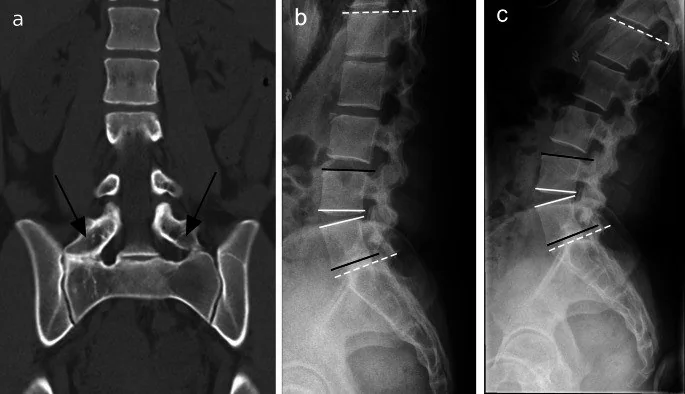
- Kontrastsız BT'de en alt lomber tip vertebranın geniş transvers çıkıntısı, sakral ala ile aradaki gerçek eklem aralığı veya kortike kemik köprüsü aksiyel ve koronal kemik rekonstrüksiyonlarında görülür. Vertebra sayısı tüm omurga görüntüsü olmadan belirsiz kalabilir; kontrast fazı kemik varyantını tanımlamak için gerekmez.
Önerilen pencereler: Kemik (G 1800 / M 400).
BT bulguları
- Geniş transvers çıkıntı — en alt lomber tip vertebranın tek veya iki taraflı transvers çıkıntısı sakral ala yönünde uzar.
- Yalancı eklem — geniş çıkıntı ile sakral ala arasında kortike, eklem benzeri temas ve aralık bulunur.
- Kemik füzyonu — transvers çıkıntı ile sakrum arasında kesintisiz kortikal-trabeküler köprü izlenir.
- Tek taraflı veya çift taraflı morfoloji — sağ ve sol geçiş bulguları ayrı değerlendirilir; Castellvi sınıflaması anatomi ve tarafı dikkate alır.
- Üst sakral segment lumbarizasyonu — S1-S2 arasında daha belirgin disk aralığı ve ayrışmış segment morfolojisi eşlik edebilir.
- Komşu segment dejenerasyonu — geçiş segmentinin üstündeki disk ve fasetlerde erken dejeneratif değişiklik görülebilir; bu görünüm tek başına semptom kaynağı kanıtı değildir.
- Yalancı eklem sklerozu — anormal temas yüzlerinde subkondral skleroz veya osteofit gelişebilir.
- Seviye numaralandırma belirsizliği — tüm omurga sayımı yoksa L5 sakralizasyonu ile S1 lumbarizasyonu yalnız sınırlı bel BT'sinde güvenle ayrılmayabilir.
Ölçütler ve sınıflamalar
- Castellvi tip I transvers çıkıntı genişliği
- Kraniyokaudal genişlik ≥19 mm
- Castellvi
- Tip I: displastik geniş transvers çıkıntı (Ia tek, Ib çift taraflı; yalancı eklem veya füzyon yok). Tip II: sakrumla inkomplet eklemleşme (IIa tek, IIb çift taraflı). Tip III: sakrumla tam kemik füzyonu (IIIa tek, IIIb çift taraflı). Tip IV: bir tarafta tip II, karşı tarafta tip III morfolojisi.
Normalde
Normal lumbosakral bileşkede L5 transvers çıkıntıları sakral ala ile yalancı eklem veya kemik füzyonu yapmaz; L5-S1 disk aralığı ile sakral ala konturları belirgin geçiş köprüsü olmadan izlenir. Sağ-sol çıkıntı genişliğini, ala ile temas yüzlerini ve kortikal devamlılığı karşılaştırın; ayrıca numaralandırmayı mümkünse üst omurga ve kaburga sayımıyla doğrulayın.
Normal BT ile kıyasla
Aynı bölgenin, kaynak veri setinde patoloji içermediği belirtilen bir BT incelemesini kesit kesit kaydırın; organ sınırlarını açarak anatomiyi hasta görüntüsüyle karşılaştırın. Küçük rastlantısal bulgular tümüyle dışlanmamıştır.
Kaynak: TotalSegmentator v2.01 veri seti (Wasserthal ve ark., Radiology: Artificial Intelligence 2023), CC BY 4.0, vaka s1338; organ sınırları veri setinin otomatik segmentasyonundan, birleştirilerek sadeleştirildi. Görüntüler 2,5 mm kesit aralığına yeniden örneklendi.
Ayırıcı tanı
- İzole geniş lomber transvers çıkıntı
- çıkıntı belirgin olsa da sakral ala ile eklemleşme veya füzyon yoksa geçiş varyantı sınıflaması dikkatle yapılır.
- İyileşmiş transvers çıkıntı kırığı ile lumbosakral geçiş omurunun ayrımında, geçiş omurunda transvers çıkıntı ile sakrum arasında yalancı eklem veya kemik birleşmesi bulunabileceğini göz önünde bulundurun.
- Önceki lumbosakral füzyon cerrahisi varsa BT, kemik füzyonunu ve cerrahi materyali değerlendirmeye yardımcı olur.
- Lumbarizasyonlu S1
- ek segment ve S1-S2 disk morfolojisiyle sakralize L5'ten farklı numaralandırma gerektirir; bütün omurga sayımı yardımcıdır.
Tuzaklar
- Castellvi tipi morfolojiyi tanımlar ancak ilgili vertebra seviyesini tüm omurgada numaralandırmaz; sınırlı lomber incelemede seviye belirsizliğini açıkça belirtin.
- Yalnız geniş transvers çıkıntı görmek Bertolotti sendromu tanısı koydurmaz; sendrom, varyantla ilişkili klinik ağrıyı ifade eder.
- Tip II geçişte transvers çıkıntı ile sakrum arasında yalancı eklem bulunur; sınıflamada bu eklemleşmenin varlığını değerlendirin.
- Sakral ala ile gerçek kemik füzyonunu üst üste binen korteks sanmayın; ardışık kesitlerde kesintisiz trabeküler köprüyü doğrulayın.
Kendini dene
L5 transvers çıkıntısı sakral ala ile kortike yalancı eklem yapıyor; kemik köprüsü yok. Hangi Castellvi tipi?
Cevabı göster
Tip II. Sakral ala ile inkomplet artikülasyon Castellvi tip II'dir. Tip I geniş çıkıntıda sakrumla artikülasyon yoktur; tip III tam füzyondur; tip IV bir tarafta II, diğerinde III birlikteliğidir.
Geçiş omuru BT'de görülüyor ancak tüm omurga görüntüsü yok. Seviyeyi kesin saymanın temel kısıtı nedir?
Cevabı göster
L5-S1 sayımı belirsizliği. Lokal lumbosakral morfoloji, L5 sakralizasyonu ile S1 lumbarizasyonunu her zaman ayırt ettirmez; güvenilir numaralandırma için üst omurgadan sayım gerekebilir. Kontrast, yumuşak doku penceresi ve dansitometri bu segment sayım sorununu çözmez.
Kaynaklar
Bu sayfadaki 43 cümle ve 2 quiz sorusu aşağıdaki kaynaklardan alıntılarla tek tek karşılaştırıldı; 8 cümle bu karşılaştırmada düzeltildi (2026-10-09).
- Radiologic Insights: Diagnosing Lumbosacral Transitional Vertebrae. Systematic Review of the Literaturepmc.ncbi.nlm.nih.gov
- A Narrative Review of Bertolotti's Syndrome: Etiology, Classification, Diagnosis, and Treatmentpmc.ncbi.nlm.nih.gov
- Anterior spinal fusion (ALIF/OLIF/LLIF) with lumbosacral transitional vertebra: A systematic review and proposed treatment algorithmpmc.ncbi.nlm.nih.gov
- Symptomatic lumbosacral transitional vertebra: a review of the current literature and clinical outcomes following steroid injection or surgical interventionpmc.ncbi.nlm.nih.gov
- Sacroiliac Joint Anatomical Variants and Their Association with Spinopelvic Alignment: A Whole-Spine CT Studypmc.ncbi.nlm.nih.gov
- Clinical assessment and management of Bertolotti Syndrome: a review of the literature.pubmed.ncbi.nlm.nih.gov
- Radiologic Insights: Diagnosing Lumbosacral Transitional Vertebrae. Systematic Review of the Literature.pubmed.ncbi.nlm.nih.gov
- Concurrent spinal meningioma and giant invasive schwannoma without neurofibromatosis in children: A case report and literature review.pubmed.ncbi.nlm.nih.gov
- Prevalence of extraforaminal nerve root compression below lumbosacral transitional vertebrae.pubmed.ncbi.nlm.nih.gov
- Evaluation of spinal-paraspinal parameters to determine segmentation of the vertebrae.pubmed.ncbi.nlm.nih.gov
- Role of iliac crest tangent in correct numbering of lumbosacral transitional vertebrae.pubmed.ncbi.nlm.nih.gov
- Is Lumbosacral Transitional Vertebra Associated with Degenerative Lumbar Spinal Stenosis?pubmed.ncbi.nlm.nih.gov
- Lumbosacral transitional vertebrae: significance of local bone marrow edema at the transverse processes.pubmed.ncbi.nlm.nih.gov
- Numeric and morphological verification of lumbosacral segments in 8280 consecutive patients.pubmed.ncbi.nlm.nih.gov
- Lumbosacral Transitional Vertebrae Cause Spinal Level Misconception in Surgeries for Degenerative Lumbar Spine Disorders.pubmed.ncbi.nlm.nih.gov
- Frequency of potential causes of lower back pain and incidental findings in patients with suspected sacroiliitis: retrospective analysis of 886 patients with negative sacroiliac MRI examination for sacroiliitis.pubmed.ncbi.nlm.nih.gov
- Operative Treatment of Bertolotti Syndrome: Resection Versus Fusion.pubmed.ncbi.nlm.nih.gov
- Lumbosacral transitional vertebrae are associated with lumbar degeneration: retrospective evaluation of 3855 consecutive abdominal CT scans.pubmed.ncbi.nlm.nih.gov
- Lumbosacral (myelo) meningoceles in dogs, related tethered cord syndrome, and their surgical management: review of the literature and clinical experience.pubmed.ncbi.nlm.nih.gov
- Chiropractic Treatment of Dairy Cows.pubmed.ncbi.nlm.nih.gov
- Müllerian anomalies and endometriosis: associations and phenotypic variations.pubmed.ncbi.nlm.nih.gov
- Sacroiliac Joint Anatomical Variants and Their Association with Spinopelvic Alignment: A Whole-Spine CT Study.pubmed.ncbi.nlm.nih.gov
- A Narrative Review of Bertolotti's Syndrome: Etiology, Classification, Diagnosis, and Treatment.pubmed.ncbi.nlm.nih.gov
- Are spinal or paraspinal anatomic markers helpful for vertebral numbering and diagnosing lumbosacral transitional vertebrae?pubmed.ncbi.nlm.nih.gov
- Assessment of lumbosacral kyphosis in spondylolisthesis: a computer-assisted reliability study of six measurement techniques.pubmed.ncbi.nlm.nih.gov
- Use of Lumbosacral Myofascial Release and Therapeutic Exercises for a Patient with Bertolotti's Syndrome: A Case Report.pubmed.ncbi.nlm.nih.gov
- Surgical Experience in Symptomatic Bertolotti Syndrome: Clinical Follow-up of Three Patients Refractory to Conservative Management.pubmed.ncbi.nlm.nih.gov
- A novel C-Arm-free navigation-guided endoscopy-assisted extraforaminal decompression for L5 radiculopathy with far-out syndrome: a surgical technical note.pubmed.ncbi.nlm.nih.gov
- A CASE REPORT OF BERTOLOTTI SYNDROME INCIDENTALLY COEXISTING IN A 12 YEAR MALE WITH HISTORY OF PERINEAL BONE TRAUMA.pubmed.ncbi.nlm.nih.gov
- Prevalence and characteristics of lumbar ribs: a meta-analysis with anatomical and clinical considerations.pubmed.ncbi.nlm.nih.gov
- Consensus practice guidelines on sacroiliac joint complex pain from a multispecialty, international working group.pubmed.ncbi.nlm.nih.gov
- Variations in the Number of Thoracic and Lumbar Vertebrae in Patients With Adolescent Idiopathic Scoliosis: A Retrospective, Observational Study.pubmed.ncbi.nlm.nih.gov
- Anterior Decompression of L5 Nerve Root for Far-Out Syndrome Due to Anterolateral Vertebral Osteophyte Formation: 2 Case Reports and Literature Review.pubmed.ncbi.nlm.nih.gov
- Congenital and Breed-Associated Vertebral Anomalies in Dogs and Cats: Developmental Anatomy, Genetic Determinants, and Biological Significance.pubmed.ncbi.nlm.nih.gov
- Atypical Morphological Variations of the Sacrum in the Korean Population: A PMCT-Based 3D Reconstruction Study.pubmed.ncbi.nlm.nih.gov
- Presence of sacralized lumbar vertebra predisposes to adjacent level lumbar disc degeneration: A cross-sectional study.pubmed.ncbi.nlm.nih.gov
- Computed Tomography-Derived Sarcopenia and Two- Versus Three-Dimensional Body Composition for Prognostication in Colorectal Cancer Patients Receiving Chemoradiotherapy: An Automated Deep Learning Segmentation Study with External Reproducibility Assessment.pubmed.ncbi.nlm.nih.gov
- Anatomy and Pathologies of the Spinous Process.pubmed.ncbi.nlm.nih.gov
- A Primer on Anatomy, Biophysics, Pathology, Imaging and Treatment of the Intervertebral Disc and the Anterior Spinal Column: The Discogenic, Intervertebral, Spinal Column (DISC) ASPN Workgroup.pubmed.ncbi.nlm.nih.gov
- The value of magnetic resonance imaging and computed tomography in the study of spinal disorders.pubmed.ncbi.nlm.nih.gov
- Discordance Between Anatomical Nerve Root Compression and Functional Radiculopathy in Patients with Lumbosacral Transitional Vertebrae.pubmed.ncbi.nlm.nih.gov
- Does fast running limit numerical variability of the vertebral column in rabbits and hares (Leporidae: Lagomorpha)?pubmed.ncbi.nlm.nih.gov
- Identification of L5 vertebra on lumbar spine radiographs using deep learning.pubmed.ncbi.nlm.nih.gov
- Three-Dimensional, Image-Based Evaluation of the L5 Vertebral Body and Its Ossification Center in Human Fetuses.pubmed.ncbi.nlm.nih.gov
- The Anterolateral Transpedicular Transverse Process Screw Technique: Surgical Techniques, Biomechanical study and Preliminary Outcomes.pubmed.ncbi.nlm.nih.gov
- Risk Factors for Proximal Junctional Kyphosis in Osteoporotic Versus Non-Osteoporotic Patients After Adult Spinal Deformity Surgery.pubmed.ncbi.nlm.nih.gov
- L5-S1 Anatomic Features Relevant to Minimally Invasive Decompression and Fusion: A Cadaveric and Imaging-Based Study.pubmed.ncbi.nlm.nih.gov
- BERTOLOTTI SYNDROME.pubmed.ncbi.nlm.nih.gov
- Multi-Detector Helical Computer Tomography, Transrectal Ultrasonography and Histology of the Lumbosacral Area: A Comparative Study in Warmblood Horse Cadavers.pubmed.ncbi.nlm.nih.gov
- Bertolotti Syndrome in the Pediatric Population: A Literature Review and Management Algorithm.pubmed.ncbi.nlm.nih.gov
- Specimen-level phylogenetic analysis and taxonomic revision of North American metoposaurids: a case for monospecific genera in vertebrate paleontology.pubmed.ncbi.nlm.nih.gov
- Prevalence, diagnosis, and clinical significance of lumbosacral transitional vertebrae: A systematic review and narrative analysis.pubmed.ncbi.nlm.nih.gov
- A Review of Symptomatic Lumbosacral Transitional Vertebrae: Bertolotti's Syndrome.pubmed.ncbi.nlm.nih.gov
- Dynamic transition from spinal arachnoid diverticulum to syringohydromyelia in a cat: Correlation between computed tomography myelography and magnetic resonance imaging.pubmed.ncbi.nlm.nih.gov
- State of the art of 18 F-FDG PET/CT application in inflammation and infection: a guide for image acquisition and interpretation.pubmed.ncbi.nlm.nih.gov
- Long-term safety and effectiveness of the "OptEase" vena cava filter.pubmed.ncbi.nlm.nih.gov
- Bertolotti joint-related cyst: illustrative case.pubmed.ncbi.nlm.nih.gov
- The Role of Lumbar Sagittal Balance on Clinical Results after Posterior Interbody Fusion for Patients with Isthmic Spondylolisthesis: A Retrospective Study.pubmed.ncbi.nlm.nih.gov
- Automated measurement of spinopelvic alignment parameters using a spine planning software: a validation study.pubmed.ncbi.nlm.nih.gov
- Anatomical Variations That Can Lead to Spine Surgery at the Wrong Level: Part III Lumbosacral Spine.pubmed.ncbi.nlm.nih.gov
- Bilateral abducens nerve palsy following ligamentous C1-C2 distraction.pubmed.ncbi.nlm.nih.gov
- O-arm navigation-guided uni-portal non-coaxial spinal endoscopic surgery for the precise treatment of far-out syndrome-a case report and literature review.pubmed.ncbi.nlm.nih.gov
- A systematic review and bibliometric study of Bertolotti's syndrome: clinical characteristics and global trends.pubmed.ncbi.nlm.nih.gov
- Transverse Process Fractures in Athletes: Mechanisms, Management and Return-to-Play Considerations.pubmed.ncbi.nlm.nih.gov
- Sequential stress fractures of the ulna and radius in a patient with neurofibromatosis type 1: A case report.pubmed.ncbi.nlm.nih.gov
- Complex Charcot Spine Reconstruction in a Young Paraplegic Patient: A Case Report.pubmed.ncbi.nlm.nih.gov
- Imaging persistent spinal pain syndrome and spine surgery complications: an interpretation guide for radiologists.pubmed.ncbi.nlm.nih.gov
- Radiomics in spinal research: a narrative review.pubmed.ncbi.nlm.nih.gov
- Principles Underlying Surgical Decision-Making in Lowest-Instrumented Vertebra Level Selection of Adolescent Idiopathic Scoliosis: From Traditional Landmarks to Emerging Predictive Models-A Narrative Review.pubmed.ncbi.nlm.nih.gov
- Posterior deformity correction and internal fixation for sagittal imbalance due to spontaneous fusion in lumbar spondylolisthesis: A case report.pubmed.ncbi.nlm.nih.gov
- Techniques for restoring optimal spinal biomechanics to alleviate symptoms in Bertolotti syndrome: illustrative case.pubmed.ncbi.nlm.nih.gov
Eğitim amaçlıdır; hasta başında tanı ve tedavi kararının yerine geçmez. İçerik yapay zekâ destekli bir süreçle güncel literatüre göre cümle cümle kaynaklarla karşılaştırıldı, ancak uzman radyolog incelemesinden geçmedi. Klinik kararlarda güncel kılavuzları, kurum protokollerini ve radyoloji raporunu esas alın.