Özet
Vertebra kompresyon kırığında benign osteoporotik çökme ile tümör ilişkili kırık aynı yükseklik kaybını oluşturabildiği için ayrım morfoloji ve çevre kemik/yumuşak doku bulgularına dayanır. Kontrastsız BT kırık morfolojisini ve kortikal kemik bütünlüğünü değerlendirmede yararlıdır. MR, kemik iliği infiltrasyonu, tümör yayılımı ve epidural uzanımı değerlendirmede BT'ye tamamlayıcı bilgi sağlar. Tek bir bulguyu kesin kabul etmek yanlış benignlik veya malignlik sınıflamasına ve altta yatan hastalığın geç tanınmasına yol açabilir.
Faz ve pencere
- Kontrastsız tanısal
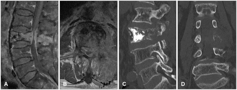
- Korpusun trabeküler ve kortikal yapısında keskin kırık hattı, posterior duvardan kanala doğru köşeli ve keskin sınırlı kemik fragmanı retropulsiyonu, intravertebral gaz cleft’i ve impaksiyon benign örüntüyü destekler; retropulsiyon tek başına ayırt ettirici değildir. Kemik yıkımı, korpus ekspansiyonu ve epidural/paravertebral yumuşak doku kitlesi maligniteyi destekler. BT, kırık morfolojisini ve kortikal kemik bütünlüğünü değerlendirmede kullanılır.
- Kemik algoritması tanısal
- Sagittal ve aksiyel ince kesit rekonstrüksiyonlarında kırık kenarlarının keskin mi destrüktif mi olduğu, posterior duvar fragmanının şekli, pedikül bütünlüğü ve kortikal genişleme değerlendirilir.
Önerilen pencereler: Kemik (G 1800 / M 400), Yumuşak doku (G 400 / M 40).
BT bulguları
- Benign lehine keskin kırık çizgisi — düzenli sınırlı kırık aralığı ve eşlik eden osteolitik kemik kaybı olmaması.
- Retropulse kortikal fragman — posterior duvar parçası kanala doğru köşeli biçimde yer değiştirir; vertebra dış konturunun düzgün yaygın kabarmasından ayrılır.
- İntravertebral vakum cleft'i — kırık korpusu içindeki gaz yarığı benign kırık/instabiliteyle ilişkili olabilir ancak mutlak dışlama bulgusu değildir.
- Malignite lehine kemik destrüksiyonu — trabeküler veya kortikal yıkımın kırık hattı dışına taşması.
- Korpus ekspansiyonu — vertebra arka konturunun yaygın dışbükeyleşmesi ya da gövdenin tümöral genişlemesi.
- Epidural/paravertebral kitle — kırık korpusun ötesinde kanal veya paravertebral alana uzanan fokal yumuşak doku komponenti.
- Çok seviyeli kemik lezyonları — diğer vertebralardaki tekli veya çoklu lezyonların sayısı ve morfolojisi ayırıcı değerlendirmede dikkate alınır; tek başına kökeni belirlemez.
Normalde
Normal korpusun trabeküler dokusu ve kortikal konturu kesintisizdir; posterior sınır spinal kanala doğru düzgün ve hafif konkav konturunu korur. Aynı düzey ve komşu vertebralarla kıyaslarken sınırlı keskin kırık kenarını, retropulse fragmanın şeklini, kemik yıkımı/ekspansiyonu ve kemik dışı kitleyi ayrı ayrı değerlendirin.
Normal BT ile kıyasla
Aynı bölgenin, kaynak veri setinde patoloji içermediği belirtilen bir BT incelemesini kesit kesit kaydırın; organ sınırlarını açarak anatomiyi hasta görüntüsüyle karşılaştırın. Küçük rastlantısal bulgular tümüyle dışlanmamıştır.
Kaynak: TotalSegmentator v2.01 veri seti (Wasserthal ve ark., Radiology: Artificial Intelligence 2023), CC BY 4.0, vaka s1338; organ sınırları veri setinin otomatik segmentasyonundan, birleştirilerek sadeleştirildi. Görüntüler 2,5 mm kesit aralığına yeniden örneklendi.
Ayırıcı tanı
- Benign osteoporotik kompresyon
- keskin kırık kenarı, son plak impaksiyonu, retropulse fragman veya intravertebral vakum görülebilir; destrüktif odak ve kitle yokluğu destekleyicidir.
- Malign patolojik kompresyon
- fokal kemik yıkımı, korpus ekspansiyonu, pedikül tutulumu ve epidural/paravertebral kitle maligniteyi destekler.
- Vertebral osteomiyelit
- komşu son plaklarda ve disk aralığında inflamatuvar destrüksiyon, paravertebral flegmon veya koleksiyonla ayrılır.
- Vertebral hemanjiyom
- kaba, dikey yönelimli trabeküler kalınlaşma ve aksiyel polka-dot görünümü tipik ipuçlarıdır; agresif lezyonlar yumuşak doku uzanımı yapabilir.
- Vertebral osteonekroz
- intravertebral gaz veya sıvı cleft'i gösterebilir; klinik zaman çizgisi ve MR bulguları benign kırık spektrumunda değerlendirilmeyi gerektirir.
Tuzaklar
- Posterior duvar çıkıntısı tek başına malignite değildir; keskin ve açılı retropulse kemik fragmanı benign kırıkta görülebilir, tümöral ekspansiyon ise daha yaygın dışbükey kontur oluşturur.
- Vakum cleft’i vertebral osteonekroz/Kümmell hastalığıyla ilişkilendirilmiştir; ancak bu bulgunun tek başına maligniteyi dışladığı sonucuna varılamaz.
- Pedikül tutulumu maligniteyi destekler fakat akut kırıkta reaktif değişiklik olabilir; bulguyu yumuşak doku kitlesi, diğer kemik odakları ve MR ile birlikte tartın.
- BT'de kesin ayrım yapılamıyorsa belirsizliği koruyun; MR kemik iliği sinyali ve epidural yayılım için daha güçlü tamamlayıcı yöntemdir.
Kendini dene
Kompresyon kırığında arka korteksten kanala doğru köşeli kemik parçası uzanıyor; korpus ekspansiyonu ve yumuşak doku kitlesi yok. Hangi örüntü daha olasıdır?
Cevabı göster
Benign osteoporotik kırık. Köşeli retropulse kortikal fragman ve ekspansiyon/kitle olmaması benign kırık örüntüsünü destekler; tek başına kesin dışlama sağlamaz. Malign kırıkta destrüksiyon/ekspansiyon, enfeksiyonda disk-son plak değişikliği, agresif hemanjiyomda kaba trabeküler yapı aranır.
Korpus çökmesine eşlik eden posterior vertebral tutulum, benign ve malign kırık ayrımında hangi yönde değerlendirilmelidir? Posterior vertebral tutulum, çalışmada bağımsız bir öngördürücü olarak bildirilmiştir; bu bulgu hangi kırık grubuyla ilişkilidir?
Cevabı göster
Malign kompresyon. Çalışmada posterior vertebral tutulum, benign-malign vertebral kompresyon kırığı ayrımında bağımsız bir klinik öngördürücü olarak bildirilmiştir; bu bulgu tek başına kesin tanı koydurmaz. Benign ve malign kompresyon kırıklarının görüntüleme bulguları örtüşebilir; bu nedenle tek bir işaret kesin ayrım sağlamaz.
İlgili konular
Kaynaklar
Bu sayfadaki 40 cümle ve 2 quiz sorusu aşağıdaki kaynaklardan alıntılarla tek tek karşılaştırıldı; 10 cümle bu karşılaştırmada düzeltildi (2026-10-09).
- Radiomics in spinal research: a narrative reviewpmc.ncbi.nlm.nih.gov
- Quantification in bone SPECT/CTpmc.ncbi.nlm.nih.gov
- Metastatic Retroperitoneal Rhabdomyosarcoma in a Child: A Case Report Highlighting the Role of Imaging in Diagnosis, Staging, and Follow-Uppmc.ncbi.nlm.nih.gov
- Management of aneurysmal bone cysts of the spine and sacrum: A narrative reviewpmc.ncbi.nlm.nih.gov
- Differential diagnosis of benign and malignant vertebral compression fractures based on CT radiomics modelpmc.ncbi.nlm.nih.gov
- ESR Essentials: bone marrow MRI in oncology-practice recommendations by the European Society of Musculoskeletal Radiology.pubmed.ncbi.nlm.nih.gov
- Beyond Chordoma: A Comprehensive Review of Sacral Lesions.pubmed.ncbi.nlm.nih.gov
- Comprehensive Review of the Utility of Dynamic Contrast-Enhanced MRI for the Diagnosis and Treatment Assessment of Spinal Benign and Malignant Osseous Disease.pubmed.ncbi.nlm.nih.gov
- Spinal lesions: a comprehensive radiologic overview.pubmed.ncbi.nlm.nih.gov
- A pictorial essay of thoracic wall diseases: multiple pathologies in the same anatomical site.pubmed.ncbi.nlm.nih.gov
- Diffusion-Weighted Imaging in the Musculoskeletal System: Evolving Role in Modern Imaging Practice.pubmed.ncbi.nlm.nih.gov
- 2015 American Thyroid Association Management Guidelines for Adult Patients with Thyroid Nodules and Differentiated Thyroid Cancer: The American Thyroid Association Guidelines Task Force on Thyroid Nodules and Differentiated Thyroid Cancer.pubmed.ncbi.nlm.nih.gov
- Identifying osteoporotic vertebral endplate and cortex fractures.pubmed.ncbi.nlm.nih.gov
- Primary Spinal Marginal Zone Lymphoma Presenting With Spinal Cord Compression: A Case Report.pubmed.ncbi.nlm.nih.gov
- Cervical spine chordoma: Imaging features and diagnostic challenges of a rare C6 localization.pubmed.ncbi.nlm.nih.gov
- Unveiling Primary Bone Tumors of the Spine: A Review of Essential Imaging Clues.pubmed.ncbi.nlm.nih.gov
- Artificial Intelligence in Spine Neuroimaging: Diagnostic and Prognostic Utility of Novel Biomarkers in Lower Back Pain.pubmed.ncbi.nlm.nih.gov
- Multimodal imaging in neuro-ophthalmology.pubmed.ncbi.nlm.nih.gov
- Deep learning and hybrid architectures for atypical and complex bone fracture diagnosis: a systematic review of performance and clinical validity.pubmed.ncbi.nlm.nih.gov
- Anatomical and biological reconstruction of traumatic complete burst fractures of the thoracolumbar vertebrae using pedicle fixation combined with stent-armed kyphoplasty and intracorporeal bone grafting: technique rationale, 7-year experience and clinical, imaging, and histological outcomes.pubmed.ncbi.nlm.nih.gov
- Aggressive lumbar vertebral hemangioma presenting with foot drop mimicking spinal tuberculosis: A case report.pubmed.ncbi.nlm.nih.gov
- Intrabody Cage Augmentation in Kümmell Disease and Osteoporotic Burst Fractures: Technical Insights and Narrative Review of Current Evidence.pubmed.ncbi.nlm.nih.gov
- Clavicular Gorham-Stout disease: a rare case report and literature review.pubmed.ncbi.nlm.nih.gov
- Non-Infectious Spinal Diseases Mimicking Infection: A Pictorial Essay.pubmed.ncbi.nlm.nih.gov
- Comparison of Artificial Intelligence Models Using CT Radiomics for Predicting Post-Vertebral Augmentation Residual Back Pain in Osteoporotic Vertebral Compression Fractures.pubmed.ncbi.nlm.nih.gov
- Review of the Imaging Features of Benign Osteoporotic and Malignant Vertebral Compression Fractures.pubmed.ncbi.nlm.nih.gov
- Kümmell's Disease: Clarifying the Mechanisms and Patients' Inclusion Criteria.pubmed.ncbi.nlm.nih.gov
- Paget's Disease of the Spine as a Catalyst for Osteosarcoma: A Narrative Review.pubmed.ncbi.nlm.nih.gov
- Radiomics in spinal research: a narrative review.pubmed.ncbi.nlm.nih.gov
- Imaging of ewing sarcoma: an updated analysis including presenting features, prognostic imaging biomarkers, and treatment response assessment.pubmed.ncbi.nlm.nih.gov
- Bone Metastases Are Measurable: The Role of Whole-Body MRI and Positron Emission Tomography.pubmed.ncbi.nlm.nih.gov
- Axial Spondyloarthritis: Mimics and Pitfalls of Imaging Assessment.pubmed.ncbi.nlm.nih.gov
- Pathological Vertebral Fractures Misdiagnosed as Osteoporotic Vertebral Fractures: A Case Series of Four Patients and Diagnostic Strategies.pubmed.ncbi.nlm.nih.gov
- Case Report: Diagnostic and therapeutic challenges of secondary spinal tuberculosis following percutaneous vertebroplasty: a case of staged surgery with bone cement retention report.pubmed.ncbi.nlm.nih.gov
- An overview of the tumors affecting the spine-inside to out.pubmed.ncbi.nlm.nih.gov
- Long-term safety and effectiveness of the "OptEase" vena cava filter.pubmed.ncbi.nlm.nih.gov
- The strain distribution in the lumbar anterior longitudinal ligament is affected by the loading condition and bony features: An in vitro full-field analysis.pubmed.ncbi.nlm.nih.gov
- Statistical shape modelling of the human lumbar spine: a scoping review characterising the research landscape and potential clinical applications.pubmed.ncbi.nlm.nih.gov
- Oral Food Supplement with Bio-Activated Silicium and Vitamins D3 and K2 in the Conservative Management of Osteoporotic Vertebral Compression Fractures.pubmed.ncbi.nlm.nih.gov
- Spinal granulocytic sarcoma with both extradural and intradural involvement: a case report.pubmed.ncbi.nlm.nih.gov
- Acute Cauda Equina Syndrome Following Transpedicular Biopsy of a Lumbar Vertebral Lesion: A Case Report of Spinal Non-Hodgkin's Lymphoma.pubmed.ncbi.nlm.nih.gov
- Differentiating Tuberculous and Pyogenic Spondylodiscitis: Part II: MRI/CT Features, Prediction Models, and the Role of PET/CT-A Narrative Review.pubmed.ncbi.nlm.nih.gov
- Purely osteolytic bone lesions in the early osteoarticular phase of synovitis, acne, pustulosis, hyperostosis and osteitis syndrome.pubmed.ncbi.nlm.nih.gov
- Streptomyces morookaense spinal suppurative infection: a case report.pubmed.ncbi.nlm.nih.gov
- Aggressive vertebral hemangiomas contain no adipose tissue resulting in thoracic spine kyphosis: A case report.pubmed.ncbi.nlm.nih.gov
- Advances in Bone Marrow Imaging: Strengths and Limitations from a Clinical Perspective.pubmed.ncbi.nlm.nih.gov
- What is the Current Evidence on Vertebral Body Osteonecrosis?: A Systematic Review of the Literature.pubmed.ncbi.nlm.nih.gov
- The value of magnetic resonance imaging and computed tomography in the study of spinal disorders.pubmed.ncbi.nlm.nih.gov
- Discrimination between Malignant and Benign Vertebral Fractures Using Magnetic Resonance Imaging.pubmed.ncbi.nlm.nih.gov
- Focal nodular marrow hyperplasia: Imaging features of 53 cases.pubmed.ncbi.nlm.nih.gov
- Vertebral Sarcoidosis Mimicking Metastatic Disease in a Patient With Prior Melanoma: A Diagnostic Challenge.pubmed.ncbi.nlm.nih.gov
- Routine MRI for opportunistic osteoporosis screening: diagnostic accuracy of vertebral bone quality and a novel femoral bone quality score compared with DXA.pubmed.ncbi.nlm.nih.gov
- Generation of Vertebra Micro-CT-like Image from MDCT: A Deep-Learning-Based Image Enhancement Approach.pubmed.ncbi.nlm.nih.gov
- Myeloma bone disease imaging on a 1st-generation clinical photon-counting detector CT vs. 2nd-generation dual-source dual-energy CT.pubmed.ncbi.nlm.nih.gov
- Application of tomosynthesis for vertebral compression fracture diagnosis and bone healing assessment in fracture liaison services.pubmed.ncbi.nlm.nih.gov
- Clinical outcomes and complications after pedicle subtraction osteotomy for fixed sagittal imbalance patients : a long-term follow-up data.pubmed.ncbi.nlm.nih.gov
- Current State-of-the-Art 3D MRI Sequences for Assessing Bone Morphology with Emphasis on Cranial and Spinal Imaging: A Narrative Review.pubmed.ncbi.nlm.nih.gov
- Expert consensus on a multidisciplinary approach for the management of multiple myeloma-related bone disease.pubmed.ncbi.nlm.nih.gov
- A transformer-based deep learning algorithm for diagnosing spinal infections on axial non-contrast computed tomography images: a dual-center retrospective study.pubmed.ncbi.nlm.nih.gov
- Role of 3D Printing in Preoperative Planning for Spine Surgery: A Scoping Review.pubmed.ncbi.nlm.nih.gov
- The role of radiography in the study of spinal disorders.pubmed.ncbi.nlm.nih.gov
- Development and validation of a multiparametric MRI-based radiomics nomogram for the tripartite discrimination of primary benign, primary malignant, and metastatic lumbar spinal tumors.pubmed.ncbi.nlm.nih.gov
- Romosozumab Versus Teriparatide for Fracture Prevention, Cardiovascular Events, and Mortality in Osteoporosis: A Propensity Score-Matched Real-World Cohort Study.pubmed.ncbi.nlm.nih.gov
- Nonprostatic diseases on PSMA PET imaging: a spectrum of benign and malignant findings.pubmed.ncbi.nlm.nih.gov
- Cervical Pyogenic Spondylitis: A Comprehensive Review of Diagnosis and Treatment Strategy.pubmed.ncbi.nlm.nih.gov
- The American Society of Pain and Neuroscience (ASPN) Evidence-Based Clinical Guideline of Interventional Treatments for Low Back Pain.pubmed.ncbi.nlm.nih.gov
- Pathological Fracture of Thoracic Spine Aneurysmal Bone Cyst: A Case With Successful Outcome Following Debulking, Posterior Instrumentation, and Allogenic Bone Impaction Grafting.pubmed.ncbi.nlm.nih.gov
- Unusual Presentation of Thoracic Chordoma with Spinal Epidural Hematoma: A Rare Case Report and PRISMA-Driven Systematic Review.pubmed.ncbi.nlm.nih.gov
- The Role of CT and MR Imaging in Stereotactic Body Radiotherapy of the Spine: From Patient Selection and Treatment Planning to Post-Treatment Monitoring.pubmed.ncbi.nlm.nih.gov
- Quantification in bone SPECT/CT.pubmed.ncbi.nlm.nih.gov
- Metastatic Retroperitoneal Rhabdomyosarcoma in a Child: A Case Report Highlighting the Role of Imaging in Diagnosis, Staging, and Follow-Up.pubmed.ncbi.nlm.nih.gov
- Geriatric spine fractures - Demography, changing trends, challenges and special considerations: A narrative review.pubmed.ncbi.nlm.nih.gov
- Radiologic-Histopathologic Correlation of Adult Spinal Tumors: A Retrospective Study.pubmed.ncbi.nlm.nih.gov
- An exploratory multimodal imaging and clinicopathological strategy for suspected Kümmell's disease in the absence of MRI.pubmed.ncbi.nlm.nih.gov
- Deep learning magnetic resonance imaging algorithm for differentiating metastatic vertebral fractures.pubmed.ncbi.nlm.nih.gov
- Preoperative Cobb angle-guided surgical decision-making between percutaneous kyphoplasty and short-segment fixation with vertebroplasty in stage III Kümmell disease without neurological deficits: a two-center retrospective study.pubmed.ncbi.nlm.nih.gov
- Three-dimensional computed tomography morphometric analysis of lumbar endplates in Chinese young adults.pubmed.ncbi.nlm.nih.gov
- Arthroscopic capsular release combined with Latarjet procedure for chronic locked anterior shoulder dislocation secondary to epilepsy: a case report with one-year follow-up.pubmed.ncbi.nlm.nih.gov
- High-Riding Vertebral Artery in Cervical Spine Surgery: A Review of Preoperative Identification and Surgical Implications.pubmed.ncbi.nlm.nih.gov
- Diagnostic decision-making, vertebral augmentation, and osteoporosis therapy in vertebral compression fractures: a narrative review.pubmed.ncbi.nlm.nih.gov
- The orthopaedic management of osteopetrosis in paediatric populations: a narrative review of the literature.pubmed.ncbi.nlm.nih.gov
- Imaging intraosseous hemangiomas: Typical and atypical features across modalities.pubmed.ncbi.nlm.nih.gov
- Aggressive thoracic vertebral hemangioma managed with preoperative embolization and surgical decompression: A case report.pubmed.ncbi.nlm.nih.gov
- Paraspinal Muscle Fat Infiltration as a Key Predictor of Symptomatic Intravertebral Vacuum Cleft: A Machine Learning Approach.pubmed.ncbi.nlm.nih.gov
- Multidisciplinary management of spinal metastases: what the radiologist needs to know.pubmed.ncbi.nlm.nih.gov
- Multivariate Risk Factor Analysis and Clinical Prediction Model Construction of Intercostal Neuralgia Following Single-Segment Osteoporotic Thoracic Vertebral Compression Fractures.pubmed.ncbi.nlm.nih.gov
- Combine Short-Segment Fixation and Leverage Reduction Technique to Correct Kyphotic Angle in Kümmell's Disease.pubmed.ncbi.nlm.nih.gov
- Manifestations of radiation toxicity in the head, neck, and spine: An image-based review.pubmed.ncbi.nlm.nih.gov
- Current trends in mini-invasive management of spine metastases.pubmed.ncbi.nlm.nih.gov
- CT and MR imaging of odontoid abnormalities: A pictorial review.pubmed.ncbi.nlm.nih.gov
- Understanding Photon-Counting CT: Physics, Detector Technology, and Image Reconstructions.pubmed.ncbi.nlm.nih.gov
- Global Vascular Guidelines on the Management of Chronic Limb-Threatening Ischemia.pubmed.ncbi.nlm.nih.gov
- Global vascular guidelines on the management of chronic limb-threatening ischemia.pubmed.ncbi.nlm.nih.gov
- Applicability of RECIST1.1, MDACC and SPINO Tumor Response Criteria After Adjuvant Stereotactic Spinal Radiosurgery in Surgically Treated Spinal Metastases.pubmed.ncbi.nlm.nih.gov
- Case review of 3 patients with musculoskeletal manifestation of human immunodeficiency virus and highly active antiretroviral therapy in the form of osteopenia and insufficiency fractures.pubmed.ncbi.nlm.nih.gov
- Multiple Lumbar Compression Fractures Following a New-Onset Seizure: A Case Report.pubmed.ncbi.nlm.nih.gov
Eğitim amaçlıdır; hasta başında tanı ve tedavi kararının yerine geçmez. İçerik yapay zekâ destekli bir süreçle güncel literatüre göre cümle cümle kaynaklarla karşılaştırıldı, ancak uzman radyolog incelemesinden geçmedi. Klinik kararlarda güncel kılavuzları, kurum protokollerini ve radyoloji raporunu esas alın.