Özet
Atriyal septal defekt, atriyumlar arasında doğuştan bulunan bir açıklık veya venöz giriş düzeyindeki septal gelişim anomalisi olup; uzun süreli soldan sağa şant sağ kalp hacim yüküne yol açabilir. EKG eşlemeli kontrastlı BT, özellikle sinus venosus defektinde açıklığın yerini ve eşlik eden parsiyel anormal pulmoner venöz dönüşleri anatomik olarak haritalandırır. Ekokardiyografi kardiyak CHD'de birinci basamak görüntülemedir; hemodinamik değerlendirmede CMR ve gerekirse BT tamamlayıcı bilgi sağlar. Gözden kaçan anormal pulmoner ven dönüşü, sağ kalp genişlemesinin nedeninin ve cerrahi anatominin eksik tanımlanmasına neden olabilir.
Faz ve pencere
- BTA tanısal
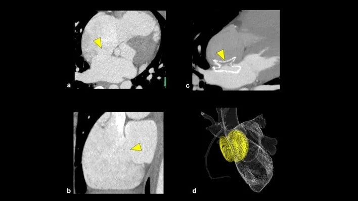
- EKG eşlemeli kontrastlı kardiyak BT’de fossa ovalis, atriyoventriküler bileşke ve vena kava-sağ atriyum birleşimindeki septal devamlılık değerlendirilir; sinus venosus tipinde üst veya alt kavoatriyal bileşkede açıklık ve pulmoner venlerin SVC, IVC ya da sağ atriyuma anormal drenajı izlenebilir. Sağ atriyum ve sağ ventrikül genişlemesi, sol-sağ şanta bağlı hacim yükünün varlığını gösterir.
Önerilen pencereler: Mediasten (G 350 / M 50), Yumuşak doku (G 400 / M 40), Anjiyo (G 600 / M 150).
BT bulguları
- Ostium secundum defektinde fossa ovalis düzeyinde interatriyal septumun orta bölümünde açıklık veya doku eksikliği.
- Ostium primum defektinde atriyoventriküler kapaklara komşu alt septumda açıklık; eşlik eden atriyoventriküler septal bileşke anomalisi.
- Sinus venosus defektinde SVC-sağ atriyum veya daha seyrek IVC-sağ atriyum birleşiminde septumun üst ya da alt kenarında açıklık.
- Sağ üst pulmoner venin veya diğer sağ pulmoner venlerin sol atriyum yerine SVC ya da sağ atriyuma bağlandığı parsiyel anormal pulmoner venöz dönüş.
- Uzun süreli hacim yüküyle uyumlu sağ atriyum ve sağ ventrikül genişlemesi; interventriküler septumda düzleşme görülebilmesi.
- Koroner sinüs çatısındaki defektin sol atriyumla koroner sinüs arasında bağlantı oluşturması.
- İnteratriyal kontrast opasifikasyon farkı tek başına şant akımını göstermez; kontrastlı BT şant akımını değerlendiremez.
Ölçütler ve sınıflamalar
- Anatomik ASD tipleri
- Ostium secundum: fossa ovalis düzeyinde orta septum. Ostium primum: atriyoventriküler kapaklara komşu alt septum. Sinus venosus: SVC-RA veya IVC-RA bileşkesinde, septumun üst/alt kenarında. Koroner sinüs tipi: koroner sinüs çatısının kısmen veya tamamen yokluğu ile sol atriyum-koroner sinüs iletişimi.
Normalde
Normal interatriyal septum fossa ovalisi çevresinde kesintisiz ince doku olarak sağ ve sol atriyumu ayırır; pulmoner venler sol atriyuma açılır ve sağ kalp boşlukları sol tarafa göre belirgin hacim genişlemesi göstermez. Septumu fossa ovalis, AV kapak düzlemi ve vena kava birleşimleri boyunca izleyin; pulmoner venlerin her birinin sol atriyumla bağlantısını aynı kontrastlı hacimde doğrulayın.
Normal BT ile kıyasla
Aynı bölgenin, kaynak veri setinde patoloji içermediği belirtilen bir BT incelemesini kesit kesit kaydırın; organ sınırlarını açarak anatomiyi hasta görüntüsüyle karşılaştırın. Küçük rastlantısal bulgular tümüyle dışlanmamıştır.
Kaynak: TotalSegmentator v2.01 veri seti (Wasserthal ve ark., Radiology: Artificial Intelligence 2023), CC BY 4.0, vaka s0977; organ sınırları veri setinin otomatik segmentasyonundan, birleştirilerek sadeleştirildi. Görüntüler 2,5 mm kesit aralığına yeniden örneklendi.
Ayırıcı tanı
- Patent foramen ovale
- üst üste binen septum primum ve secundum arasında oblik bir tünel bulunur; gerçek bir septal açıklık (doku kaybı) değil, örtüşen yaprakların oluşturduğu geçicidir.
- Sinus venosus tipi parsiyel anormal pulmoner venöz bağlantı
- SVC veya IVC’ye açılan pulmoner venler, septal defekt gözden kaçsa bile birlikte bulunması sinus venosus tipini düşündürür.
- Sağ ventrikül hacim yükünün diğer nedenleri
- pulmoner kapak yetersizliği veya başka soldan sağa şantlar sağ boşlukları genişletebilir; interatriyal açıklık ve venöz bağlantılar ayrıca aranır.
- Atriyal septal anevrizma, interatriyal septum anomalileri arasında yer alır.
- Koroner sinüs çatısı defekti
- klasik fossa ovalis açıklığı yerine sol atriyum ile genişlemiş koroner sinüs arasında bağlantı izlenir.
Tuzaklar
- İnteratriyal septum anatomisini BT verilerinden dikkatle yeniden yapılandırılmış çok düzlemli görüntülerde değerlendirin.
- Kontrastlı BT septal şant akımını değerlendiremez; opasifikasyon farkı tek başına şantın varlığını göstermez.
- Sinus venosus defekti çoğu zaman fossa ovalis içinde değil kavoatriyal bileşkededir; pulmoner venlerin tamamını SVC ve IVC boyunca ayrı ayrı izleyin.
- Sağ kalp genişlemesi ASD’ye özgü değildir; doğrudan septal bulgu ve pulmoner ven bağlantıları gösterilmeden yalnızca hacim artışı bulgusundan defekt tipi çıkarılmamalıdır.
Kendini dene
Sağ ventrikül geniş; sağ üst pulmoner ven SVC’ye açılıyor ve açıklık üst kavoatriyal sınırda. En olası defekt tipi nedir?
Cevabı göster
Sinus venosus. Üst kavoatriyal açıklıkla SVC’ye anormal pulmoner venöz dönüş sinus venosus tipini destekler. Secundum defekt fossa ovalis merkezindedir; primum alt AV bileşkeye komşudur; patent foramen ovale ise örtüşen septal yaprakların tünelidir.
Fossa ovalis düzeyinde örtüşen ince septal yapraklar arasında oblik tünel izleniyor; doku kaybı yok. En olası yapı nedir?
Cevabı göster
Patent foramen ovale. Örtüşen septum primum ve secundum arasındaki oblik tünel patent foramen ovale görünümüdür. Secundum ASD gerçek orta septal doku açıklığıdır; sinus venosus kavoatriyal bileşkededir; primum defekt alt septumda AV kapaklara komşudur.
İlgili konular
Kaynaklar
Bu sayfadaki 42 cümle ve 2 quiz sorusu aşağıdaki kaynaklardan alıntılarla tek tek karşılaştırıldı; 11 cümle bu karşılaştırmada düzeltildi (2026-10-09).
- Life-Threatening Aorto-Atrial Erosion Following Transcatheter Ostium Secundum Atrial Septal Defect Closure: A Case-Based Reviewpmc.ncbi.nlm.nih.gov
- Breaking the Barriers: Transseptal Puncture in Patients with Atrial Septal Defect Closure Devicespmc.ncbi.nlm.nih.gov
- Atrial Septal Defect and Heart Rhythm Disorders: Physiopathological Linkage and Clinical Perspectivespmc.ncbi.nlm.nih.gov
- Anti-Thrombotic Therapy Following Transcatheter Structural Heart Interventionpmc.ncbi.nlm.nih.gov
- Cardiac Computed Tomography in Structural Heart Interventions: From Preprocedural Planning to Procedural Strategypmc.ncbi.nlm.nih.gov
- Common Left-to-Right Shunts.pubmed.ncbi.nlm.nih.gov
- Imaging congenital heart disease in adults.pubmed.ncbi.nlm.nih.gov
- Vici syndrome in siblings born to consanguineous parents.pubmed.ncbi.nlm.nih.gov
- Clinical Relevance of Computed Tomography Pulmonary Venography.pubmed.ncbi.nlm.nih.gov
- The role of cardiovascular magnetic resonance in adults with congenital heart disease.pubmed.ncbi.nlm.nih.gov
- Multidetector CT of left atrium.pubmed.ncbi.nlm.nih.gov
- Multimodality imaging anatomy of interatrial septum and mitral annulus.pubmed.ncbi.nlm.nih.gov
- Complex atrial septal defect closure in children.pubmed.ncbi.nlm.nih.gov
- Demonstration of living anatomy clarifies the morphology of interatrial communications.pubmed.ncbi.nlm.nih.gov
- Assessing potential divers with a history of congenital heart disease.pubmed.ncbi.nlm.nih.gov
- Patent foramen ovale and thromboembolic complications.pubmed.ncbi.nlm.nih.gov
- Unilateral absence of pulmonary artery: pathophysiology, symptoms, diagnosis and current treatment.pubmed.ncbi.nlm.nih.gov
- Tricuspid Valve Imaging and Intervention in Pediatric and Adult Patients With Congenital Heart Disease.pubmed.ncbi.nlm.nih.gov
- Cardiovascular Imaging for Guiding Interventional Therapy in Structural Heart Diseases.pubmed.ncbi.nlm.nih.gov
- AMPLATZER Septal Occluder failure resulting in paradoxical cerebral embolism.pubmed.ncbi.nlm.nih.gov
- Staged Therapeutic Approach to Concomitant Severe Aortic Stenosis and Atrial Septal Defect: Addressing Dual Mechanisms of Heart Failure-Case Report and Literature Review.pubmed.ncbi.nlm.nih.gov
- Imaging the Adult With Secundum Atrial Septal Defect: A Practical Echocardiographic Approach From Diagnosis to Device Closure.pubmed.ncbi.nlm.nih.gov
- Long-Term Outcome After Pulsed Field Ablation for Atrial Fibrillation Across an Atrial Septal Defect Closure Device: A 20-Month Follow-Up Case Report.pubmed.ncbi.nlm.nih.gov
- Inferior Sinus Venosus Defect: Inconclusive Transesophageal Echocardiography Clarified by Computed Tomography and Intracardiac Echocardiography.pubmed.ncbi.nlm.nih.gov
- The Initial Diagnostic Approach to Adult Congenital Heart Disease: A Practical Imaging Pathway for General Cardiologists and Cardiac Imagers.pubmed.ncbi.nlm.nih.gov
- Cardiovascular Computed Tomography Angiographic Assessment of Simple Cardiac Shunts in Adults.pubmed.ncbi.nlm.nih.gov
- Society for Cardiovascular Magnetic Resonance 2022 Cases of SCMR case series.pubmed.ncbi.nlm.nih.gov
- Early Diagnosis of Cardiovascular Diseases in Workers: Role of Standard and Advanced Echocardiography.pubmed.ncbi.nlm.nih.gov
- Anomalous Coronary Venous Drainage to the Right Atrium: A Case Report.pubmed.ncbi.nlm.nih.gov
- Raghib Syndrome Physiology Revealed by Multimodality Cardiac Imaging.pubmed.ncbi.nlm.nih.gov
- Surgical Correction of a Sinus Venosus Atrial Septal Defect with Partial Anomalous Pulmonary Venous Connections Using Cardiac Computed Tomography Imaging and a 3D-Printed Model.pubmed.ncbi.nlm.nih.gov
- Delayed Diagnosis of Congenital Heart Diseases in Adults: Opportunities for Improving Diagnostic Strategies.pubmed.ncbi.nlm.nih.gov
- Supracardiac Total Anomalous Pulmonary Venous Return With Extrinsic Compression of the Vertical Vein by the Left Pulmonary Artery: A Case Report.pubmed.ncbi.nlm.nih.gov
- Urgent sutureless repair of supracardiac total anomalous pulmonary venous return coexisting with Ebstein's anomaly in a female neonate: a rare case report.pubmed.ncbi.nlm.nih.gov
- Transseptal puncture in cardiovascular interventions: a clinical consensus statement of the European Heart Rhythm Association, the Heart Failure Association, the European Association of Percutaneous Cardiovascular Interventions, the European Association of Cardiovascular Imaging of the ESC, and the Association for European Paediatric and Congenital Cardiology.pubmed.ncbi.nlm.nih.gov
- Diagnosis and Management of Patients with Patent Foramen Ovale: Current Evidence and Future Perspective.pubmed.ncbi.nlm.nih.gov
- Multimodality echocardiography in structural heart disease: the evolving role of the interventional imager.pubmed.ncbi.nlm.nih.gov
- The Congenital Malformation of the Interatrial Septum-A Review of Its Development and Embryology with Clinical Implications.pubmed.ncbi.nlm.nih.gov
- Same-same, but different? Postoperative rhythm disturbances and other analysis after rotational Z-flap vs. patch repair of sinus venosus atrial defects.pubmed.ncbi.nlm.nih.gov
- Transesophageal Echocardiography and Clinical Significance of Giant Eustachian Valve during Cardiac Surgery: A Narrative Review.pubmed.ncbi.nlm.nih.gov
- Sequential transcatheter closure of sinus venosus and secundum atrial septal defects using covered stents and a septal occluder, a two-step strategy: a case report.pubmed.ncbi.nlm.nih.gov
- Congenital Heart Disease in a Paralympic Athlete With Klinefelter Syndrome.pubmed.ncbi.nlm.nih.gov
- Reversible coma and elevated central venous pressure: warning signs of acute superior vena cava graft thrombosis following Warden procedure: a case report.pubmed.ncbi.nlm.nih.gov
- Beyond the right ventricle: assessment of the left ventricular function in pulmonary hypertension.pubmed.ncbi.nlm.nih.gov
- Unrepaired Congenital Shunt Lesions in Adults: Principles for Clinical Management.pubmed.ncbi.nlm.nih.gov
- Beyond the right ventricle: left heart involvement in pulmonary arterial hypertension.pubmed.ncbi.nlm.nih.gov
- Electrosurgery in pediatric catheter interventions.pubmed.ncbi.nlm.nih.gov
- Inferior Left Atrial Diverticulum Communicating with the Right Atrium or Inferior Vena Cava: Prevalence and CT Features.pubmed.ncbi.nlm.nih.gov
- Crossing the Septum: Step-by-Step Anatomy, Technique, and Enabling Technologies in Transseptal Puncture for Atrial Fibrillation and Electrophysiological Procedures.pubmed.ncbi.nlm.nih.gov
- First- and Second-Trimester Cardiovascular Anomalies in Trisomy 21 Fetuses: Anatomy, Embryology, Genetics and Imaging.pubmed.ncbi.nlm.nih.gov
- Atrial wall dissection: a contemporary overview for a proper decision-making approach.pubmed.ncbi.nlm.nih.gov
- Cardiac Computed Tomography in Structural Heart Interventions: From Preprocedural Planning to Procedural Strategy.pubmed.ncbi.nlm.nih.gov
- Atrial Septal Defects: From Embryology to Pediatric Pulmonary Hypertension.pubmed.ncbi.nlm.nih.gov
- When Palpitations Unmask Crista Terminalis Hypertrophy: A Case Report and Review of Current Literature.pubmed.ncbi.nlm.nih.gov
- When an Atrial Septal Defect Is No Longer Silent: A Case Report of a Large Symptomatic Defect Presenting in Adulthood.pubmed.ncbi.nlm.nih.gov
- Transcatheter Closure of Patent Ductus Arteriosus in Adults.pubmed.ncbi.nlm.nih.gov
- Pulmonary Hypertension Workup Unraveling a Rare Anomaly in Turner Syndrome.pubmed.ncbi.nlm.nih.gov
- Congenital heart disease: types, pathophysiology, diagnosis, and treatment options.pubmed.ncbi.nlm.nih.gov
- A review of the oral health of individuals with disabilities in Puerto Rico and among U.S. Hispanics.pubmed.ncbi.nlm.nih.gov
- Society for Cardiovascular Magnetic Resonance 2024 Cases of SCMR case series.pubmed.ncbi.nlm.nih.gov
- Nongated Photon-Counting Computed Tomography Angiography Detects Cardioembolic Stroke Sources and Thoracic Pathology: A Retrospective Cohort Study.pubmed.ncbi.nlm.nih.gov
- Ferumoxytol-enhanced cardiovascular magnetic resonance imaging: applications and technical advances.pubmed.ncbi.nlm.nih.gov
- 2024 European Heart Rhythm Association/Heart Rhythm Society/Asia Pacific Heart Rhythm Society/Latin American Heart Rhythm Society expert consensus statement on catheter and surgical ablation of atrial fibrillation.pubmed.ncbi.nlm.nih.gov
- Congenital Pericardial Agenesis: An Innocent Finding or Clinically Significant Condition? A Case Series and Literature Review.pubmed.ncbi.nlm.nih.gov
- How to Do Echo in Left Ventricular Hypertrophy: A Consensus Statement of the Italian Society of Echocardiography and Cardiovascular Imaging.pubmed.ncbi.nlm.nih.gov
- Inferior vena cava anomalous drainage into the left atrium with severe pulmonary arterial hypertension: a case report and review of management strategies.pubmed.ncbi.nlm.nih.gov
- Long-term safety and effectiveness of the "OptEase" vena cava filter.pubmed.ncbi.nlm.nih.gov
- Anatomy of the atrial septum and interatrial communications.pubmed.ncbi.nlm.nih.gov
- Cardiac conduction system: delineation of anatomic landmarks with multidetector CT.pubmed.ncbi.nlm.nih.gov
- A rare congenital triad: ebsteinoid tricuspid valve, giant right atrial aneurysm, and atrial septal defect.pubmed.ncbi.nlm.nih.gov
- First Reported Concomitant Pulsed-Field Ablation and Left Atrial Appendage Occlusion in a Patient With Prior Atrial Septal Defect Closure Device.pubmed.ncbi.nlm.nih.gov
- Progressive Enlargement of a Coronary Artery Communication With an Atrial Septal Defect Closure Device.pubmed.ncbi.nlm.nih.gov
- 2019 HRS/EHRA/APHRS/LAHRS expert consensus statement on catheter ablation of ventricular arrhythmias.pubmed.ncbi.nlm.nih.gov
- 2019 HRS/EHRA/APHRS/LAHRS expert consensus statement on catheter ablation of ventricular arrhythmias.pubmed.ncbi.nlm.nih.gov
- The Central Role of Echocardiography in Mavacamten Therapy for Obstructive Hypertrophic Cardiomyopathy: A Comprehensive Review.pubmed.ncbi.nlm.nih.gov
- A novel ventricular septal reconstruction device for post-myocardial infarction ventricular septal rupture: preclinical and first-in-human study results.pubmed.ncbi.nlm.nih.gov
- The pivotal role of echocardiography in structural heart interventions: from planning to success.pubmed.ncbi.nlm.nih.gov
- The Next Frontiers in Stroke Prevention: Optimizing Left Atrial Appendage Closure in Atrial Fibrillation: Current Devices, Imaging-Guided Strategies, and Emerging Approaches.pubmed.ncbi.nlm.nih.gov
- Right-to-left shunt and transcranial Doppler as a diagnostic tool: when and how to run it. Position statement by the Italian society of neurosonology and cerebral haemodynamics.pubmed.ncbi.nlm.nih.gov
- Multimodality Imaging in Hypertrophic Cardiomyopathy.pubmed.ncbi.nlm.nih.gov
- Atrial Septal Aneurysms: The Heart's Hidden Bulge.pubmed.ncbi.nlm.nih.gov
- Case Report: Multimodal ultrasound diagnosis of traumatic right coronary sinus rupture combined with atrial septal tears.pubmed.ncbi.nlm.nih.gov
- Acute Hypoxic Respiratory Failure Secondary to an Intermittent Interatrial Shunt.pubmed.ncbi.nlm.nih.gov
- Coronary computed tomography angiography for clinical practice.pubmed.ncbi.nlm.nih.gov
- Stress Cardiovascular Magnetic Resonance Imaging for the Detection of Coronary Artery Disease.pubmed.ncbi.nlm.nih.gov
- Aortic Injury and Hemopericardium: Key Findings and Evaluation.pubmed.ncbi.nlm.nih.gov
- 4-D Reconstruction of Fetal Left Ventricle From Echocardiography via 2.5-D Radial Segmentation and Graph-Fourier Reconstruction.pubmed.ncbi.nlm.nih.gov
- Technical Recommendations for Computed Tomography Guidance of Intervention in the Right Ventricular Outflow Tract: Native RVOT, Conduits and Bioprosthetic Valves: A White Paper of the Society of Cardiovascular Computed Tomography (SCCT), Congenital Heart Surgeons' Society (CHSS), and Society for Cardiovascular Angiography & Interventions (SCAI).pubmed.ncbi.nlm.nih.gov
- Technical Recommendations for Computed Tomography Guidance of Intervention in the Right Ventricular Outflow Tract: Native RVOT, Conduits, and Bioprosthetic Valves.pubmed.ncbi.nlm.nih.gov
- What Is the Heart? Anatomy, Function, Pathophysiology, and Misconceptions.pubmed.ncbi.nlm.nih.gov
- Diagnostic Value and Operational Recommendations for Late Iodine Enhancement and ECV Quantification in Single-Energy Computed Tomography: A Narrative Review.pubmed.ncbi.nlm.nih.gov
- Patent Foramen Ovale: Epidemiology, Risk Factors, Pathophysiology, Clinical Features, Diagnosis, and Management.pubmed.ncbi.nlm.nih.gov
- Coronary computed tomography angiography findings of anomalous left hepatic vein drainage into the coronary sinus with complex anomalies: a case report.pubmed.ncbi.nlm.nih.gov
- Conservative management of iatrogenic pulmonary vein obstruction following transcatheter sinus venosus ASD repair: a case insight and literature review.pubmed.ncbi.nlm.nih.gov
- Case Report: Interventricular septal hematoma after ventricular septal defect repair with persistent left ventricular communication: 10-year follow-up.pubmed.ncbi.nlm.nih.gov
- Fetal dextro-transposition of the great arteries: a narrative review of management and neurodevelopmental outcomes.pubmed.ncbi.nlm.nih.gov
- Multimodal imaging of infantile scimitar syndrome with Eisenmenger physiology: A case report of complex intracardiac and extracardiac shunts.pubmed.ncbi.nlm.nih.gov
- A Delayed Presentation of a Sinus Venosus Atrial Septal Defect.pubmed.ncbi.nlm.nih.gov
- Teaching points in anesthetic management for transcatheter pulmonary valve replacement: a case series.pubmed.ncbi.nlm.nih.gov
- Pulsed-Field Ablation for Atrial Fibrillation in Partial Anomalous Pulmonary Venous Return With Right Atrial Drainage of Both Right Pulmonary Veins.pubmed.ncbi.nlm.nih.gov
- Beyond Tunnel Length: Tunnel Width-Derived Cover Index Improves Device Sizing and Predicts Outcomes After Transcatheter Patent Foramen Ovale Closure.pubmed.ncbi.nlm.nih.gov
- Impact of Patent Foramen Ovale Anatomical Complexity on Fluoroscopy Time During Transcatheter Closure.pubmed.ncbi.nlm.nih.gov
- BATMAN-Assisted Valve-in-Valve TMVR With Planned Hemodynamic Support in Severe Pulmonary Hypertension.pubmed.ncbi.nlm.nih.gov
- Novel compound heterozygous variants in the <i>PLD1</i> gene causing cardiac valve dysplasia 1 complicated with dilated cardiomyopathy: a case report.pubmed.ncbi.nlm.nih.gov
- Case Report: Higher modelled pulmonary wall shear stress in an adult with Celoria-Patton type B aortic interruption, ventricular septal defect, and patent ductus arteriosus.pubmed.ncbi.nlm.nih.gov
- Case series: the right parasternal view and its prognostic value in detecting pathological abnormalities on transthoracic echocardiography.pubmed.ncbi.nlm.nih.gov
Eğitim amaçlıdır; hasta başında tanı ve tedavi kararının yerine geçmez. İçerik yapay zekâ destekli bir süreçle güncel literatüre göre cümle cümle kaynaklarla karşılaştırıldı, ancak uzman radyolog incelemesinden geçmedi. Klinik kararlarda güncel kılavuzları, kurum protokollerini ve radyoloji raporunu esas alın.