Özet
Kardiyak tamponad, perikard içi basıncın kalbin doluşunu ve dolaşımı bozduğu klinik-hemodinamik acildir; hızlı birikmiş küçük efüzyon da etkili olabilir. BT, perikard sıvısını ve sağ kalp boşluklarında dıştan bası, vena kava genişliği ya da venöz kontrast reflüsü gibi şüphe uyandıran morfolojik ipuçlarını gösterir. Bu bulgular tarama veya başka bir acil inceleme sırasında önemlidir, ancak tek bir statik BT hemodinamik tamponadı doğrulamaz ya da dışlamaz; klinik değerlendirme ve ekokardiyografi belirleyicidir. Şüpheli BT işaretlerinin tamponad diye kesin raporlanması da, belirgin odacık basısı varken uyarının atlanması da hastanın acil yönetimini geciktirebilir.
Faz ve pencere
- Portal tanısal
- Kontrastlı toraks BT’de perikardiyal sıvının hacmi/dağılımı, sağ atriyum veya sağ ventrikülde dıştan düzleşme ya da içe çökme, IVC ve hepatik venlerde genişleme incelenir. Hepatik ven/IVC yönünde kontrast reflüsü sistemik venöz basınç artışını destekleyebilir; tek başına tamponad kanıtı değildir.
- BTA
- Aortik acil neden araştırılırken elde edilen BTA’da da perikard sıvısı, odacık deformasyonu ve kontrast reflüsü görülebilir; BTA’nın bu morfolojik bulguları hemodinamik tamponadı ölçmez. Aort diseksiyonu şüphesinde intravenöz kontrastlı BT tanı ve sınıflamada sık kullanılan yöntemdir; tamponadın hemodinamik değerlendirmesinde ekokardiyografi gerekir.
Önerilen pencereler G genişlik, M merkez; değerler Hounsfield birimi (HU)
- MediastenG 350 / M +50
- AnjiyoG 600 / M +150
BT bulguları
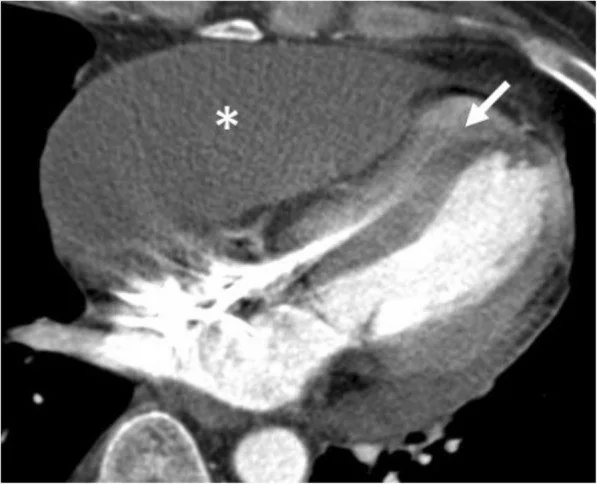
- Perikardiyal koleksiyon ve kalp şekli — sıvının çevresel ya da loküle oluşu ile kalp odacıklarında dıştan basının birlikte bulunması.
- Sağ atriyum konturunda düzleşme/içe çökme — efüzyona komşu duvar deformasyonu; kalp döngüsü ve inceleme tekniğinden etkilenir.
- Loküle perikardiyal efüzyon sağ ventriküle bası yapabilir; sağ ventrikül komşuluğundaki koleksiyonlarda bu bası olasılığı değerlendirilmelidir.
- Geniş IVC ve hepatik venler — sağ kalbe venöz dönüşte basınç artışıyla uyumlu dolgunluk; volüm durumu ve sağ kalp hastalığı da benzer görünüm yapabilir.
- IVC veya hepatik venlere kontrast reflüsü, kontrastlı toraks BT’de görülebilir.
- Eşlik eden plevral sıvı ve portal ven genişlemesi gibi bulgular, sistemik venöz konjesyonun eşlik ettiğini düşündürebilir.
- Ekokardiyografide sağ atriyumun sistolik, sağ ventrikülün diyastolik çökmesi ve transvalvüler akımda solunumsal değişiklikler tamponad fizyolojisini destekler; bulgular klinik tabloyla birlikte yorumlanır.
Normalde
Perikardiyal hastalık şüphesinde BT, klinik senaryoya göre ekokardiyografiyle birlikte kullanılan tamamlayıcı bir görüntüleme yöntemidir.
Normal BT ile kıyasla
Aynı bölgenin, kaynak veri setinde patoloji içermediği belirtilen bir BT incelemesini kesit kesit kaydırın; organ sınırlarını açarak anatomiyi hasta görüntüsüyle karşılaştırın. Küçük rastlantısal bulgular tümüyle dışlanmamıştır.
Kaynak: TotalSegmentator v2.01 veri seti (Wasserthal ve ark., Radiology: Artificial Intelligence 2023), CC BY 4.0, vaka s0977; organ sınırları veri setinin otomatik segmentasyonundan, birleştirilerek sadeleştirildi. Görüntüler 2,5 mm kesit aralığına yeniden örneklendi.
Ayırıcı tanı
- Konstriktif perikardit
- venöz konjesyon yapabilir; perikard kalınlaşması/kalsifikasyonu ve tübüler ventrikül konturu tamponad efüzyonundan ayrılmaya yardım eder.
- Sağ kalp yetmezliği
- IVC/hepatik ven genişliği ve kontrast reflüsü yapabilir; belirgin perikardiyal koleksiyonla ilişkili odacık basısı beklenmez.
- Hipovolemi
- küçük kalp boşlukları oluşturabilir, ancak gergin perikard sıvısı ve venöz dolgunluk birlikteliği tipik değildir.
- Pulmoner hipertansiyon
- sağ ventrikül genişlemesi ve septal şekil değişikliği yapabilir; perikardiyal efüzyonun bası etkisiyle karıştırılmamalıdır.
- Loküle postoperatif hematom
- sağ kalp boşluğuna bölgesel bası uygulayabilir; sıvı içi pıhtı ve cerrahi anatomi koleksiyonun doğasını açıklar.
Tuzaklar
- Sağ atriyumun sistolik fazda geçici biçim değişikliği statik BT’de fizyolojik tamponad göstergesi sayılamaz; kalp döngüsü ve klinik/eko korelasyonu gereklidir.
- IVC’ye kontrast reflüsü hızlı enjeksiyon veya sağ kalp hastalığıyla da görülebilir; tek başına tamponad tanısı koydurmaz.
- Pulmoner hipertansiyon veya sağ ventrikül hipertrofisi, tamponadda beklenen sağ boşluk çökmesini maskeleyebilir; çökme olmaması tamponadı kesin dışlamaz.
- Özellikle postoperatif adezyonlarda loküle sıvı, toplam miktar az olsa bile tek bir odacığı sıkıştırabilir; yalnız çevresel efüzyon hacmine odaklanmayın.
Kendini dene
Perikardiyal sıvıya komşu sağ atriyumda düzleşme, geniş IVC ve venöz kontrast reflüsü görülüyor. BT’de en uygun yorum hangisidir?
Cevabı göster
Tamponad şüphesi. Efüzyon, sağ boşluklarda dıştan bası ve venöz konjesyon birlikteliği tamponad şüphesini artırır. BT statik morfoloji sağlar; hemodinamik tamponad tanısı klinik ve ekokardiyografik değerlendirmeyle konur.
BT’de perikardiyal sıvı ve sağ boşluklarda dıştan bası görüldüğünde hemodinamik tamponadı değerlendirmek için öncelikle hangi inceleme kullanılır?
Cevabı göster
Ekokardiyografi. BT morfolojik şüphe bulgularını gösterir; tamponad fizyolojisi klinik bulgular ve ekokardiyografiyle değerlendirilir.
İlgili konular
Kaynaklar
Bu sayfadaki 36 cümle ve 2 quiz sorusu aşağıdaki kaynaklardan alıntılarla otomatik olarak karşılaştırıldı; 5 cümle bu karşılaştırmada düzeltildi (2026-10-09).
- Cardiac Tamponade Secondary to Anterior Mediastinal Masspmc.ncbi.nlm.nih.gov
- Spontaneous Rectus Sheath Hematoma Mimicking Bladder Tamponade: A Case Reportpmc.ncbi.nlm.nih.gov
- Subacute normotensive cardiac tamponade as the initial presentation of metastatic lung adenocarcinoma: a case reportpmc.ncbi.nlm.nih.gov
- Primary cardiac angiosarcoma mimicking minoxidil-related pericarditis: a case reportpmc.ncbi.nlm.nih.gov
- Electrical Alternans as an Early Clue to a Rapidly Evolving Large Pericardial Effusion: The Role of Point-of-Care Ultrasoundpmc.ncbi.nlm.nih.gov
- Multimodality imaging of pericardial disease.pubmed.ncbi.nlm.nih.gov
- A 63-year-old woman with multiple secondary tumours.pubmed.ncbi.nlm.nih.gov
- Screening, evaluation, and early management of acute aortic dissection in the ED.pubmed.ncbi.nlm.nih.gov
- Optimal insertion depth of central venous catheters--is a formula required? A prospective cohort study.pubmed.ncbi.nlm.nih.gov
- Cardiac tamponade from peripherally-inserted central venous catheters in neonates: Three case reports.pubmed.ncbi.nlm.nih.gov
- Prevalence and prognosis of pericardial effusion in patients affected by pectus excavatum: A case-control study.pubmed.ncbi.nlm.nih.gov
- Diagnostic echocardiography in an unstable intensive care patient.pubmed.ncbi.nlm.nih.gov
- Is routine echocardiography necessary after catheter ablation of atrioventricular nodal re-entrant tachycardia?pubmed.ncbi.nlm.nih.gov
- Purulent Pericarditis: An Uncommon Presentation of a Common Organism.pubmed.ncbi.nlm.nih.gov
- Challenging diagnosis of a coronary arteriovenous malformation: a case report.pubmed.ncbi.nlm.nih.gov
- Delayed diagnosis of traumatic ventricular septal perforation in penetrating chest injury: hematoma formation in the ventricular septum in CT suggests perforation.pubmed.ncbi.nlm.nih.gov
- Postmortem contrast-enhanced computed tomography via direct large-vessel puncture.pubmed.ncbi.nlm.nih.gov
- Routine postoperative computed tomography is superior to cardiac ultrasonography for predicting delayed cardiac tamponade.pubmed.ncbi.nlm.nih.gov
- Electrical Alternans as an Early Clue to a Rapidly Evolving Large Pericardial Effusion: The Role of Point-of-Care Ultrasound.pubmed.ncbi.nlm.nih.gov
- Cardiogenic Versus Obstructive Shock as a Consequence of Road Traffic Accidents: A Case Report on Approach.pubmed.ncbi.nlm.nih.gov
- A Near-Fatal Leak: Acute Cardiac Tamponade From Contrast Extravasation via a Thrombosed Port-a-Cath.pubmed.ncbi.nlm.nih.gov
- Role of Multimodal Cardiac Imaging in Pericardial Effusions and Tamponade.pubmed.ncbi.nlm.nih.gov
- Intracardiac migration of a left renal vein stent after endovascular treatment of nutcracker-associated pelvic congestion syndrome: a case report.pubmed.ncbi.nlm.nih.gov
- Pathophysiology and management of right ventricular failure in critically ill patients: A narrative review.pubmed.ncbi.nlm.nih.gov
- Pericardial Diseases in Cardiovascular Medicine: Contemporary Diagnosis, Risk Stratification, Multimodality Imaging, and Targeted Anti-Inflammatory Therapy.pubmed.ncbi.nlm.nih.gov
- Subacute normotensive cardiac tamponade as the initial presentation of metastatic lung adenocarcinoma: a case report.pubmed.ncbi.nlm.nih.gov
- Transdiaphragmatic Extension of a Pyogenic Liver Abscess Causing Purulent Pericarditis With Cardiac Tamponade in Septic Shock: A Case Report.pubmed.ncbi.nlm.nih.gov
- Purulent Pericardial Effusion: An Elusive Diagnosis.pubmed.ncbi.nlm.nih.gov
- Aortic Injury and Hemopericardium: Key Findings and Evaluation.pubmed.ncbi.nlm.nih.gov
- Diagnostic performance of third-generation sequencing for tuberculous pericarditis: a systematic review and meta-analysis protocol.pubmed.ncbi.nlm.nih.gov
- Retained central line guidewire traversing the heart: An unusual case of iatrogenic pericarditis-case report.pubmed.ncbi.nlm.nih.gov
- Pathway for the Diagnosis and Management of Pericardial Disease: Contemporary Perspective.pubmed.ncbi.nlm.nih.gov
- Heterotopic Caval Valve Implantation for Severe Tricuspid Regurgitation: A Systematic Review and Recommendations for Implantation and Futility.pubmed.ncbi.nlm.nih.gov
- Assessment of Congestion in Heart Failure Using VExUS: Current Evidence, Limitations and Clinical Perspectives.pubmed.ncbi.nlm.nih.gov
- Assessing Venous Congestion in Acute and Chronic Heart Failure: A Review of Splanchnic, Cardiac and Pulmonary Ultrasound: Part 1: Conventional B-Mode, Colordoppler, and Vexus Protocol.pubmed.ncbi.nlm.nih.gov
- Multidetector computer tomography: evaluation of blunt chest trauma in adults.pubmed.ncbi.nlm.nih.gov
- Patterns of injury and violence in Yaoundé Cameroon: an analysis of hospital data.pubmed.ncbi.nlm.nih.gov
- Ruptured Penetrating Aortic Ulcer With Hemopericardium in a Young Adult: A Case Report.pubmed.ncbi.nlm.nih.gov
- [Spontaneous Rupture of Ascending Aorta:Report of Two Cases].pubmed.ncbi.nlm.nih.gov
- Modernizing Aortic Dissection Classification in the Era of Endovascular and Hybrid Repair: Implications for Radiology Reporting.pubmed.ncbi.nlm.nih.gov
- Cardiac Tamponade Under Positive-Pressure Ventilation: Pathophysiological Insights and Implications for Diagnosis.pubmed.ncbi.nlm.nih.gov
- Acute Encephalopathy and Cardiac Tamponade as Initial Presentations of High-Grade B-Cell Lymphoma.pubmed.ncbi.nlm.nih.gov
- Point-of-Care Transesophageal Echocardiography in Emergency and Intensive Care: An Evolving Imaging Modality.pubmed.ncbi.nlm.nih.gov
- Computed tomography and magnetic resonance imaging evaluation of pericardial disease.pubmed.ncbi.nlm.nih.gov
- Pericardial disease--anatomy and function.pubmed.ncbi.nlm.nih.gov
- Electrosurgery in pediatric catheter interventions.pubmed.ncbi.nlm.nih.gov
- The Role of PoCUS in the Diagnosis and Early Monitoring of Cardiogenic Shock in the Emergency Department: A Narrative Review.pubmed.ncbi.nlm.nih.gov
- Intermittent Echocardiographic Monitoring During Superior Vena Cava Recanalization: A Protocol for Early Recognition and Management of Hemopericardium.pubmed.ncbi.nlm.nih.gov
- Imaging in scrub typhus - a pictorial review.pubmed.ncbi.nlm.nih.gov
- Parasitic infections and the development of endomyocardial fibrosis: systematic review of case reports and case series.pubmed.ncbi.nlm.nih.gov
- Long-Term Care in Fontan Circulation: Surveillance and Management of Fontan-Associated Liver Disease and Protein-Losing Enteropathy.pubmed.ncbi.nlm.nih.gov
- Transcatheter closure of patent ductus venosus in an adolescent using a KONAR-Multifunctional Occluder device.pubmed.ncbi.nlm.nih.gov
- Multimodal imaging of infantile scimitar syndrome with Eisenmenger physiology: A case report of complex intracardiac and extracardiac shunts.pubmed.ncbi.nlm.nih.gov
- Pediatric scimitar syndrome with an intrahepatic venous-venous shunt: A rare imaging case report.pubmed.ncbi.nlm.nih.gov
- A Stepwise Approach to Computed Tomography Imaging of Pulmonary Hypertension.pubmed.ncbi.nlm.nih.gov
- Case Report: Higher modelled pulmonary wall shear stress in an adult with Celoria-Patton type B aortic interruption, ventricular septal defect, and patent ductus arteriosus.pubmed.ncbi.nlm.nih.gov
- Cardiac Angiosarcoma as an Unexpected Cause of Palpitations: Case Report, Differential Diagnosis, and Current Therapeutic Approach to Cardiac Tumors.pubmed.ncbi.nlm.nih.gov
- Purulent Pericarditis Complicated With Cardiac Tamponade, Multiorgan Failure, and Constrictive Pericarditis.pubmed.ncbi.nlm.nih.gov
- Cardiac disease in systemic sclerosis: a narrative review.pubmed.ncbi.nlm.nih.gov
- Ultrasonic manifestations of intravascular leiomyomatosis involving the right atrium: a case report.pubmed.ncbi.nlm.nih.gov
- How to Do Echo in Left Ventricular Hypertrophy: A Consensus Statement of the Italian Society of Echocardiography and Cardiovascular Imaging.pubmed.ncbi.nlm.nih.gov
- Heart in Focus: Advancing Pericardial Effusion Diagnosis With Point-of-Care Ultrasound.pubmed.ncbi.nlm.nih.gov
- Critical Care Ultrasound in Shock: A Comprehensive Review of Ultrasound Protocol for Hemodynamic Assessment in the Intensive Care Unit.pubmed.ncbi.nlm.nih.gov
- Society for Cardiovascular Magnetic Resonance 2024 Cases of SCMR case series.pubmed.ncbi.nlm.nih.gov
- Women's heart and echocardiography.pubmed.ncbi.nlm.nih.gov
- Echocardiographic Assessment Before, During, and After Impella Positioning: State of the Art.pubmed.ncbi.nlm.nih.gov
- The New Era of Pulmonary Hypertension: The Dawn of Disease Modification & Therapeutic Modalities.pubmed.ncbi.nlm.nih.gov
- Late-Onset Type I Left Ventricular Rupture Following Double Valve Replacement: An Unexpected Cause of Cardiac Tamponade.pubmed.ncbi.nlm.nih.gov
- Non-pulmonary postoperative complications of cardiothoracic surgery.pubmed.ncbi.nlm.nih.gov
- Evolution of a Pseudoaneurysm Due to Microvascular Obstruction in Myocardial Infarction Assessed by Cardiac Magnetic Resonance.pubmed.ncbi.nlm.nih.gov
- Radiologic Interventions for Gastrointestinal Bleeding.pubmed.ncbi.nlm.nih.gov
- 2024 European Heart Rhythm Association/Heart Rhythm Society/Asia Pacific Heart Rhythm Society/Latin American Heart Rhythm Society expert consensus statement on catheter and surgical ablation of atrial fibrillation.pubmed.ncbi.nlm.nih.gov
- 2024 European Heart Rhythm Association/Heart Rhythm Society/Asia Pacific Heart Rhythm Society/Latin American Heart Rhythm Society expert consensus statement on catheter and surgical ablation of atrial fibrillation.pubmed.ncbi.nlm.nih.gov
- Preoperative initiation of peripheral veno-arterial extracorporeal membrane oxygenation for a complex case of cardiac tamponade.pubmed.ncbi.nlm.nih.gov
- 2019 HRS/EHRA/APHRS/LAHRS expert consensus statement on catheter ablation of ventricular arrhythmias.pubmed.ncbi.nlm.nih.gov
- 2019 HRS/EHRA/APHRS/LAHRS expert consensus statement on catheter ablation of ventricular arrhythmias.pubmed.ncbi.nlm.nih.gov
- Extensive Right Ventricular Involvement With Thrombus and Myocarditis: An Unanticipated Discovery in Advanced Hepatocellular Carcinoma.pubmed.ncbi.nlm.nih.gov
- Aneurysmal and obstructive lesions of the left ventricular outflow: evaluation on multidetector computed tomography angiography.pubmed.ncbi.nlm.nih.gov
- Tricuspid regurgitation: standardized stepwise work-up from referral to expert heart valve centre.pubmed.ncbi.nlm.nih.gov
- Adding Estimates of Central Venous Pressure Boosts the Performance of Non-Invasive Assessment of the Portosystemic Gradient Prior to TIPS Implantation.pubmed.ncbi.nlm.nih.gov
- Usefulness of Bedside Point-of-care Ultrasounds in Intensive Care Unit for Septic Patients: A Consensus Statement of the Italian Society of Echocardiography and Cardiovascular Imaging.pubmed.ncbi.nlm.nih.gov
- Japanese clinical practice guidelines for vascular tumors, vascular malformations, lymphatic malformations, and lymphangiomatosis 2022.pubmed.ncbi.nlm.nih.gov
- Japanese Clinical Practice Guidelines for Vascular Tumors, Vascular Malformations, Lymphatic Malformations, and Lymphangiomatosis 2022.pubmed.ncbi.nlm.nih.gov
- Point-of-Care Ultrasound in Nephrology: Beyond Kidney Ultrasound.pubmed.ncbi.nlm.nih.gov
- Rapid Diagnosis and Surgical Management of Catastrophic Stanford Type A Acute Aortic Dissection Resulting in Successful Lung Donation: A Case Report.pubmed.ncbi.nlm.nih.gov
- Cardiac Tamponade in Late Pregnancy Caused by <i>Corynebacterium amycolatum</i> Pericarditis and Managed by a Surgical Pleuro-Pericardial Window.pubmed.ncbi.nlm.nih.gov
- Chylopericardium in a Dog with Concurrent Chylothorax, Mesothelioma, and Bilateral Brachiocephalic Vein Thrombosis: A Case Report.pubmed.ncbi.nlm.nih.gov
- Right Heart Evaluation: A Tough Challenge for Clinicians.pubmed.ncbi.nlm.nih.gov
- Point-of-care ultrasound to evaluate volume status in congestive heart failure.pubmed.ncbi.nlm.nih.gov
- The use of bedside echocardiography in the care of critically ill patients - a joint consensus document of the Associação de Medicina Intensiva Brasileira, Associação Brasileira de Medicina de Emergência and Sociedade Brasileira de Medicina Hospitalar. Part 2 - Technical aspects.pubmed.ncbi.nlm.nih.gov
- Extravertebral low back pain: a scoping review.pubmed.ncbi.nlm.nih.gov
- Imaging of thoracic hernias: types and complications.pubmed.ncbi.nlm.nih.gov
- Radiological Insights Into the Pericardium: A Comprehensive Review.pubmed.ncbi.nlm.nih.gov
- Unusual presentation and delayed diagnosis of cardiac angiosarcoma.pubmed.ncbi.nlm.nih.gov
- Pediatric emergency medicine point-of-care ultrasound: summary of the evidence.pubmed.ncbi.nlm.nih.gov
- Cardiac Tamponade Secondary to Anterior Mediastinal Mass: A Cardiology-Centered Approach to Diagnosis and Intervention.pubmed.ncbi.nlm.nih.gov
- Management of Post-cardiotomy Shock.pubmed.ncbi.nlm.nih.gov
- Risks and complications of coronary angiography: a comprehensive review.pubmed.ncbi.nlm.nih.gov
- Hemodynamic Effects of Positive Airway Pressure: A Cardiologist's Overview.pubmed.ncbi.nlm.nih.gov
- In-hospital outcomes of cardiac tamponade in patients with pulmonary hypertension: A contemporary analysis.pubmed.ncbi.nlm.nih.gov
- Expanding the role of ultrasonography in cardiopulmonary assessment in dromedary camels.pubmed.ncbi.nlm.nih.gov
- Acute Stanford Type A Aortic Dissection Mimicking Acalculous Cholecystitis: A Case Report.pubmed.ncbi.nlm.nih.gov
- Incremental Value of Magnetic Resonance Imaging in Further Characterizing Hypodense Mediastinal and Paracardiac Lesions Identified on Computed Tomography.pubmed.ncbi.nlm.nih.gov
- Pictorial essay: Non-coronary applications of cardiac CT.pubmed.ncbi.nlm.nih.gov
- Pathophysiological management of gastric varices: From hemodynamics to targeted therapies.pubmed.ncbi.nlm.nih.gov
- Double-J stent traversing the inferior vena cava into the right atrium: a rare cause of bacteremia after chronic stent retention.pubmed.ncbi.nlm.nih.gov
- Mass Encroaching on the Right Atrium: An Unusual Echocardiography Finding.pubmed.ncbi.nlm.nih.gov
- A cheesy exudate: a case report on management of pericardial disease through multimodal cardiac imaging.pubmed.ncbi.nlm.nih.gov
- Parasternal Pericardiocentesis for Methicillin-Resistant Staphylococcus aureus Pericarditis Complicated by Abscess and Tamponade.pubmed.ncbi.nlm.nih.gov
- Coronary computed tomography angiography findings of anomalous left hepatic vein drainage into the coronary sinus with complex anomalies: a case report.pubmed.ncbi.nlm.nih.gov
- Right atrial extension of inferior vena cava thrombosis after microwave ablation of colorectal liver metastasis: a case report.pubmed.ncbi.nlm.nih.gov
- Deer Horn Sign in Congestive Hepatopathy Due to Heart Failure.pubmed.ncbi.nlm.nih.gov
- Multimodality imaging of acute and chronic pulmonary thromboembolic disease.pubmed.ncbi.nlm.nih.gov
- Non-invasive Multimodality Cardiovascular Imaging of the Right Heart and Pulmonary Circulation in Pulmonary Hypertension.pubmed.ncbi.nlm.nih.gov
- Staged visceral revascularization prior to central aortic repair in acute type A aortic dissection complicated by mesenteric malperfusion: a case report.pubmed.ncbi.nlm.nih.gov
Eğitim amaçlıdır; hasta başında tanı ve tedavi kararının yerine geçmez, tıbbi tavsiye değildir. Metinler yapay zekâ desteğiyle yazıldı ve otomatik bir adımda kaynak alıntılarıyla karşılaştırıldı; bu bir tıbbi doğrulama değildir ve içerik uzman radyolog incelemesinden geçmedi. Klinik kararlarda güncel kılavuzları, kurum protokollerini ve radyoloji raporunu esas alın.