Özet
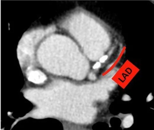
Rutin, EKG eşzamanlamasız toraks BT'de koroner arter duvarı boyunca kalsifiye aterosklerotik plak sık rastlanan ve klinik açıdan anlamlı bir tesadüfi bulgudur. Kalsiyum kontrastsız incelemede yüksek atenüasyonlu odaklar olarak seçilebilir; non-gated görüntüde varlığı raporlanabilir, ancak bu inceleme lümen darlığını güvenilir biçimde derecelendiren koroner BT anjiyografi veya standart Agatston kalsiyum skoru değildir. Koroner kalsifikasyonun saptanması önceden bilinmeyen koroner arter hastalığının fark edilmesine ve kardiyovasküler risk faktörlerinin değerlendirilmesine katkı sağlayabilir; kalsiyum varlığı stenoz şiddetini göstermez.
Faz ve pencere
- Kontrastsız tanısal
- EKG eşzamanlamasız toraks BT'de LAD, sirkumfleks arter veya RCA seyri boyunca damar duvarına uyan kaba ya da noktasal yüksek atenüasyonlu kalsifiye plak odakları görülür; ölçüm eşiği bu tarama bulgusu için kullanılmaz.
Önerilen pencereler: Mediasten (G 350 / M 50), Yumuşak doku (G 400 / M 40).
BT bulguları
- Koroner arterlerin seyri boyunca yüksek atenüasyonlu materyali arayın; hasta bazlı görsel değerlendirmede tüm koroner arterlerdeki kalsiyumun toplam yükünü özetleyin.
- Çok damarlı kalsiyum — birden fazla koroner arter dağılımında kalsifiye plak bulunması yaygın koroner ateroskleroz yükünü gösterir.
- Bulanık küçük odak — kalın kesitli nongated toraks BT’de koroner kalsiyum skorunun yanlış sınıflandırılma olasılığı artabilir.
- Lümen derecesi belirsizliği — kalsiyumun varlığı plak yükü göstergesidir, ancak nongated rutin toraks BT tek başına stenoz yüzdesi vermez.
Ölçütler ve sınıflamalar
- Görsel hasta bazlı koroner kalsiyum derecesi
- Koroner kalsiyum hasta bazında yok, hafif, orta veya şiddetli olarak görsel sınıflandırılabilir; kategori plakların toplam görsel yükünü özetler ve rutin nongated BT’de stenoz yüzdesi anlamına gelmez.
Normalde
Normal koroner arterlerde damar duvarı boyunca belirgin kalsifiye yüksek atenüsyon odağı bulunmaz; kalsifikasyon koroner arter yolu boyunca izlenir. Şüpheli odağı bu anatomik hatlara göre takip edin ve aort kapağı, mitral anülüs, perikard ile aort duvarı kalsifikasyonlarından ayırın; kontrastsız ve yumuşak doku/mediasten görüntülerini karşılaştırın. Görsel koroner kalsiyum değerlendirmesi kontrastsız ve kontrastlı görüntülerde uygulanabilir; şüpheli odağı koroner arter seyri ve komşu anatomik yapılarla birlikte değerlendirin.
Normal BT ile kıyasla
Aynı bölgenin, kaynak veri setinde patoloji içermediği belirtilen bir BT incelemesini kesit kesit kaydırın; organ sınırlarını açarak anatomiyi hasta görüntüsüyle karşılaştırın. Küçük rastlantısal bulgular tümüyle dışlanmamıştır.
Kaynak: TotalSegmentator v2.01 veri seti (Wasserthal ve ark., Radiology: Artificial Intelligence 2023), CC BY 4.0, vaka s0977; organ sınırları veri setinin otomatik segmentasyonundan, birleştirilerek sadeleştirildi. Görüntüler 2,5 mm kesit aralığına yeniden örneklendi.
Ayırıcı tanı
- Aort kapağı kalsifikasyonu
- yoğunluk koroner olukta değil, aort kökü çıkışındaki kapak yaprakçıklarında merkezlenir.
- Mitral anüler kalsifikasyon
- posterior mitral kapak anülüsünü izleyen kavisli kalsifiye bant görünümündedir.
- Perikardiyal kalsifikasyon
- kalbin perikardında görülen, koroner arter kalsifikasyonundan ayrı bir kardiyak kalsifikasyon bulgusudur.
- Aort duvarı ateromu
- kalsifikasyon koroner oluk yerine çıkan aorta veya arkus duvarının konturuna oturur.
- Koroner stent
- önceki girişime bağlı stenti tanıyın; görüntülemede stent veya bypass grefti görülüyorsa kaynak önerisi uyarınca tesadüfi koroner kalsifikasyonu ayrıca değerlendirmeniz gerekmez, çünkü koroner arter hastalığı zaten belirlenmiştir.
Tuzaklar
- Sağ atriyoventriküler olukta RCA'yı izlerken komşu kapak/anülüs kalsiyumunu koroner plak diye işaretlemeyin; damarın seyriyle sürekliliği arayın.
- Kesit kalınlığı koroner kalsiyum skorlamasını etkileyebilir; değerlendirmede rekonstrüksiyon kesit kalınlığını göz önünde bulundurun.
- Koroner kalsiyum varlığından lümen darlığı veya tıkalı damar sonucu çıkarmayın; non-gated rutin toraks BT kalsifikasyonu saptar, stenozu güvenilir ölçmez.
- Aort kökü çevresindeki parlak odakları koroner diye adlandırmadan önce LAD, sirkumfleks ve RCA'nın anatomik oluklarını ayrı ayrı izleyin.
Kendini dene
EKG eşzamanlamasız toraks BT'de koroner arter seyri boyunca yüksek atenüasyonlu odaklar görülüyor. Bu bulgu en iyi neyi gösterir?
Cevabı göster
Koroner kalsifiye aterosklerotik plak. Koroner arter seyri içindeki yüksek atenüasyonlu materyal koroner kalsifikasyonla uyumludur ve ateroskleroz yükünün bir belirtecidir. EKG eşzamanlamasız toraks BT'de kalsiyum varlığı darlığın derecesini göstermez. Aort kapağı, mitral anülüs ve perikard kalsifikasyonları farklı anatomik yapılardadır.
EKG eşzamanlamasız toraks BT’de koroner arter kalsifikasyonu en iyi neyi gösterir?
Cevabı göster
Koroner ateroskleroz yükü için bir belirteci. Koroner arter kalsifikasyonu ateroskleroz yükünün bir belirtecidir; tek başına darlığın şiddetini veya akımı kısıtlayıp kısıtlamadığını göstermez.
Kaynaklar
Bu sayfadaki 35 cümle ve 2 quiz sorusu aşağıdaki kaynaklardan alıntılarla otomatik olarak karşılaştırıldı; 12 cümle bu karşılaştırmada düzeltildi (2026-10-09).
- Reporting incidental coronary, aortic valve and cardiac calcification on non-gated thoracic computed tomography, a consensus statement from the BSCI/BSCCT and BSTIpmc.ncbi.nlm.nih.gov
- Anatomical References to Evaluate Thoracic Aorta Calcium by Computed Tomography.pubmed.ncbi.nlm.nih.gov
- Coronary artery calcium clinical utilization: An update.pubmed.ncbi.nlm.nih.gov
- Automated coronary calcium scoring using deep learning with multicenter external validation.pubmed.ncbi.nlm.nih.gov
- Coronary artery calcium score may be a novel predictor of COVID-19 prognosis: a retrospective study.pubmed.ncbi.nlm.nih.gov
- Identifying Coronary Artery Calcification on Non-gated Computed Tomography Scans.pubmed.ncbi.nlm.nih.gov
- Comparison of ordinal versus Agatston coronary calcification scoring for cardiovascular disease mortality in community-living individuals.pubmed.ncbi.nlm.nih.gov
- Incidental coronary calcifications on routine chest CT: Clinical implications.pubmed.ncbi.nlm.nih.gov
- The Future of Concurrent Automated Coronary Artery Calcium Scoring on Screening Low-Dose Computed Tomography.pubmed.ncbi.nlm.nih.gov
- Reporting incidental coronary, aortic valve and cardiac calcification on non-gated thoracic computed tomography, a consensus statement from the BSCI/BSCCT and BSTI.pubmed.ncbi.nlm.nih.gov
- Hyperacute Incidental Late Myocardial Enhancement in Ischemic Stroke Using Chest Spectral CT: Relationship with Etiology.pubmed.ncbi.nlm.nih.gov
- Impact of Deep Learning-Based Image Conversion on Fully Automated Coronary Artery Calcium Scoring Using Thin-Slice, Sharp-Kernel, Non-Gated, Low-Dose Chest CT Scans: A Multi-Center Study.pubmed.ncbi.nlm.nih.gov
- DINO-LG: Enhancing vision transformers with label guidance for coronary artery calcium detection.pubmed.ncbi.nlm.nih.gov
- Age-Dependent Efficacy of Incidental Coronary Artery Calcium on Pre-CAG Chest CT: Potentially Missed Prevention Opportunities in Chinese Population.pubmed.ncbi.nlm.nih.gov
- Coronary artery calcium score-emerging role for opportunistic screening on thoracic CT and novel applications.pubmed.ncbi.nlm.nih.gov
- Massive mitral annular calcification with valvular involvement: Multimodality and longitudinal imaging correlation.pubmed.ncbi.nlm.nih.gov
- State-of-the-Art Imaging Biomarkers in Screening of Atherosclerotic Cardiovascular Disease.pubmed.ncbi.nlm.nih.gov
- Emerging quantitative CCTA imaging biomarkers for cardiovascular risk stratification: a narrative review.pubmed.ncbi.nlm.nih.gov
- Coronary artery calcium and thoracic aortic calcification on non-ECG-gated chest CT in a large community-based cohort of Chinese men and women.pubmed.ncbi.nlm.nih.gov
- Agreement and workflow efficiency of AI-based coronary artery calcification quantification in lung cancer screening: Comparison with semi-automated and visual assessment.pubmed.ncbi.nlm.nih.gov
- Association between pericoronary CT fat attenuation index and coronary artery spasm.pubmed.ncbi.nlm.nih.gov
- Coronary Dominance on Coronary CT Angiography: Prevalence and Potential Implications for Electrocardiographic Correlation.pubmed.ncbi.nlm.nih.gov
- Motion correction algorithm improves right coronary artery image quality at heart rates > 75 bpm in non-ECG-gated coronary CT angiography.pubmed.ncbi.nlm.nih.gov
- Coronary artery disease imaging with total-body PET.pubmed.ncbi.nlm.nih.gov
- Understanding Photon-Counting CT: Physics, Detector Technology, and Image Reconstructions.pubmed.ncbi.nlm.nih.gov
- Spectral computed tomography characterization of coronary atherosclerotic plaque: principles, imaging biomarkers, and clinical significance.pubmed.ncbi.nlm.nih.gov
- Clinical Applications of Artificial Intelligence in Cardiac CT: From Coronary CT Angiography to CT-Derived Fractional Flow Reserve and Myocardial Perfusion Imaging.pubmed.ncbi.nlm.nih.gov
- Fractional Flow Reserve Measurement Using Dynamic CT Perfusion Imaging in Patients with Heavy Coronary Calcification and Stent.pubmed.ncbi.nlm.nih.gov
- Coronary Computed Tomography Angiography for the Diagnosis and Revascularization Guidance of Coronary Bifurcation Lesions: A Contemporary Review.pubmed.ncbi.nlm.nih.gov
- Integrating Invasive and Noninvasive Imaging for Coronary Atherosclerosis: A Systematic Review With Pragmatic Algorithms From Anatomy to Physiology.pubmed.ncbi.nlm.nih.gov
- Role of Cardiac CT in the Evaluation of Valvular Heart Disease.pubmed.ncbi.nlm.nih.gov
- Management of Idiopathic Outflow Tract Ventricular Arrhythmias.pubmed.ncbi.nlm.nih.gov
- Society for Cardiovascular Magnetic Resonance 2024 Cases of SCMR case series.pubmed.ncbi.nlm.nih.gov
- Prognostication and clinical opportunities with AI for coronary artery calcium: a scoping review.pubmed.ncbi.nlm.nih.gov
- Serum Alpha-Klotho and Coronary Atherosclerosis: Associations with Coronary CT Angiography Findings and Atherogenic Indices.pubmed.ncbi.nlm.nih.gov
- Coronary Artery Calcium Score and Carotid Intima-Media Thickness As Markers of Computed Tomography Angiographic Severity in Coronary Artery Disease: A Cross-Sectional Study.pubmed.ncbi.nlm.nih.gov
- Longitudinal Changes in Coronary Artery Calcium Distribution and Atherosclerotic Cardiovascular Disease Risk.pubmed.ncbi.nlm.nih.gov
- Beyond the Agatston calcium score: role of calcium density and other calcified plaque markers for cardiovascular disease prediction.pubmed.ncbi.nlm.nih.gov
- Clinical Applications of Cardiac Computed Tomography: A Focused Review for the Clinical Cardiologists.pubmed.ncbi.nlm.nih.gov
- Lipoprotein(a) and calcific aortic stenosis: from inherited risk marker to therapeutic target.pubmed.ncbi.nlm.nih.gov
- Aortic valve disease: Novel animal models for advancing our understanding of the underlying mechanisms.pubmed.ncbi.nlm.nih.gov
- Concomitant Aortic Valve Neocuspidization and Resection of a Cardiac Calcified Amorphous Tumor in a Hemodialysis Patient: A Case Report.pubmed.ncbi.nlm.nih.gov
- Myval Beyond the Aortic Valve: Device-Anatomy Interaction, Procedural Strategy, and Clinical Evidence in Mitral, Tricuspid, and Pulmonary Positions.pubmed.ncbi.nlm.nih.gov
- Transcatheter Aortic Valve Migration and Embolization During Transcatheter Aortic Valve Replacement: A Contemporary Management Review With a Practical Bailout Algorithm.pubmed.ncbi.nlm.nih.gov
- Mesenchymal Stem Cells of Epicardial Adipose Tissue Show Differences in Immunophenotype and Osteogenic Potential in Patients with Coronary and Non-Coronary Heart Disease.pubmed.ncbi.nlm.nih.gov
- NaF-PET Imaging for Detection of Early Arterial Microcalcification and Monitoring of Targeted Therapy: A Narrative Review.pubmed.ncbi.nlm.nih.gov
- From incidental positron emission uptake to <i>in-vivo</i> phenotyping: a short history of positron emission tomography in the study of atherosclerosis and the vulnerable plaque.pubmed.ncbi.nlm.nih.gov
- Advanced quantitative CT for coronary artery disease: integrating stenosis quantification, plaque quantification, and fractional flow reserve-CT.pubmed.ncbi.nlm.nih.gov
- Pictorial review: a step-by-step guide to coronary CT angiography with photon-counting detector CT.pubmed.ncbi.nlm.nih.gov
- Technical advances and clinical impact of photon-counting computed tomography angiography for peripheral artery disease.pubmed.ncbi.nlm.nih.gov
- Moderate aortic stenosis: diagnostic pitfalls and the role of multimodality imaging in risk stratification and clinical management.pubmed.ncbi.nlm.nih.gov
- Computed Tomography Imaging of Thoracic Aortic Surgery: Distinguishing Life-Saving Repairs from Life-Threatening Complications.pubmed.ncbi.nlm.nih.gov
- Application of postmortem imaging modalities in cases of sudden death due to cardiovascular diseases-current achievements and limitations from a pathology perspective : Endorsed by the Association for European Cardiovascular Pathology and by the International Society of Forensic Radiology and Imaging.pubmed.ncbi.nlm.nih.gov
- Association between fibroblast growth factor 23 and coronary artery calcification in patients with chronic kidney disease: a meta-analysis.pubmed.ncbi.nlm.nih.gov
- Balloon-Expandable Versus Self-Expandable Transcatheter Aortic Valve Implantation Devices: A Contemporary Systematic Review.pubmed.ncbi.nlm.nih.gov
- Association of Composite Metabolic Indices and Oxidative Stress Biomarkers with Coronary Atherosclerosis Detected by CT Angiography in Premenopausal Women with Metabolic Syndrome.pubmed.ncbi.nlm.nih.gov
- Computed tomography pitfalls and diagnostic value in a patient with post-infarction ventricular septal defect, cardiogenic shock, and iatrogenic type B aortic dissection during extracorporeal membrane oxygenation support: a case report.pubmed.ncbi.nlm.nih.gov
- When Palpitations Unmask Crista Terminalis Hypertrophy: A Case Report and Review of Current Literature.pubmed.ncbi.nlm.nih.gov
- A difference that matters: the aortic root anatomy of large animal models vs. humans.pubmed.ncbi.nlm.nih.gov
- Cardiovascular CT in Bicuspid Aortic Valve Disease: A State-of-the-Art Narrative Review of Advances, Clinical Integration, and Future Directions.pubmed.ncbi.nlm.nih.gov
- Arterial Supply to the Atrioventricular Node: Anatomical Variability and Clinical Implications.pubmed.ncbi.nlm.nih.gov
- A Rare Vascular Tangle Unraveled: Multimodal Imaging in Giant Coronary Arteriovenous Fistula.pubmed.ncbi.nlm.nih.gov
- Incidental Finding of a Superdominant Left Anterior Descending Artery in an 80-Year-Old Woman: Case Report and Literature Review.pubmed.ncbi.nlm.nih.gov
- A Giant in Plain Sight: Multimodality Imaging of a Left Ventricular Fibroma.pubmed.ncbi.nlm.nih.gov
- An Elegant Approach for Complete Revascularization of the Circumflex Territory.pubmed.ncbi.nlm.nih.gov
- Atrial wall dissection: a contemporary overview for a proper decision-making approach.pubmed.ncbi.nlm.nih.gov
- Photon-Counting CT in Cardiovascular Imaging: Clinical Applications.pubmed.ncbi.nlm.nih.gov
- A Rare Case of Isolated Single Coronary Artery, Lipton's Type R-IIB in an Elderly Woman With Recurrent Chest Pain and Left Bundle Branch Block: A Case Report.pubmed.ncbi.nlm.nih.gov
- Coronary Artery Anomalies Revisited: Description of the Types, Pathophysiology and Treatment Options Based on Latest Guidelines.pubmed.ncbi.nlm.nih.gov
- Commentary: an eye on PET quantification.pubmed.ncbi.nlm.nih.gov
- Computed Tomography 2.0: New Detector Technology, AI, and Other Developments.pubmed.ncbi.nlm.nih.gov
- Iron Oxide Nanoparticles in Regenerative Medicine and Tissue Engineering.pubmed.ncbi.nlm.nih.gov
- Padua Days on Muscle and Mobility Medicine, March 25-29, 2025, Hotel Petrarca, Euganean Thermae, Italy: Program and Abstracts.pubmed.ncbi.nlm.nih.gov
- High-affinity alphavbeta3 integrin targeted optical probe as a new imaging biomarker for early atherosclerosis: initial studies in Watanabe rabbits.pubmed.ncbi.nlm.nih.gov
- Patterns of injury and violence in Yaoundé Cameroon: an analysis of hospital data.pubmed.ncbi.nlm.nih.gov
- The Shifting Boundary Between Invasive and Non-Invasive Angiographic Investigation in Contemporary Cardiology and Cardiac Surgery: An Up-to-Date Narrative Review.pubmed.ncbi.nlm.nih.gov
- Developing a calibration method to utilize low-dose chest CT for assessment of coronary artery calcification score.pubmed.ncbi.nlm.nih.gov
- High-Z Contrast Media for Coronary Photon-counting Detector CT Angiography: Improved Quantification of Calcified Stenoses.pubmed.ncbi.nlm.nih.gov
- Impact of scanning parameters on deep learning coronary artery calcium scoring in non‑gated chest CT.pubmed.ncbi.nlm.nih.gov
- Optimizing coronary computed tomography angiography image quality with motion-compensated reconstruction: a prospective electrocardiogram-triggered mode with second-generation dual-layer spectral detector computed tomography.pubmed.ncbi.nlm.nih.gov
- Effect of a breathing exercise program on coronary artery motion artifacts in 256 rows of coronary computed tomography angiography in patients with different heart rate variability.pubmed.ncbi.nlm.nih.gov
- Virtual Non-Iodine Coronary Calcium Scoring on Photon-Counting CT: Patient- and Plaque-Level Analysis.pubmed.ncbi.nlm.nih.gov
- Cardiac CT in the Era of Precision Cardiology: From Calcium Scoring to Comprehensive Risk Profiling.pubmed.ncbi.nlm.nih.gov
- Obstructive Sleep Apnea Risk and Incidental Coronary Artery Calcification on Routine Chest Computed Tomography.pubmed.ncbi.nlm.nih.gov
- Radiation dose optimization in coronary CT angiography clinical application of absolute phase gating in patients with atrial fibrillation.pubmed.ncbi.nlm.nih.gov
- Patient-centric performance and interpretation of positron emission tomography /computed tomography myocardial perfusion imaging: a clinical consensus statement of the European Association of Cardiovascular Imaging of the European Society of Cardiology.pubmed.ncbi.nlm.nih.gov
- Cardiac Computed Tomography in Structural Heart Interventions: From Preprocedural Planning to Procedural Strategy.pubmed.ncbi.nlm.nih.gov
- Evaluation of Coronary Artery Disease Plaques Using Coronary CT Angiography in a High-Risk Population.pubmed.ncbi.nlm.nih.gov
- Association Between Pericoronary Fat Attenuation Index and High-Risk Plaque Features Assessed by Photon-Counting CT.pubmed.ncbi.nlm.nih.gov
- High-Risk Vascular Remodeling in Takayasu Arteritis: Descending Aortic Multilobulated Saccular Aneurysms and Aortic Coarctation.pubmed.ncbi.nlm.nih.gov
- Cardiac computed tomography for cardiac masses: a necessity or a luxury?pubmed.ncbi.nlm.nih.gov
- Imaging of the Acute and Chronic Cardiovascular Complications of Radiation Therapy.pubmed.ncbi.nlm.nih.gov
- Coronary artery calcium identified on non-gated chest computed tomography guides downstream coronary computed tomography angiography.pubmed.ncbi.nlm.nih.gov
- Coronary Microvascular Dysfunction Is Present Among Well-Treated Asymptomatic Persons With HIV and Similar to Those With Diabetes.pubmed.ncbi.nlm.nih.gov
- Imaging Advances in Chronic Obstructive Pulmonary Disease. Insights from the Genetic Epidemiology of Chronic Obstructive Pulmonary Disease (COPDGene) Study.pubmed.ncbi.nlm.nih.gov
- Cardiac Angiosarcoma as an Unexpected Cause of Palpitations: Case Report, Differential Diagnosis, and Current Therapeutic Approach to Cardiac Tumors.pubmed.ncbi.nlm.nih.gov
- Evaluation of causal heart diseases in cardioembolic stroke by cardiac computed tomography.pubmed.ncbi.nlm.nih.gov
- Pre- and post-procedural cardiac imaging (computed tomography and magnetic resonance imaging) in electrophysiology: a clinical consensus statement of the European Heart Rhythm Association and European Association of Cardiovascular Imaging of the European Society of Cardiology.pubmed.ncbi.nlm.nih.gov
- Materials Advances in Devices for Heart Disease Interventions.pubmed.ncbi.nlm.nih.gov
- Rheumatic and Degenerative Mitral Stenosis: From an Iconic Clinical Case to the Literature Review.pubmed.ncbi.nlm.nih.gov
- Photon-counting CT versus energy-integrating detectors for cardiac imaging: a systematic review of evidence from in vivo human studies on image quality and radiation dose.pubmed.ncbi.nlm.nih.gov
- Innovative Integration of 4D Cardiovascular Reconstruction and Hologram: Framework Development of a New Visualization Tool for Coronary Artery Bypass Grafting Planning.pubmed.ncbi.nlm.nih.gov
- Early Echocardiography and ECG Changes Following Radiotherapy in Patients with Stage II-III HER2-Positive Breast Cancer Treated with Anthracycline-Based Chemotherapy with or without Trastuzumab-Based Therapy.pubmed.ncbi.nlm.nih.gov
- SCMR expert consensus statement for cardiovascular magnetic resonance of acquired and non-structural pediatric heart disease.pubmed.ncbi.nlm.nih.gov
- Analysis of epicardial adipose tissue in relation to arterial hypertension using radiomics in photon-counting CT.pubmed.ncbi.nlm.nih.gov
- How to Individualize Coronary Assessment and Revascularization in Severe AS Patients Undergoing TAVI in the Era of Lifetime Management?pubmed.ncbi.nlm.nih.gov
- Predictive value of coronary artery calcification for osteoporosis in COPD patients: a retrospective AI-based study.pubmed.ncbi.nlm.nih.gov
- The Weston Score in coronary artery calcification among patients with End-Stage Renal Disease.pubmed.ncbi.nlm.nih.gov
- Enabling Transfemoral TAVI Through a Hostile Abdominal Aortic Anatomy and a Migrated Mesenteric Stent Fragment: Review of the Literature and a Case Presentation.pubmed.ncbi.nlm.nih.gov
- Prognostic Value of CT-Based Coronary Imaging for Perioperative Cardiovascular Risk Stratification Before Noncardiac Surgery: An Updated Systematic Review and Meta-Analysis.pubmed.ncbi.nlm.nih.gov
- Assessing Coronary Calcium Thresholds on Attenuation-Correction CT With Myocardial Perfusion Imaging Equating to Secondary Prevention.pubmed.ncbi.nlm.nih.gov
Eğitim amaçlıdır; hasta başında tanı ve tedavi kararının yerine geçmez, tıbbi tavsiye değildir. Metinler yapay zekâ desteğiyle yazıldı ve otomatik bir adımda kaynak alıntılarıyla karşılaştırıldı; bu bir tıbbi doğrulama değildir ve içerik uzman radyolog incelemesinden geçmedi. Klinik kararlarda güncel kılavuzları, kurum protokollerini ve radyoloji raporunu esas alın.