Özet
Siyanozlu çocukta Fallot tetralojisi, malalignment VSD, aortanın septum üzerine ata binmesi, sağ ventrikül çıkış yolu obstrüksiyonu ve sağ ventrikül hipertrofisiyle tanınan konotrunkal bir anomalidir. BT anjiyografi, ekokardiyografinin sınırlı kaldığı pulmoner arter dalları, aortopulmoner kollateraller, koroner seyir ve onarım materyalinin anatomik haritasını sunar. Onarılmış hastada RV-PA konduiti, transannüler yama, pulmoner arter darlığı ve yama psödoanevrizması geç dönemde yeniden değerlendirme gerektirebilir. BT anatomiyi gösterir ancak pulmoner kapak yetersizliği ve ventrikül işlevini tek başına nicelleştirmez; yapısal bulguyu işlevsel sonuçla eşitlemek doğru değildir.
Faz ve pencere
- BTA tanısal
- Kardiyak hareket azaltılmış kontrastlı hacimde sağ ventrikül çıkış yolunu, ana ve dal pulmoner arterleri, aort kökü/arkı, MAPCA'ları ve ameliyat sonrası konduit-yama hattını opaklaştır; koroner çıkışlar veya kompleks anatomi sorusu varsa EKG eşzamanlaması yararlı olabilir.
Önerilen pencereler: Anjiyo (G 600 / M 150), Mediasten (G 350 / M 50).
BT bulguları
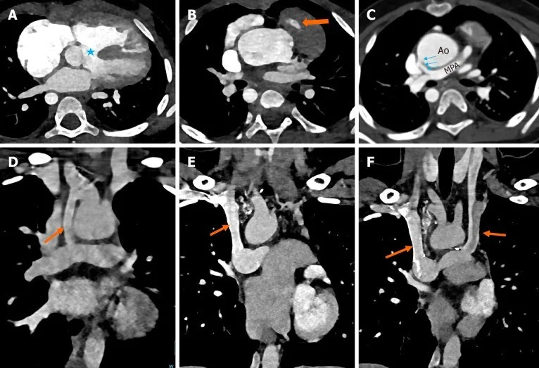
- Malalignment VSD — outlet septumun yer değiştirmesiyle oluşan geniş interventriküler açıklık ve aort köküne uzanım gösterilebilir.
- Aortik overriding — aort kökü ventriküler septumun üzerine yerleşerek her iki ventrikülden kan alır; yalnız çıkış damarının konumu değil septumla ilişkisi tanımlanır.
- Sağ ventrikül çıkış yolu darlığı — infundibüler kas demetleri, pulmoner kapak veya ana pulmoner arter düzeyinde daralma olabilir.
- Sağ ventrikül hipertrofisi — serbest duvar kalınlaşması morfolojik olarak görülür; BT tek başına basınç ya da kasılma işlevini belirlemez.
- Pulmoner arter hipoplazisi veya ayrıklığı — ana, sağ ve sol dalların kalibresi, konfluens ve hiler devamlılığı haritalanır.
- MAPCA — inen aorta veya ark dallarından akciğere uzanan sistemik kaynaklı kollateral arterler ve besledikleri akciğer bölgeleri izlenir.
- Onarım sonrası konduit veya yama — RV-PA bağlantısında daralma, kalsifikasyon, deformasyon ya da psödoanevrizma aranır.
- Pulmoner arter ve koroner komplikasyonu — yama veya stent düzeyinde dal darlığı ve çıkış yolu komşuluğunda seyreden anormal koroner arter tanımlanır.
Normalde
Normal kalpte interventriküler septum kesintisizdir, aort kökü sol ventrikülden çıkar ve sağ ventrikül çıkış yolu pulmoner kapağa doğru daralmadan devam eder; pulmoner arter gövdesi sağ ve sol dallara olağan biçimde ayrılır. Aynı düzeylerde VSD ile aort kökünün septuma göre konumunu, RVOT lümenini ve iki pulmoner arterin sürekliliğini kıyasla; ameliyatlı hastada bu referansa cerrahi yama ve konduit geometrisini ekle.
Normal BT ile kıyasla
Aynı bölgenin, kaynak veri setinde patoloji içermediği belirtilen bir BT incelemesini kesit kesit kaydırın; organ sınırlarını açarak anatomiyi hasta görüntüsüyle karşılaştırın. Küçük rastlantısal bulgular tümüyle dışlanmamıştır.
Kaynak: TotalSegmentator v2.01 veri seti (Wasserthal ve ark., Radiology: Artificial Intelligence 2023), CC BY 4.0, vaka s0977; organ sınırları veri setinin otomatik segmentasyonundan, birleştirilerek sadeleştirildi. Görüntüler 2,5 mm kesit aralığına yeniden örneklendi.
Ayırıcı tanı
- Çift çıkışlı sağ ventrikül
- her iki büyük arterin önemli bölümü morfolojik sağ ventrikülden çıkar; yalnız overriding aorta değil, her iki çıkış bağlantısı belirlenmelidir.
- Pulmoner atrezi-VSD
- RVOT boyunca patent dar çıkış yerine pulmoner kapak veya ana arter düzeyinde devamlı lümen yoktur; akciğer kanlanması MAPCA'lara bağlı olabilir.
- Trunkus arteriozus
- tek ortak arteriyel gövde pulmoner ve sistemik dolaşıma dallanır; ayrı aort ve pulmoner kökler bulunmaz.
- Onarım sonrası pulmoner arter darlığı
- konduit kalsifikasyonu veya degenerasyona bağlı fokal daralma, üç boyutlu kesitsel imajing teknikleriyle primer doğumsal merkezi darlıktan ayırt edilmelidir.
Tuzaklar
- Geniş bir aort kökünün septumu örtmesini tek başına overriding kabul etme; çıkış kökünün septumla anatomik ilişkisini farklı düzlemlerde doğrula.
- Postoperatif yama çıkıntısını psödoanevrizma saymadan önce kontrast lümeninin kese ile devamını ve eski görüntülerdeki değişimi ara.
- Pulmoner regürjitasyon şiddetini BT morfolojisinden çıkarsama; akım ve sağ ventrikül hacim işlevi için CMR veya ekokardiyografi gerekir.
Kendini dene
Geniş outlet VSD, septum üzerinde ata binen aort ve infundibüler daralma birlikte görülüyor. En olası tanı?
Cevabı göster
Fallot tetralojisi. Malalignment VSD, aortik overriding ve RVOT obstrüksiyonunun birlikteliği klasik Fallot morfolojisidir. Çift çıkışlı sağ ventrikülde iki arteriyel kökün kaynağı sağ ventriküldür; trunkusta tek ortak arter bulunur; izole pulmoner stenoz Fallot çıkış ilişkilerini içermez.
Geniş transannüler yama içeren eski bir Fallot onarımından sonra, doğal RVOT duvarından değil yama dokusundan gelişebilen geç komplikasyon hangisidir?
Cevabı göster
RVOT psödoanevrizması. RVOT psödoanevrizması, geniş transannüler yama içeren onarımlardan sonra görülebilir; psödoanevrizma doğal RVOT duvarından değil yama dokusundan oluşur.
İlgili konular
Kaynaklar
Bu sayfadaki 34 cümle ve 2 quiz sorusu aşağıdaki kaynaklardan alıntılarla otomatik olarak karşılaştırıldı; 2 cümle bu karşılaştırmada düzeltildi (2026-10-09).
- Essentials of Imaging the Repaired Tetralogy of Fallot Patientpmc.ncbi.nlm.nih.gov
- Computed tomography in tetralogy of Fallot: pre- and postoperative imaging evaluation.pubmed.ncbi.nlm.nih.gov
- Double Outlet Right Ventricle: In-Depth Anatomic Review Using Three-Dimensional Cardiac CT Data.pubmed.ncbi.nlm.nih.gov
- Congenital heart disease and multi-modality imaging.pubmed.ncbi.nlm.nih.gov
- Cardiovascular imaging approach in pre and postoperative tetralogy of Fallot.pubmed.ncbi.nlm.nih.gov
- Computed tomography for evaluating right ventricle and pulmonary artery in pediatric tetralogy of Fallot: correlation with post-operative pulmonary regurgitation.pubmed.ncbi.nlm.nih.gov
- Post Blalock-Taussig shunt mediastinal mass - a single shadow with two different destinies.pubmed.ncbi.nlm.nih.gov
- A Right Upper Lobe Mass in the Thorax After Modified Blalock-Taussig Shunt.pubmed.ncbi.nlm.nih.gov
- Three-Year Outcomes From the Harmony Native Outflow Tract Early Feasibility Study.pubmed.ncbi.nlm.nih.gov
- Case report: inflammatory pseudotumor in the lung parenchyma caused by a medical suture originating from a cardiac surgery 35 years ago.pubmed.ncbi.nlm.nih.gov
- Unexpected migration of a sternal pin: A word of caution.pubmed.ncbi.nlm.nih.gov
- Cardiac CT to assess the risk of coronary compression in patients evaluated for percutaneous pulmonary valve implantation.pubmed.ncbi.nlm.nih.gov
- Stroke Volume Ratio Predicts Redilatation of the Right Ventricle After Pulmonary Valve Replacement.pubmed.ncbi.nlm.nih.gov
- Imaging considerations in tetralogy of Fallot: A comprehensive review.pubmed.ncbi.nlm.nih.gov
- Modelling Tetralogy of Fallot: insights and limitations of animal models.pubmed.ncbi.nlm.nih.gov
- A Rare Case of Uncorrected Tetralogy of Fallot With Right Hemitruncus in an Adult Survivor Highlighting the Role of Cardiac Computed Tomography.pubmed.ncbi.nlm.nih.gov
- CCTA of Pediatric Congenital Right Heart Obstructive Lesions: A Pictorial Review.pubmed.ncbi.nlm.nih.gov
- Late-onset hypercyanotic spells in duct-dominant Tetralogy of Fallot with pulmonary atresia from juxtaductal PDA-LPA stenosis: CT angiography for preoperative mapping.pubmed.ncbi.nlm.nih.gov
- Cardiovascular magnetic resonance in Tetralogy of Fallot-state of the art.pubmed.ncbi.nlm.nih.gov
- Multimodality Imaging Evaluation of Diseases of the Pulmonic Valve and Right Ventricular Outflow Tract for the Adult Cardiologist.pubmed.ncbi.nlm.nih.gov
- Essentials of Imaging the Repaired Tetralogy of Fallot Patient.pubmed.ncbi.nlm.nih.gov
- A rare congenital triad: ebsteinoid tricuspid valve, giant right atrial aneurysm, and atrial septal defect.pubmed.ncbi.nlm.nih.gov
- Ebstein Anomaly: Imaging, Pathophysiology, and Therapeutics.pubmed.ncbi.nlm.nih.gov
- The Initial Diagnostic Approach to Adult Congenital Heart Disease: A Practical Imaging Pathway for General Cardiologists and Cardiac Imagers.pubmed.ncbi.nlm.nih.gov
- Role of CT in the Pre- and Postoperative Assessment of Conotruncal Anomalies.pubmed.ncbi.nlm.nih.gov
- Multi-detector computed tomography angiographic evaluation of right ventricular outflow tract obstruction and other associated cardiovascular anomalies in tetralogy of Fallot patients.pubmed.ncbi.nlm.nih.gov
- EDUCATIONAL SERIES IN CONGENITAL HEART DISEASE: Cardiovascular MRI and CT in congenital heart disease.pubmed.ncbi.nlm.nih.gov
- Delayed Diagnosis of Congenital Heart Diseases in Adults: Opportunities for Improving Diagnostic Strategies.pubmed.ncbi.nlm.nih.gov
- First- and Second-Trimester Cardiovascular Anomalies in Trisomy 21 Fetuses: Anatomy, Embryology, Genetics and Imaging.pubmed.ncbi.nlm.nih.gov
- Double-Outlet Left Ventricle With Leftward Anterior Aorta in Infants.pubmed.ncbi.nlm.nih.gov
- Adapting to Constraints: Successful Repair of a Challenging Tetralogy of Fallot Case.pubmed.ncbi.nlm.nih.gov
- Recognizing Cyanotic Congenital Heart Disease Masked by Infant Pneumonia in a Resource-Limited Setting: A Somali Case Report of Tetralogy of Fallot.pubmed.ncbi.nlm.nih.gov
- Repair of Double-Chamber Right Ventricle and Subvalvular Pulmonic Stenosis in a 78-Year-Old With Repaired Tetralogy of Fallot: Anesthetic Considerations and Review of the Literature.pubmed.ncbi.nlm.nih.gov
- Cerebral thrombosis and abscess: A child with 2 rare complications of tetralogy of Fallot.pubmed.ncbi.nlm.nih.gov
- Repaired tetralogy of Fallot across adulthood: from right ventricular remodelling to targeted intervention.pubmed.ncbi.nlm.nih.gov
- The Three-Y and Trachea View (3Y + T): A Conceptual Extension of the Three-Vessel and Trachea View for Comprehensive Fetal Echocardiographic Assessment.pubmed.ncbi.nlm.nih.gov
- Congenital extrahepatic portosystemic shunts (Abernethy malformation): A report of two cases.pubmed.ncbi.nlm.nih.gov
- Major Aortopulmonary Collateral Artery as a Rare Cause of Heart Failure in a Neonate with Transposition of the Great Arteries: An Unexpected Turn in an Otherwise Routine Case.pubmed.ncbi.nlm.nih.gov
- Dramatic Remodeling of Hypoplastic Pulmonary Arteries by Pulmonary Valve Opening, Right Ventricular Outflow Stenting, and Aortopulmonary Collateral Lung Territory Incorporation.pubmed.ncbi.nlm.nih.gov
- Aneurysmal systemic-to-pulmonary arterial connection mimicking MAPCA in a structurally normal adult: computed tomography angiography diagnosis and endovascular exclusion.pubmed.ncbi.nlm.nih.gov
- Guidelines for physical activity in children with heart disease.pubmed.ncbi.nlm.nih.gov
- Management of Idiopathic Outflow Tract Ventricular Arrhythmias.pubmed.ncbi.nlm.nih.gov
- Intracardiac Echocardiography during Invasive Electrophysiological Procedures: a Scientific Statement of the European Heart Rhythm Association of the ESC, and the European Association of Percutaneous Cardiovascular Interventions of the ESC.pubmed.ncbi.nlm.nih.gov
- A novel GDF1 frameshift mutation in a young adult with right coronary artery hypoplasia and myocardial bridging: a case report.pubmed.ncbi.nlm.nih.gov
- A Retrospective Analysis of Cardiac Anatomy in Patients Undergoing the Fontan Operation.pubmed.ncbi.nlm.nih.gov
- Aortic Atresia With Ventricular Septal Defect and Normal-Sized Left Ventricle Mimicking Truncus Arteriosus.pubmed.ncbi.nlm.nih.gov
- Cyanotic Heart Disease.pubmed.ncbi.nlm.nih.gov
- Clinical presentation and genotype-phenotype correlation of a novel pure duplication of 6p25.2-p22.3: a case report and literature review.pubmed.ncbi.nlm.nih.gov
- Outcomes following repair of truncus arteriosus type A3.pubmed.ncbi.nlm.nih.gov
- Progress With New Challenges: Self-Expanding Transcatheter Pulmonary Valve Endocarditis.pubmed.ncbi.nlm.nih.gov
- Improvement of left ventricular systolic function after intervention on right ventricular hemodynamics.pubmed.ncbi.nlm.nih.gov
- Perioperative Point-of-Care Ultrasound Utilization in Abdominal Organ Transplantation. Part II: Postoperative Care.pubmed.ncbi.nlm.nih.gov
- Aortic Dissection Mimicry Under Extracorporeal Membrane Oxygenation (ECMO) After Cardiac Arrest: A Case Report of Emergency Imaging Dilemmas.pubmed.ncbi.nlm.nih.gov
- Society for Cardiovascular Magnetic Resonance 2024 Cases of SCMR case series.pubmed.ncbi.nlm.nih.gov
- A step-wise approach for analysis of the mouse embryonic heart using 17.6Tesla MRI.pubmed.ncbi.nlm.nih.gov
- A Case of Chronic Expanding Hematoma Presenting as a Subcutaneous Mass 24 Years after Konno Procedure.pubmed.ncbi.nlm.nih.gov
- A retrospective observational study of tetralogy of fallot with pulmonary atresia and ductal-dependent pulmonary circulation focusing on the staged transannular patch procedure: a single-center study.pubmed.ncbi.nlm.nih.gov
- Precision-guided heart failure interventions: The expanding role of advanced cardiac imaging and artificial intelligence.pubmed.ncbi.nlm.nih.gov
- Multidimensional cardiac functional phenotyping beyond ejection fraction: a multimodality imaging framework for cardiovascular care.pubmed.ncbi.nlm.nih.gov
- Case Report: Higher modelled pulmonary wall shear stress in an adult with Celoria-Patton type B aortic interruption, ventricular septal defect, and patent ductus arteriosus.pubmed.ncbi.nlm.nih.gov
- Coronary Arterial Anomalies in Adults with Congenital Heart Disease: From Detection to Management Strategies.pubmed.ncbi.nlm.nih.gov
- The heterogeneous nature of atrioventricular conduction tissues in tetralogy of Fallot demonstrated by hierarchical phase-contrast tomography.pubmed.ncbi.nlm.nih.gov
- Cardiovascular Computed Tomography Angiographic Assessment of Simple Cardiac Shunts in Adults.pubmed.ncbi.nlm.nih.gov
- Mitral Stenosis in the Multimodality Imaging Era: Pitfalls, Stress Echocardiography, and Integrated Therapeutic Assessment.pubmed.ncbi.nlm.nih.gov
- Continuous-wave Doppler interrogation in valvular heart disease: pearls and pitfalls.pubmed.ncbi.nlm.nih.gov
- Main branches pulmonary artery stenting in congenital heart diseases: a case series.pubmed.ncbi.nlm.nih.gov
- Unifocalization of Major Aortopulmonary Collateral Arteries (MAPCAs) and Native Pulmonary Arteries in Infancy-Application of 3D Printing and Virtual Reality.pubmed.ncbi.nlm.nih.gov
- Society for Cardiovascular Magnetic Resonance 2023 Cases of SCMR case series.pubmed.ncbi.nlm.nih.gov
- A Rare Triad of Giant Occipital Encephalocele with Lipomyelomeningocele, Tetralogy of Fallot, and Situs Inversus.pubmed.ncbi.nlm.nih.gov
- Clinical Course and Surgical Outcomes of Patients with Tetralogy of Fallot with Absent Pulmonary Valve Syndrome: A Single-Center Retrospective Study.pubmed.ncbi.nlm.nih.gov
- Large aneurysm of the right atrial and the tetralogy of fallot: a rare case report and literature review.pubmed.ncbi.nlm.nih.gov
- Society for Cardiovascular Magnetic Resonance/European Society of Cardiovascular Imaging/American Society of Echocardiography/Society for Pediatric Radiology/North American Society for Cardiovascular Imaging Guidelines for the use of cardiovascular magnetic resonance in pediatric congenital and acquired heart disease : Endorsed by The American Heart Association.pubmed.ncbi.nlm.nih.gov
- Society for Cardiovascular Magnetic Resonance/European Society of Cardiovascular Imaging/American Society of Echocardiography/Society for Pediatric Radiology/North American Society for Cardiovascular Imaging Guidelines for the Use of Cardiac Magnetic Resonance in Pediatric Congenital and Acquired Heart Disease: Endorsed by The American Heart Association.pubmed.ncbi.nlm.nih.gov
- 2019 HRS/EHRA/APHRS/LAHRS expert consensus statement on catheter ablation of ventricular arrhythmias.pubmed.ncbi.nlm.nih.gov
- Ruptured infective sinus of valsalva aneurysm complicated by peripheral mycotic pulmonary artery aneurysm: a case report.pubmed.ncbi.nlm.nih.gov
- LACHT syndrome with branchial cyst in a female infant; case report and review of literature.pubmed.ncbi.nlm.nih.gov
- Utility of prenatal ultrasound in diagnosis and prognosis of aortic arch obstruction: a 10-year single-center retrospective study.pubmed.ncbi.nlm.nih.gov
- Life-Threatening Hemoptysis Secondary to a Pulmonary Artery Retained Calcified Fibrin Sheath Embolus Related to a Central Venous Catheter: A Case Report.pubmed.ncbi.nlm.nih.gov
- Ultra-high spatial resolution at photon-counting computed tomography: technical insights and sustainable applications in cardiothoracic imaging.pubmed.ncbi.nlm.nih.gov
- When the Heart's Blueprint Goes Awry: Exploring Congenital Absence of the Left Circumflex Artery in a Case of Chronic Dyspnea.pubmed.ncbi.nlm.nih.gov
- Transforming Cardiac Imaging: Can CT Angiography Replace Interventional Angiography in Tetralogy of Fallot?pubmed.ncbi.nlm.nih.gov
- Transcatheter Pulmonary Valve Replacement (TPVR): A Saudi Consensus Statement.pubmed.ncbi.nlm.nih.gov
- Review of D-Shape Left Ventricle Seen on Magnetic Resonance Imaging (MRI) or Computed Tomography (CT), Similar to the Movahed Sign Seen on Cardiac Gated Single-Photon Emission Computed Tomography (SPECT) as an Indicator for Right Ventricular Overload.pubmed.ncbi.nlm.nih.gov
- Cardiovascular imaging in children with cardiac implantable electronic devices.pubmed.ncbi.nlm.nih.gov
- Spectrum of Congenital Heart Diseases in Iraqi Patients.pubmed.ncbi.nlm.nih.gov
- Confounder-Matched Deep Learning on Cardiac CT for the Diagnosis of Tetralogy of Fallot: A Proof of Concept.pubmed.ncbi.nlm.nih.gov
- Computed Tomography Coronary Angiography as a Gatekeeper for Invasive Coronary Assessment Before Transcatheter Aortic Valve Implantation.pubmed.ncbi.nlm.nih.gov
- Double trouble in tetralogy: Rare sequential pseudoaneurysms managed by distinct surgical and endovascular approaches.pubmed.ncbi.nlm.nih.gov
- Isolated native pulmonary valve endocarditis on a bicuspid valve with large vegetation and septic pulmonary emboli: a multimodality imaging case report.pubmed.ncbi.nlm.nih.gov
- BATMAN-Assisted Valve-in-Valve TMVR With Planned Hemodynamic Support in Severe Pulmonary Hypertension.pubmed.ncbi.nlm.nih.gov
- Impact of Left Ventricular Mass Index and Geometric Pattern on Right Ventricular Dimensions and Function in Hypertensive Patients Using Dual-Source Computed Tomography.pubmed.ncbi.nlm.nih.gov
- Hypertrophic cardiomyopathy: state of the art on diagnosis and current therapeutic approach.pubmed.ncbi.nlm.nih.gov
- p.(V142I)-Associated Variant Transthyretin Amyloid Cardiomyopathy Complicating Complex Congenital Heart Disease: A Lethal Combination.pubmed.ncbi.nlm.nih.gov
- Unveiling the uncommon: hypoplasia of external iliac artery-a case report and literature review.pubmed.ncbi.nlm.nih.gov
- Multiple Major Aortopulmonary Collateral Arteries With Tetralogy of Fallot and Other Congenital Cardiac Disorders Detected in the Fourth Decade: A Report of a Rare Case.pubmed.ncbi.nlm.nih.gov
- Canadian Cardiovascular Society 2009 Consensus Conference on the management of adults with congenital heart disease: outflow tract obstruction, coarctation of the aorta, tetralogy of Fallot, Ebstein anomaly and Marfan's syndrome.pubmed.ncbi.nlm.nih.gov
Eğitim amaçlıdır; hasta başında tanı ve tedavi kararının yerine geçmez, tıbbi tavsiye değildir. Metinler yapay zekâ desteğiyle yazıldı ve otomatik bir adımda kaynak alıntılarıyla karşılaştırıldı; bu bir tıbbi doğrulama değildir ve içerik uzman radyolog incelemesinden geçmedi. Klinik kararlarda güncel kılavuzları, kurum protokollerini ve radyoloji raporunu esas alın.