Özet
Karotis BT anjiyografisinde kalsifikasyonun lümeni olduğundan dar göstermesi, kontrast karışımının yalancı dolum kusuru oluşturması ve metal çizgilenmesi damar açıklığını değerlendirmeyi bozabilir. Artefaktı tanımak, özellikle inme incelemesinde gerçek kritik stenoz veya oklüzyon varmış gibi yanlış yorumlamayı önler. Kaynak aksiyel kesitler, damar eksenine dik reformasyonlar, iki taraflı opasifikasyon ve komşu seviyelerde lümen devamlılığı birlikte incelenmelidir. Bilgisayarlı tomografi ölçümlerinin doğruluğu görüntü kalitesine bağlıdır; diş protezi veya diğer kaynaklı artefaktlar ölçümleri bozabileceğinden, stenoz derecesi ve plak tanımı artefakt kısıtlılıkları belirtilerek raporlanmalıdır.
Okuma sırası
- Karotis bulbusu. Karotis plağının eksantrik morfolojisini değerlendirin; stenoz derecesini damarın uzun eksenine dik düzlemde ölçün.
- Blooming komşuluğundaki lümen. Stent strutlarına bağlı blooming ölçümü etkileyebilir. Karotis stent içi darlık değerlendirmesinde CTA, metalik stent materyaline bağlı blooming nedeniyle darlığı fazla gösterebilir; CTA ölçümleri DSA ile karşılaştırılarak yorumlanabilir.
- ICA kaynak kesitleri. Her iki tarafı ve internal karotis arterlerin intrakraniyal segmentler dâhil tüm seyrini inceleyin; damar lümenindeki değişiklikleri değerlendirin.
- Çizgilenme alanı. Omuz düzeyindeki ışın sertleşmesi artefaktlarının, kafa tabanına yakınlığın ve dental donanım kaynaklı çizgilenmelerin damar değerlendirmesine etkisini inceleyin.
- Distal ICA ve intrakraniyal arterler. Proksimal ICA’daki yalancı ve gerçek oklüzyonu ayırt etmek için CTA’da proksimal segmentin opaklaşan uzunluğunu ve kontrastın kademeli azalıp azalmadığını değerlendirin.
Faz ve pencere
- BTA tanısal
- Arteriyel CTA’da kalsifiye bulb plağının çevresindeki blooming/parsiyel hacim parlaklığı lümen sınırını örter; kontrast karışımı veya yavaş akım damarda geçici düşük opasite, omuz-dental metal ve kafa tabanı kemikleri ise çizgisel foton açlığı/beam-hardening bantları oluşturabilir. Bu artefaktların değerlendirildiği tanısal görüntüleme yöntemi arteriyel faz bilgisayarlı tomografi anjiyografisidir (CTA).
Önerilen pencereler: Anjiyo (G 600 / M 150), Yumuşak doku (G 400 / M 40), Kemik (G 1800 / M 400).
BT bulguları
- Kalsifiye plak çevresindeki parlak blooming, komşu opak lümene taşarak bulb stenozunu olduğundan ileri gösterebilir.
- Plak kalsifikasyonu damar duvarına tutunur ve ardışık kesitlerde aynı anatomik düzeyde izlenir; kalsifikasyonun yüksek yoğunluğu nedeniyle oluşan blooming, kullanılan enerji seviyesi ve rekonstrüksiyon parametrelerine bağlı olarak lümen sınırını farklı gösterebilir.
- Bolus karışımı veya yavaş akım, özellikle omuz kuşağı girişinde ve tek damar segmentinde yama tarzı düşük kontrastlı alan ya da yalancı kesilme oluşturabilir.
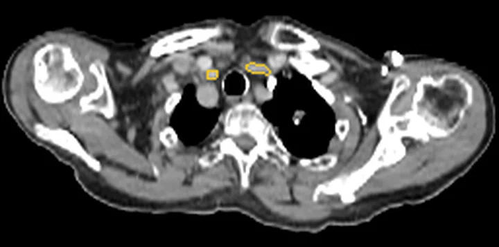
- Omuzlar arasındaki foton açlığı, subklavyen çıkışları ve alt boyun karotis/vertebral arterlerini çapraz koyu-açık çizgilerle örtebilir.
- Dental metal çizgileri mandibula komşuluğundaki karotis bifurkasyonu ve üst servikal damarları radyal biçimde kesebilir.
- Kafa tabanı kemiği yakınındaki beam-hardening çizgileri distal ICA opasifikasyonunu veya damar duvarı kenarını belirsizleştirir.
- Yalancı dolum kusuru ince kaynak kesitlerde azalıyor veya kayboluyorsa, gerçek duvara tutunan plak/trombüs yerine parsiyel hacim ya da akım artefaktını destekler.
Normalde
Normal kontrastlı karotis bifurkasyonunda ortak karotis, internal ve eksternal dallara ayrılır; damar lümenleri düzgün opaklaşır ve kalsiyum lümen sınırını kapatmaz. Aynı seviyede iki tarafın opak lümenini, komşu kesitlerdeki devamlılığı ve kemik/dental/omuz çizgilerinin yönünü karşılaştır; gerçek lezyon damara bağlı kalırken artefakt damar dışı yapılardan uzanan bant veya kısa süreli kontrast heterojenliği olarak izlenir.
Normal BT ile kıyasla
Aynı bölgenin, kaynak veri setinde patoloji içermediği belirtilen bir BT incelemesini kesit kesit kaydırın; organ sınırlarını açarak anatomiyi hasta görüntüsüyle karşılaştırın. Küçük rastlantısal bulgular tümüyle dışlanmamıştır.
Kaynak: TotalSegmentator v2.01 veri seti (Wasserthal ve ark., Radiology: Artificial Intelligence 2023), CC BY 4.0, vaka s0622; organ sınırları veri setinin otomatik segmentasyonundan, birleştirilerek sadeleştirildi. Görüntüler 2,5 mm kesit aralığına yeniden örneklendi.
Ayırıcı tanı
- Gerçek kalsifiye aterosklerotik stenoz
- duvara yerleşen plak ve damar uzun eksenine dik çok düzlemli reformasyonda da belirgin lümen daralması devam eder.
- Karotis trombozu veya oklüzyonu
- lümen kesilmesi ardışık kaynak görüntülerde ve damar merkez hattında sürer; distal yeniden dolum ayrıca araştırılır.
- Karotis diseksiyonu
- duvar boyunca uzanan eksantrik daralma, flep veya dış kontur değişikliği farklı düzlemde de doğrulanabilir.
- Karotis web
- bulbun posterior duvarına tutunan sabit ince raf, aksiyel ve oblik sagittal planda aynı yerde izlenir.
- Akım kaynaklı yalancı oklüzyon
- distal tıkanmaya bağlı yavaş dolan proksimal servikal ICA’da tek fazlı incelemede kontrast sütunu sönük kalabilir.
Tuzaklar
- Kalsifikasyonun parlak blooming sınırını gerçek lümen duvarı sayma; pencere genişliğini artırıp kontrastlı lümeni ince kaynak kesitlerde ve damara dik MPR’de yeniden değerlendir.
- Tek bir MIP üzerindeki çizgisel kesintiyi oklüzyon kabul etme; dental metal veya omuz foton açlığı kesintiyi damarın dışından da geçirir.
- Karşı tarafla kontrast yoğunluğu farklı diye tek başına stenoz tanısı koyma; enjeksiyon tarafı, venöz reflü ve bolus karışımının tüm damar yatağındaki dağılımını incele.
- Diş protezi veya kafa tabanı kaynaklı artefaktlar tanısal doğruluğu önemli ölçüde bozabileceğinden, görüntüleme hataları açısından sistematik ve dikkatli bir inceleme zorunludur.
- Tek fazlı BTA'da distal ICA oklüzyonuna bağlı durağan akım, proksimal servikal ICA'da yalancı oklüzyon görünümü oluşturabilir; proksimal opaklaşmanın uzunluğu ve kontrastın kademeli azalması bu görünümün gerçek oklüzyondan ayrımına yardımcı olur.
Kendini dene
Kalsifikasyon veya metal artefaktı lümen sınırını bozduğunda, stenozu değerlendirmek için hangi görüntüleme yaklaşımı uygundur?
Cevabı göster
Damar eksenine dik düzlemlerde ve ince kesitlerde değerlendirme. Dental çalışma veya başka artefaktlar BT ölçümlerini bozabilir; stenozu değerlendirmek için damarın uzun eksenine dik düzlemde çok düzlemli reformasyon (CMPR) kullanın. İntralüminal trombüs, çok düzlemli reformasyonlarda lümen kontrastıyla çevrili dolum kusuru olarak görülebilir; akım artefaktları ise diseksiyon veya damar oklüzyonunu taklit edebilir.
Dental metal artefaktı, karotis BT anjiyografisinde damarın yapay olarak kesintili görünmesine yol açabilir. En olası neden nedir?
Cevabı göster
Metal kaynaklı çizgilenme artefaktı. Dental donanım damar lümeninde yapay kesintilere yol açabilir. Karotis webi, proksimal servikal internal karotis arter lümenine kalsifikasyon olmadan uzanan raf benzeri bir çıkıntıdır; akım artefaktları ise diseksiyon veya damar oklüzyonunu taklit edebilir.
İlgili konular
Kaynaklar
Bu sayfadaki 39 cümle ve 2 quiz sorusu aşağıdaki kaynaklardan alıntılarla otomatik olarak karşılaştırıldı; 15 cümle bu karşılaştırmada düzeltildi (2026-10-09).
- State-of-the-art CT and MR imaging and assessment of atherosclerotic carotid artery disease: the reporting—a consensus document by the European Society of Cardiovascular Radiology (ESCR)pmc.ncbi.nlm.nih.gov
- Analysis of carotid artery deformation in different head and neck positions for maxillofacial catheter navigation in advanced oral cancer treatment.pubmed.ncbi.nlm.nih.gov
- Differentiating pseudo-occlusion from true occlusion of proximal internal carotid artery in acute ischemic stroke on CT angiography.pubmed.ncbi.nlm.nih.gov
- Neurologic applications of whole-brain volumetric multidetector computed tomography.pubmed.ncbi.nlm.nih.gov
- Dual-Source Dual-Energy CT Angiography of the Supra-Aortic Arteries with Tin Filter: Impact of Tube Voltage Selection.pubmed.ncbi.nlm.nih.gov
- Beyond the collaterals: Additional value of multiphase CTA in acute ischemic stroke evaluation.pubmed.ncbi.nlm.nih.gov
- Time-Resolved 4-Dimensional Computed-Tomography Angiography Can Correctly Identify Carotid Pseudo-Occlusion.pubmed.ncbi.nlm.nih.gov
- Four-dimensional computed tomography angiography analysis of internal carotid arteries opacification at the skull base to detect delayed cerebral ischemia: a feasibility study.pubmed.ncbi.nlm.nih.gov
- Sixty-four-section multidetector CT angiography of carotid arteries: a systematic analysis of image quality and artifacts.pubmed.ncbi.nlm.nih.gov
- Carotid CTA at the Lowest Tube Voltage (70 kV) in Comparison with Automated Tube Voltage Adaption.pubmed.ncbi.nlm.nih.gov
- Interpretation errors in CT angiography of the head and neck and the benefit of double reading.pubmed.ncbi.nlm.nih.gov
- Carotid CT angiography: comparison of image quality for left versus right arm injections.pubmed.ncbi.nlm.nih.gov
- Dual energy CTA of the supraaortic arteries: technical improvements with a novel dual source CT system.pubmed.ncbi.nlm.nih.gov
- Thrombus Characteristics Are Related to Collaterals and Angioarchitecture in Acute Stroke.pubmed.ncbi.nlm.nih.gov
- Impact of metal artifact reduction software on image quality of gemstone spectral imaging dual-energy cerebral CT angiography after intracranial aneurysm clipping.pubmed.ncbi.nlm.nih.gov
- Plaque-Adapted Virtual Monoenergetic Image Selection for Carotid Plaque Visualization Using Photon-Counting CT.pubmed.ncbi.nlm.nih.gov
- Association between carotid atherosclerotic plaque characteristics and white matter hyperintensity in cerebral small vessel disease.pubmed.ncbi.nlm.nih.gov
- Duplex ultrasound in severe carotid stenosis: diagnostic performance for differentiating near-occlusion subtypes from conventional high-grade stenosis.pubmed.ncbi.nlm.nih.gov
- A Bilateral Carotid Web Presenting as Recurrent Transient Ischemic Attacks With Negative Initial Stroke Imaging: A Case Report.pubmed.ncbi.nlm.nih.gov
- Multimodal imaging in neuro-ophthalmology.pubmed.ncbi.nlm.nih.gov
- A Metallic Screw Gun-Traumatic Vertebral Artery Dissection Injury: A Case Report and Literature Review.pubmed.ncbi.nlm.nih.gov
- Evaluation of Atherosclerotic Changes in the Superior Mesenteric Artery Using MDCT.pubmed.ncbi.nlm.nih.gov
- Local Recurrence after Microsurgical Clipping of Unruptured Intracranial Aneurysms: Incidence, Timing, and Risk Factors over 20 Years.pubmed.ncbi.nlm.nih.gov
- State-of-the-art CT and MR imaging and assessment of atherosclerotic carotid artery disease: the reporting-a consensus document by the European Society of Cardiovascular Radiology (ESCR).pubmed.ncbi.nlm.nih.gov
- Clinical implications of vessel wall imaging-State-of-the-art review.pubmed.ncbi.nlm.nih.gov
- Spectral CT and Photon-counting Detector CT: Principles and Applications in Neuroradiology.pubmed.ncbi.nlm.nih.gov
- Extra-Carotid Arterial Webs: A Single-Center Series and Systematic Review.pubmed.ncbi.nlm.nih.gov
- Technical advances and clinical impact of photon-counting computed tomography angiography for peripheral artery disease.pubmed.ncbi.nlm.nih.gov
- Study Protocol for the Evaluation of Morphologic and Imaging Remodeling of Atherosclerotic Plaque Following Intravascular Lithotripsy in Peripheral Artery Disease.pubmed.ncbi.nlm.nih.gov
- Beyond visual inspection: the deep learning revolution in quantitative cerebrovascular imaging.pubmed.ncbi.nlm.nih.gov
- Operative Procedures for Ultrasound Assessment of Extracranial Artery Disease: A Narrative Review by the Italian Society for Vascular Investigation (SIDV).pubmed.ncbi.nlm.nih.gov
- Optimization of head and neck vascular CT angiography using variable rate bolus tracking technique and third-generation dual-source CT dual-energy scanning.pubmed.ncbi.nlm.nih.gov
- Optimizing low contrast volume thoracic CT angiography: From the basics to the advanced.pubmed.ncbi.nlm.nih.gov
- Treatment of Ruptured and Nonruptured Aneurysms Using a Semisolid Iodinated Embolic Agent.pubmed.ncbi.nlm.nih.gov
- Understanding Photon-Counting CT: Physics, Detector Technology, and Image Reconstructions.pubmed.ncbi.nlm.nih.gov
- Single rotation CTA of extracranial carotids integrated with cerebral CTP provides sufficient quality for decision making in patients with ischaemic stroke.pubmed.ncbi.nlm.nih.gov
- Effects of iterative metal artifact reduction techniques on diagnostic performance in patients with dental artifacts on carotid computed tomography angiography.pubmed.ncbi.nlm.nih.gov
- Role of metal artifact reduction software in computed tomography angiography of lower limb metallic prosthesis: A retrospective study.pubmed.ncbi.nlm.nih.gov
- Congenital Anomalies and Variations of the Carotid and Vertebral Arteries: A Case-Based Imaging Review.pubmed.ncbi.nlm.nih.gov
- Diagnostic Evaluation and Surgical Management of Self-Inflicted Trans-Frontal Sinus Penetrating Brain Injury in a Patient with Schizophrenia.pubmed.ncbi.nlm.nih.gov
- Study on the combined application of selected virtual monoenergetic imaging, virtual non-contrast imaging, and adaptive statistical iterative reconstruction-V in head and neck CT angiography using 256-slice wide-body CT.pubmed.ncbi.nlm.nih.gov
- Clinical benefits and current challenges of photon-counting detector CT in vascular imaging.pubmed.ncbi.nlm.nih.gov
- Styloidogenic Internal Carotid Artery Dissection Presenting as Severe New-Onset Headache in a Young Adult.pubmed.ncbi.nlm.nih.gov
- Multimodal Imaging of a Post-Traumatic Cervical Arteriovenous Fistula with Concomitant Pseudoaneurysm After a Gunshot Wound Treated with a Covered Stent-Graft.pubmed.ncbi.nlm.nih.gov
- A rare case report of vascular Ehlers-Danlos syndrome as a pulsatile neck mass with an internal carotid artery aneurysm.pubmed.ncbi.nlm.nih.gov
- Clinical analysis of 48 patients with carotid web diagnosed by digital subtraction angiography.pubmed.ncbi.nlm.nih.gov
- Hybrid revascularization for acute common carotid artery occlusion after transcarotid transcatheter aortic valve implantation: illustrative case.pubmed.ncbi.nlm.nih.gov
- Coronary Calcium Scoring as Prediction of Coronary Artery Diseases with Low-Dose Dual-Source CT.pubmed.ncbi.nlm.nih.gov
- TEVAR for acute type B aortic dissection in pregnant women (35 weeks gestation) with Takayasu's arteritis after cesarean section: a rare case report and literature review.pubmed.ncbi.nlm.nih.gov
- Case Report: Intravascular lithotripsy combined with a new-generation 3-in-1 carotid stent for calcified carotid artery stenosis.pubmed.ncbi.nlm.nih.gov
- Hybrid repair of Kommerell diverticulum associated with severe aberrant left subclavian artery stenosis and poststenotic aneurysmal degeneration: 2-year outcomes.pubmed.ncbi.nlm.nih.gov
- Acute middle cerebral artery thrombosis in the early postoperative period after total knee arthroplasty under general anesthesia: A case report.pubmed.ncbi.nlm.nih.gov
- Evolving cerebrovascular pathology in fibromuscular dysplasia leading to aneurysmal subarachnoid hemorrhage.pubmed.ncbi.nlm.nih.gov
- Vertebral Artery Pseudoaneurysm in a Single-Vessel Posterior Circulation With Chronic Contralateral Occlusion and Recent Methamphetamine Use.pubmed.ncbi.nlm.nih.gov
- Initial magnetic resonance imaging/magnetic resonance angiography may miss intracranial artery dissection in young adults with stroke: the role of subsequent imaging.pubmed.ncbi.nlm.nih.gov
- Clinical and CT angiography characteristics of Chinese stroke patients with carotid web: a retrospective study.pubmed.ncbi.nlm.nih.gov
- Contrast Dynamics on Multiphase CT Angiography in Carotid Webs and Their Association with Ischemic Stroke.pubmed.ncbi.nlm.nih.gov
- Treatment of a Symptomatic Carotid Web: A Case Report and Review of the Literature.pubmed.ncbi.nlm.nih.gov
- True versus pseudo-occlusion of the cervical internal carotid artery in acute stroke: A multicenter MR angiography study.pubmed.ncbi.nlm.nih.gov
- Distinction between acute and chronic carotid disease based on computed tomography angiography in acute ischemic stroke patients with carotid artery occlusion.pubmed.ncbi.nlm.nih.gov
- Cranial anatomy, palaeoneurology, palaeobiology and stratigraphic age of the large-bodied ornithopod, <i>Muttaburrasaurus langdoni</i> Bartholomai and Molnar, 1981, from the mid-Cretaceous of Australia.pubmed.ncbi.nlm.nih.gov
- Imaging and Management of Blunt Cerebrovascular Injury.pubmed.ncbi.nlm.nih.gov
- Quality of extracranial carotid evaluation with 256-section CT.pubmed.ncbi.nlm.nih.gov
- Spontaneous bacteremia and spontaneous bacterial peritonitis share similar prognosis in patients with cirrhosis: a cohort study.pubmed.ncbi.nlm.nih.gov
- A comparison of magnetocardiography with noninvasive cardiac testing in the evaluation of patients with chest pain.pubmed.ncbi.nlm.nih.gov
- Proceedings of the 15th International Congress on Circumpolar Health.pubmed.ncbi.nlm.nih.gov
- Patterns of injury and violence in Yaoundé Cameroon: an analysis of hospital data.pubmed.ncbi.nlm.nih.gov
- Mapping the lingual artery: a scoping review and proposed imaging-informed risk classification for surgical and radiological practice.pubmed.ncbi.nlm.nih.gov
- Case Report: Carotid body paraganglioma in a 20-year-old woman: multimodal imaging, vessel-preserving surgical excision, and long-term clinical follow-up.pubmed.ncbi.nlm.nih.gov
- Carotid body measurement by computed tomography angiography: a cross-sectional population-based study.pubmed.ncbi.nlm.nih.gov
- Mixed coronary plaque phantom analysis by photon-counting CT: impact of calcium and iodine on low-attenuation plaque detection.pubmed.ncbi.nlm.nih.gov
- Dark-blood" Imaging in Coronary CT Angiography Using Dual layer Detector Spectral CT: Effect on Image Quality and Vessel Wall Visibilitypubmed.ncbi.nlm.nih.gov
- Mechanisms of aortic dissection: From pathological changes to experimental and <i>in silico</i> models.pubmed.ncbi.nlm.nih.gov
- Outer Wall Segmentation of Abdominal Aortic Aneurysm by Variable Neighborhood Search Through Intensity and Gradient Spaces.pubmed.ncbi.nlm.nih.gov
- Restoration of brain circulation and cellular functions hours post-mortem.pubmed.ncbi.nlm.nih.gov
- Magnetic resonance angiography detection of abnormal carotid artery plaque in patients with cryptogenic stroke.pubmed.ncbi.nlm.nih.gov
- Thrombus enhancement sign predicts distal embolization in anterior circulation stroke treated with endovascular thrombectomy.pubmed.ncbi.nlm.nih.gov
- Bilateral retinal artery occlusions with carotid artery occlusions-ocular and cerebral hemodynamic changes.pubmed.ncbi.nlm.nih.gov
- State of the art: noninvasive imaging and management of neurovascular trauma.pubmed.ncbi.nlm.nih.gov
- Spontaneous Intracranial Vertebral Artery Dissection: A Rare Cause of Ischemic Stroke.pubmed.ncbi.nlm.nih.gov
- CT Imaging Findings of Pulmonary Artery Stenosis: A Pictorial Review.pubmed.ncbi.nlm.nih.gov
- Assessment of the thoracic aorta after aortic root replacement and/or ascending aortic surgery using 3D relaxation-enhanced angiography without contrast and triggering.pubmed.ncbi.nlm.nih.gov
- Computed Tomography Angiography "Rim Sign" for Detecting Carotid Intraplaque Hemorrhage: A Computed Tomography Angiography-Magnetic Resonance Imaging-Histology Correlation Study.pubmed.ncbi.nlm.nih.gov
- David-V Procedure Combined with Zone 2 Arch Replacement for Acute Stanford Type A Dissection.pubmed.ncbi.nlm.nih.gov
- Endovascular technology for aortic arch repair: from innovation to integration.pubmed.ncbi.nlm.nih.gov
- Extraction of Coronary Atherosclerotic Plaques From Computed Tomography Imaging: A Review of Recent Methods.pubmed.ncbi.nlm.nih.gov
- Dual-Energy CT: What the Neuroradiologist Should Know.pubmed.ncbi.nlm.nih.gov
- Evaluation of carotid artery stenosis with multisection CT and MR imaging: influence of imaging modality and postprocessing.pubmed.ncbi.nlm.nih.gov
- Ultra-High-Resolution Photon-Counting CT Angiography for Noninvasive Detection of Neurovascular In-Stent Restenosis.pubmed.ncbi.nlm.nih.gov
- CT Angiography of the Upper Extremities: Review of Acute Arterial Entities.pubmed.ncbi.nlm.nih.gov
- Metal Artifact Reduction for Orthopedic Implants: Brain CT Angiography in Patients with Intracranial Metallic Implants.pubmed.ncbi.nlm.nih.gov
- Diagnostic yield of 90-kVp low-tube-voltage carotid and intracerebral CT-angiography: effects on radiation dose, image quality and diagnostic performance for the detection of carotid stenosis.pubmed.ncbi.nlm.nih.gov
- Commentary: an eye on PET quantification.pubmed.ncbi.nlm.nih.gov
- Optimization of Contrast Injection Protocols for Time-Resolved MRA Technique in Dogs: A Comparative Study of Vascular Signal Characteristics and Artifacts.pubmed.ncbi.nlm.nih.gov
- Overview of Imaging Modalities in Stroke.pubmed.ncbi.nlm.nih.gov
- Photon-counting detector CT vs energy-integrating detector CT for the diagnosis of arterial dissection in the cervicocerebral region: A single-center case series.pubmed.ncbi.nlm.nih.gov
- A wide-detector CT protocol for one-stop cardio-cerebrovascular angiography: a technical feasibility study comparing an integrated protocol with conventional two-scan approach.pubmed.ncbi.nlm.nih.gov
- Carotid Web: An Update Focusing on Its Relationship With Fibromuscular Dysplasia and Therapeutic Strategy.pubmed.ncbi.nlm.nih.gov
- Evaluation of Cone-Beam Computed Tomography Scans to Develop a Staging Method of External Carotid Artery Calcification.pubmed.ncbi.nlm.nih.gov
- Ischemic Heart Disease: Noninvasive Imaging Techniques and Findings.pubmed.ncbi.nlm.nih.gov
- High-affinity alphavbeta3 integrin targeted optical probe as a new imaging biomarker for early atherosclerosis: initial studies in Watanabe rabbits.pubmed.ncbi.nlm.nih.gov
- A case of carotid web with thrombosis causing stroke in a young adult.pubmed.ncbi.nlm.nih.gov
- RAPID CTA versus JLK LVO for large vessel occlusion detection: a pragmatic comparison of performance and common pitfalls.pubmed.ncbi.nlm.nih.gov
- Definition of Carotid Artery Free Floating Thrombus: A Systematic Review and Call for Standardisation of Imaging and Nomenclature.pubmed.ncbi.nlm.nih.gov
- Differentiating True Occlusion from Pseudo-Occlusion: The Role of Extended Multiphase Computed Tomography Angiography Scan Range in Internal Carotid Artery Occlusion.pubmed.ncbi.nlm.nih.gov
- True versus pseudo-occlusion of the cervical internal carotid artery in acute stroke: A multicenter MR angiography study.pubmed.ncbi.nlm.nih.gov
- Improving Measurement Accuracy of the Degree of Carotid In-Stent Stenosis Using CT Angiography: A Comparison With DSA.pubmed.ncbi.nlm.nih.gov
- Cardiovascular computed tomography in cardiovascular disease: An overview of its applications from diagnosis to prediction.pubmed.ncbi.nlm.nih.gov
- Novel Imaging-Based Biomarkers for Identifying Carotid Plaque Vulnerability.pubmed.ncbi.nlm.nih.gov
- Self-expanding open-cell carotid stent for symptomatic moderate-to-severe vertebral artery origin atherosclerotic stenosis: a single-center retrospective study.pubmed.ncbi.nlm.nih.gov
- Carotid bifurcation reconstruction as a novel CEA modification to address complex carotid lesions.pubmed.ncbi.nlm.nih.gov
Eğitim amaçlıdır; hasta başında tanı ve tedavi kararının yerine geçmez, tıbbi tavsiye değildir. Metinler yapay zekâ desteğiyle yazıldı ve otomatik bir adımda kaynak alıntılarıyla karşılaştırıldı; bu bir tıbbi doğrulama değildir ve içerik uzman radyolog incelemesinden geçmedi. Klinik kararlarda güncel kılavuzları, kurum protokollerini ve radyoloji raporunu esas alın.