Özet
Kalp hareketine bağlı artefakt, CTPA’da bronkovasküler yapılarda bulanıklık ve çift çizgi görünümü oluşturabilir. Kardiyak hareket artefaktı bronkovasküler yapılarda bulanıklık ve çift çizgi artefaktı oluşturabilir; bu artefaktlar subsegmental emboli saptanmasını etkileyebilir. Kardiyak hareket artefaktlarını azaltan yüksek pitch çift kaynaklı CTPA, subsegmental emboli saptanmasını artırabilir. Kalp hareketine bağlı artefaktların azaltılması, CTPA’da subsegmental pulmoner emboli saptanmasını artırabilir.
Faz ve pencere
- BTPA tanısal
- Kardiyak hareket artefaktı, bronkovasküler yapılarda bulanıklık ve çift çizgi görünümüne yol açabilir.
Önerilen pencereler G genişlik, M merkez; değerler Hounsfield birimi (HU)
- AnjiyoG 600 / M +150
- MediastenG 350 / M +50
- AkciğerG 1500 / M −600
BT bulguları
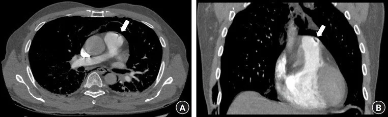
- Damar kenarında çift kontur veya basamaklanma — bronkovasküler yapılarda bulanıklık ve çift çizgi artefaktı görülebilir; bu durum özellikle kardiyak hareket kaynaklı artefaktlarla ilişkilidir.
- Hareket kaynaklı yanlış eşleşme, CTPA’da görülebilen artefakt türlerinden biridir.
- Aort, kalp ve diyafram hareketi CTPA'da çift görüntü şeklinde hareket artefaktı oluşturabilir.
- Nefesini tutmayı engelleyen KOAH veya dispne gibi durumlarda, nefes tutmalı CTPA’da belirgin solunum artefaktları görülebilir.
- Kardiyak hareket artefaktı pulmoner arterlerde bulanıklık ve bronkovasküler yapılarda çift çizgi görünümüne yol açabilir.
- Kardiyak hareket artefaktı bronkovasküler yapılarda bulanıklık ve çift çizgi görünümü oluşturabilir.
- Kardiyak hareket artefaktı, bronkovasküler yapılarda bulanıklık ve çift çizgi görünümü oluşturabilir.
Normalde
Hareket artefaktı, CTPA’da pulmoner damar bulgularının değerlendirilmesini güçleştirebilir; görüntüleri yorumlarken bu olasılığı göz önünde bulundurun.
Normal BT ile kıyasla
Aynı bölgenin, kaynak veri setinde patoloji içermediği belirtilen bir BT incelemesini kesit kesit kaydırın; organ sınırlarını açarak anatomiyi hasta görüntüsüyle karşılaştırın. Küçük rastlantısal bulgular tümüyle dışlanmamıştır.
Kaynak: TotalSegmentator v2.01 veri seti (Wasserthal ve ark., Radiology: Artificial Intelligence 2023), CC BY 4.0, vaka s1278; organ sınırları veri setinin otomatik segmentasyonundan, birleştirilerek sadeleştirildi. Görüntüler 2,5 mm kesit aralığına yeniden örneklendi.
Ayırıcı tanı
- Akut pulmoner emboli, pulmoner arter dolum kusurunun en sık nedenidir; ancak bu bulgu tek başına özgül değildir.
- Kontrast karışım artefaktı
- damar içinde bulanık yoğunluk geçişi oluşturur ancak aynı anda bronş ve plevra konturları hareketle çiftlenmez.
- SVC kaynaklı streak artefaktı
- sağ üst mediastene uzanan radyal çizgilenme ve buna komşu sağ üst lob damarında atenüasyon kaybı yapar.
- Kronik tromboemboli
- duvara yapışık web, bant, damar daralması veya eksantrik organize materyal ile birden fazla düzlemde devamlılık gösterir.
Tuzaklar
- Hareket kaynaklı çift görüntüler CTPA’da değerlendirilebilir; yüksek pitch çift kaynaklı protokol aortik, kardiyak ve diyafragmatik hareket artefaktlarını azaltabilir.
- Kardiyak hareket artefaktı, bronkovasküler yapılarda bulanıklık ve çift çizgi artefaktı oluşturabilir.
- Kardiyak hareket artefaktlarının azaltılması, CTPA’da subsegmental emboli saptanmasını artırabilir.
- Kalp hareketi, bronkovasküler yapılarda bulanıklık ve çift çizgi artefaktına yol açabilir.
- Kardiyak hareket artefaktı pulmoner arterlerde çift çizgi görünümüne yol açabilir ve subsegmental emboli saptanmasını etkileyebilir.
Kendini dene
Non-EKG-gated CTPA’da kardiyak hareket artefaktıyla hangi görüntü özelliği ilişkilidir?
Cevabı göster
Bronkovasküler yapıların bulanıklaşması ve çift kontur oluşturması. Kardiyak hareket, bronkovasküler yapılarda bulanıklık ve çift çizgi artefaktı oluşturabilir.
CTPA’da hareket artefaktlarını azaltmada hangi protokol daha başarılı bulunmuştur?
Cevabı göster
Yüksek pitch çift kaynaklı BT protokolü. Karşılaştırmalı çalışmada çift kaynaklı protokol hareket artefaktlarını tek kaynaklı tekniğe göre daha fazla azaltmıştır.
İlgili konular
Kaynaklar
Bu sayfadaki 32 cümle ve 2 quiz sorusu aşağıdaki kaynaklardan alıntılarla otomatik olarak karşılaştırıldı; 26 cümle bu karşılaştırmada düzeltildi (2026-10-09).
- Quantitative imaging features of pulmonary artery involvement in Takayasu arteritis on computed tomography pulmonary angiography in patients with and without pulmonary hypertensionpmc.ncbi.nlm.nih.gov
- Artificial Intelligence in Pulmonary Hypertension: Current State and Future Prospectspmc.ncbi.nlm.nih.gov
- Case Report: Heparin resistance as the harbinger of heparin-induced thrombocytopenia in acute pulmonary embolismpmc.ncbi.nlm.nih.gov
- Differentiating pulmonary embolism from contrast accumulation-induced beam hardening artifacts using dual-energy CT quantitative parameterspmc.ncbi.nlm.nih.gov
- The use of imaging in the diagnosis and treatment of thromboembolic pulmonary hypertensionpmc.ncbi.nlm.nih.gov
- Motion Artifact Reduction From High-Pitch Dual-Source Computed Tomography Pulmonary Angiography.pubmed.ncbi.nlm.nih.gov
- Visibility and image quality of peripheral pulmonary arteries in pulmonary embolism patients using free-breathing combined with a high-threshold bolus-triggering technique in CT pulmonary angiography.pubmed.ncbi.nlm.nih.gov
- Single-Bolus Sequential Triple-Rule-Out CT Angiography: Image Quality and Radiation Dose on Wide-Area Detector and Dual-Source CT.pubmed.ncbi.nlm.nih.gov
- Acute Traumatic Aortic Injury: What the Radiologist Needs to Know.pubmed.ncbi.nlm.nih.gov
- Ferumoxytol-enhanced cardiovascular magnetic resonance imaging: applications and technical advances.pubmed.ncbi.nlm.nih.gov
- An Integrated Approach on the Diagnosis of Cerebral Veins and Dural Sinuses Thrombosis (a Narrative Review).pubmed.ncbi.nlm.nih.gov
- Artificial intelligence in computed tomography imaging for pulmonary embolism: a narrative review from computed tomography pulmonary angiography to non-contrast computed tomography.pubmed.ncbi.nlm.nih.gov
- Can Artificial Intelligence Really Help? A Practicing Radiologist's Simplified Guide to AI, with a Critical Appraisal of the Use of AI in Cancer-Associated Thromboembolism.pubmed.ncbi.nlm.nih.gov
- Artificial Intelligence in Pulmonary Hypertension: Current State and Future Prospects.pubmed.ncbi.nlm.nih.gov
- Proceedings of the 15th International Congress on Circumpolar Health.pubmed.ncbi.nlm.nih.gov
- Society for Cardiovascular Magnetic Resonance 2024 Cases of SCMR case series.pubmed.ncbi.nlm.nih.gov
- Society for Cardiovascular Magnetic Resonance/European Society of Cardiovascular Imaging/American Society of Echocardiography/Society for Pediatric Radiology/North American Society for Cardiovascular Imaging Guidelines for the use of cardiovascular magnetic resonance in pediatric congenital and acquired heart disease : Endorsed by The American Heart Association.pubmed.ncbi.nlm.nih.gov
- Society for Cardiovascular Magnetic Resonance/European Society of Cardiovascular Imaging/American Society of Echocardiography/Society for Pediatric Radiology/North American Society for Cardiovascular Imaging Guidelines for the Use of Cardiac Magnetic Resonance in Pediatric Congenital and Acquired Heart Disease: Endorsed by The American Heart Association.pubmed.ncbi.nlm.nih.gov
- Quantification of pulmonary perfusion using LSIM-CT correlates with pulmonary hemodynamics in patients with CTEPD.pubmed.ncbi.nlm.nih.gov
- Current Management of Infective Endocarditis: A Narrative Review Focused on Unmet Clinical Needs and the Multidisciplinary Approach.pubmed.ncbi.nlm.nih.gov
- 2.5-mm articulated endoluminal forceps using a compliant mechanism.pubmed.ncbi.nlm.nih.gov
- Standardized image interpretation and post processing in cardiovascular magnetic resonance: Society for Cardiovascular Magnetic Resonance (SCMR) board of trustees task force on standardized post processing.pubmed.ncbi.nlm.nih.gov
- A scoping review of photon-counting detector computed tomography in cardiovascular imaging.pubmed.ncbi.nlm.nih.gov
- Understanding Photon-Counting CT: Physics, Detector Technology, and Image Reconstructions.pubmed.ncbi.nlm.nih.gov
- Contrast medium dose optimization in the era of multi-energy CT.pubmed.ncbi.nlm.nih.gov
- CT and MRI in the Evaluation of Thoracic Aortic Diseases.pubmed.ncbi.nlm.nih.gov
- Long-term safety and effectiveness of the "OptEase" vena cava filter.pubmed.ncbi.nlm.nih.gov
- High-affinity alphavbeta3 integrin targeted optical probe as a new imaging biomarker for early atherosclerosis: initial studies in Watanabe rabbits.pubmed.ncbi.nlm.nih.gov
- Dual-low spectral CT pulmonary angiography: a comparative study of image quality, radiation dose, and iodine intake with evaluation of pulmonary embolism detection.pubmed.ncbi.nlm.nih.gov
- Frequently Encountered Artifacts in the Application of Dual-Energy Computed Tomography to Cardiovascular Imaging for Urate Crystals in Gout: A Matched-Control Study.pubmed.ncbi.nlm.nih.gov
- A review of deep learning in medical imaging: Imaging traits, technology trends, case studies with progress highlights, and future promises.pubmed.ncbi.nlm.nih.gov
- Postoperative Pulmonary Complications: Clinical and Imaging Insightspubmed.ncbi.nlm.nih.gov
- Computed tomography and magnetic resonance imaging for pulmonary embolus evaluation in children: up-to-date review on practical imaging protocols.pubmed.ncbi.nlm.nih.gov
- The filling defect of pulmonary artery, an imaging finding what we should know.pubmed.ncbi.nlm.nih.gov
- Photon-Counting CT in Cardiovascular Imaging: Clinical Applications.pubmed.ncbi.nlm.nih.gov
- Clinical benefits and current challenges of photon-counting detector CT in vascular imaging.pubmed.ncbi.nlm.nih.gov
- The treatment of challenging visceral and peripheral arterial aneurysms with flow diversion-a German multicentric observational study.pubmed.ncbi.nlm.nih.gov
- Current Perspectives on Balloon Pulmonary Angioplasty for Chronic Thromboembolic Pulmonary Hypertension.pubmed.ncbi.nlm.nih.gov
- Optimal approach to performing and reporting computed tomography angiography for suspected acute pulmonary embolism: a clinical consensus statement of the ESC Working Group on Pulmonary Circulation & Right Ventricular Function, the Fleischner Society, the Association for Acute CardioVascular Care (ACVC) and the European Association of Cardiovascular Imaging (EACVI) of the ESC, endorsed by European Respiratory Society (ERS), Asian Society of Thoracic Radiology (ASTR), European Society of Thoracic Imaging (ESTI), and Society of Thoracic Radiology (STR).pubmed.ncbi.nlm.nih.gov
- Outcomes Associated With Catheter-Directed Therapies in Chronic Thromboembolic Pulmonary Hypertension: Cautionary Tales From a Single-Center Case Series.pubmed.ncbi.nlm.nih.gov
- Aortic Injury and Hemopericardium: Key Findings and Evaluation.pubmed.ncbi.nlm.nih.gov
- A pulsatile endobronchial lesion: A vascular mimic of airway pathology.pubmed.ncbi.nlm.nih.gov
- Anomalous Systemic Arterial Supply to the Basal Segment of the Lung Treated With Surgery Versus Transcatheter Embolisation: A Report of Two Cases.pubmed.ncbi.nlm.nih.gov
- Uni-port video-assisted thoracoscopic surgery for pulmonary arteriovenous malformation based on vascular pre-occlusion: a case report and literature review.pubmed.ncbi.nlm.nih.gov
- Endoscopic Management of Post-Bariatric Surgery Complications: Diagnostic Work-Up and Innovative Approaches for Leak, Fistula, and Stricture Management.pubmed.ncbi.nlm.nih.gov
- Mechanisms of aortic dissection: From pathological changes to experimental and <i>in silico</i> models.pubmed.ncbi.nlm.nih.gov
- Patient-Specific Inverse Modeling of In Vivo Cardiovascular Mechanics with Medical Image-Derived Kinematics as Input Data: Concepts, Methods, and Applications.pubmed.ncbi.nlm.nih.gov
- Quantitative imaging features of pulmonary artery involvement in Takayasu arteritis on computed tomography pulmonary angiography in patients with and without pulmonary hypertension.pubmed.ncbi.nlm.nih.gov
- Case Report: Heparin resistance as the harbinger of heparin-induced thrombocytopenia in acute pulmonary embolism.pubmed.ncbi.nlm.nih.gov
- Pulmonary arteries: imaging of pulmonary embolism and beyond.pubmed.ncbi.nlm.nih.gov
- Multislice CT virtual intravascular endoscopy for assessing pulmonary embolisms: a pictorial review.pubmed.ncbi.nlm.nih.gov
- Contrast opacification on thoracic CT angiography: challenges and solutions.pubmed.ncbi.nlm.nih.gov
- Commercial artificial intelligence-assisted performance and interpretation time of first-on-call radiology residents using computed tomography pulmonary angiography to detect pulmonary embolism: a multireader, multicenter study.pubmed.ncbi.nlm.nih.gov
- Clinical Impact of CT-Based FFR in Everyday Cardiology: Bridging Computation and Decision-Making.pubmed.ncbi.nlm.nih.gov
- Investigating the Influence of High-Speed Gantry Rotation in Cardiac CT on Motion Artifacts in Aortic Stenosis Patients Not Premedicated with β-Blockers: The FAST-CCT Randomized Trial Protocol.pubmed.ncbi.nlm.nih.gov
- Advancements in 3D Transoesophageal Echocardiography (TOE) and Computed Tomography (CT) for Stroke Prevention in Left Atrial Appendage Occlusion Interventions.pubmed.ncbi.nlm.nih.gov
- Multimodal imaging in neuro-ophthalmology.pubmed.ncbi.nlm.nih.gov
- When the Heart's Blueprint Goes Awry: Exploring Congenital Absence of the Left Circumflex Artery in a Case of Chronic Dyspnea.pubmed.ncbi.nlm.nih.gov
- CT-Guided Transthoracic Core-Needle Biopsy of Pulmonary Nodules: Current Practices, Efficacy, and Safety Considerations.pubmed.ncbi.nlm.nih.gov
- CT-guided percutaneous biopsy of intrathoracic lesions.pubmed.ncbi.nlm.nih.gov
- Extra-cardiac findings in cardiovascular magnetic resonance: what the imaging cardiologist needs to know.pubmed.ncbi.nlm.nih.gov
- Society for Cardiovascular Magnetic Resonance 2022 Cases of SCMR case series.pubmed.ncbi.nlm.nih.gov
- Ischemic Heart Disease: Noninvasive Imaging Techniques and Findings.pubmed.ncbi.nlm.nih.gov
- Optimizing acute chest pain diagnosis: Efficacy of 64-channel multi-slice CT with Snap-Shot Freeze technique in Triple-Rule-out CT angiography.pubmed.ncbi.nlm.nih.gov
- Novel Computed Tomography Angiography Parameter Is Associated with Low Cardiac Index in Patients with Chronic Thromboembolic Pulmonary Hypertension: A Retrospective Analysis.pubmed.ncbi.nlm.nih.gov
- Human In Vivo Cardiac Magnetic Resonance Imaging at 7 T: Feasibility, Applications, and Current Limitations-A Systematic Review.pubmed.ncbi.nlm.nih.gov
- A survey of transformer-based architectures in medical image analysis: models, applications, and challenges.pubmed.ncbi.nlm.nih.gov
- Pulmonary MRI in Newborns and Children.pubmed.ncbi.nlm.nih.gov
- Tips and Tricks in Thoracic Radiology for Beginners: A Findings-Based Approach.pubmed.ncbi.nlm.nih.gov
- Review of D-Shape Left Ventricle Seen on Magnetic Resonance Imaging (MRI) or Computed Tomography (CT), Similar to the Movahed Sign Seen on Cardiac Gated Single-Photon Emission Computed Tomography (SPECT) as an Indicator for Right Ventricular Overload.pubmed.ncbi.nlm.nih.gov
- Artificial intelligence for pulmonary embolism detection: Is it comparable to radiology residents?pubmed.ncbi.nlm.nih.gov
- Extended CTA scan range for early detection of left atrial and left atrial appendage thrombus and other filling defects in acute ischemic stroke.pubmed.ncbi.nlm.nih.gov
- ECMO-assisted catheter-directed thrombolysis for post-traumatic pulmonary embolism with bilateral atrial thrombi: a retrospective case series.pubmed.ncbi.nlm.nih.gov
- Patient-Specific Timing for Pulmonary CT Angiography: Human Validation of a Physiologically Derived Post-Trigger Delay Model.pubmed.ncbi.nlm.nih.gov
- From congenital variants to critical clues: A radiologic review of inferior vena cava pathologies.pubmed.ncbi.nlm.nih.gov
- Application of 50-keV virtual monochromatic imaging combined with a personalized contrast agent protocol in computed tomography pulmonary angiography: a randomized controlled trial.pubmed.ncbi.nlm.nih.gov
- Individualized Scan Protocol Based on Body Mass Index in Dual-Energy Computed Tomography Pulmonary Angiography to Reduce Radiation and Contrast Doses with Improved Image Quality.pubmed.ncbi.nlm.nih.gov
- Dual energy imaging in cardiothoracic pathologies: A primer for radiologists and clinicians.pubmed.ncbi.nlm.nih.gov
- Contemporary sequential segmental approach to congenital heart disease using four-dimensional magnetic resonance imaging with ferumoxytol: an illustrated editorial.pubmed.ncbi.nlm.nih.gov
- Role of CT in the Pre- and Postoperative Assessment of Conotruncal Anomalies.pubmed.ncbi.nlm.nih.gov
- SCMR expert consensus statement for cardiovascular magnetic resonance of acquired and non-structural pediatric heart disease.pubmed.ncbi.nlm.nih.gov
- Multimodal Management of Chronic Thromboembolic Pulmonary Hypertension: A Narrative Review of Treatment Selection and Sequencing.pubmed.ncbi.nlm.nih.gov
- Imaging in chronic thromboembolic pulmonary hypertension: review of the current literature.pubmed.ncbi.nlm.nih.gov
- Lung ultrasound in a nutshell. Lines, signs, some applications, and misconceptions from a radiologist's point of view. Part 2.pubmed.ncbi.nlm.nih.gov
- EANM/SNMMI guideline/procedure standard for [<sup>18</sup>F]FDG hybrid PET use in infection and inflammation in adults v2.0.pubmed.ncbi.nlm.nih.gov
- Guidance for clinical practice using emergency and point-of-care ultrasonography.pubmed.ncbi.nlm.nih.gov
- Caught in the Whirl: Computed Tomography Diagnosis of Transverse Colon Volvulus With Proximal Jejunal Involvement and Cecocolic Pneumatosis.pubmed.ncbi.nlm.nih.gov
- The Initial Diagnostic Approach to Adult Congenital Heart Disease: A Practical Imaging Pathway for General Cardiologists and Cardiac Imagers.pubmed.ncbi.nlm.nih.gov
- Artificial Intelligence Across the Radiology Workflow: A Nine-Stage Narrative Review.pubmed.ncbi.nlm.nih.gov
- Respiratory frequency-tunable dynamic imaging for lung function: New exam method using chest X-ray cine imaging considering various respiratory diseases.pubmed.ncbi.nlm.nih.gov
- Gross, microscopic, radiologic, echocardiographic and haematological findings in rats experimentally infected with Angiostrongylus cantonensis .pubmed.ncbi.nlm.nih.gov
- Radiological manifestations of thoracic hydatid cysts: pulmonary and extrapulmonary findings.pubmed.ncbi.nlm.nih.gov
- Clinical correlates and short-term prognostic value of CT-detected subclinical pulmonary congestion in acute STEMI without overt heart failure.pubmed.ncbi.nlm.nih.gov
- Total Pectoral Muscle Area and Prognostic Nutritional Index Provide Complementary Prognostic Information for Patients Hospitalized with HFmrEF/HFrEF.pubmed.ncbi.nlm.nih.gov
- Diagnostic Value and Operational Recommendations for Late Iodine Enhancement and ECV Quantification in Single-Energy Computed Tomography: A Narrative Review.pubmed.ncbi.nlm.nih.gov
- Multidetector Computed Tomography (MDCT) Angiography in the Pre-Procedural Assessment of Patients Undergoing Transcatheter Aortic Valve Replacement.pubmed.ncbi.nlm.nih.gov
- Intracranial vascular stenosis and occlusive disease: evaluation with CT angiography, MR angiography, and digital subtraction angiography.pubmed.ncbi.nlm.nih.gov
- When Virchow's Triad Goes on Overdrive: Catastrophic Multi-System Thrombosis in the Setting of New-Onset Heart Failure in a Previously Healthy Man.pubmed.ncbi.nlm.nih.gov
- An Unusual Case of Thromboangiitis Obliterans With Concurrent Deep Vein Thrombosis and Pulmonary Embolism.pubmed.ncbi.nlm.nih.gov
- Severe <i>Chlamydia psittaci</i> pneumonia complicated by deep vein thrombosis: a case report and literature review.pubmed.ncbi.nlm.nih.gov
- The use of imaging in the diagnosis and treatment of thromboembolic pulmonary hypertension.pubmed.ncbi.nlm.nih.gov
- Multivalvular involvement associated with Libman-Sacks endocarditis detected by multimodality imaging: A case report.pubmed.ncbi.nlm.nih.gov
- Imaging of suspected pulmonary embolism and deep venous thrombosis in obese patients.pubmed.ncbi.nlm.nih.gov
- International Society of Paediatric Surgical Oncology (IPSO) Surgical Practice Guidelines.pubmed.ncbi.nlm.nih.gov
- Meeting Abstracts - Academy of Managed Care Pharmacy Nexus 2017.pubmed.ncbi.nlm.nih.gov
- Invited review: Recommendations for reporting intervention studies on reproductive performance in dairy cattle: Improving design, analysis, and interpretation of research on reproduction.pubmed.ncbi.nlm.nih.gov
- Effect of blending Jersey and Holstein-Friesian milk on Cheddar cheese processing, composition, and quality.pubmed.ncbi.nlm.nih.gov
- Left Main Coronary Artery Fistula Presenting as Heart Failure: A Case Report.pubmed.ncbi.nlm.nih.gov
- Practical protocol for lung magnetic resonance imaging and common clinical indications.pubmed.ncbi.nlm.nih.gov
- External validation of a commercial AI system for pulmonary embolism detection on chest CTPA: a multicenter study.pubmed.ncbi.nlm.nih.gov
- Diagnosis and Management of Patients with Patent Foramen Ovale: Current Evidence and Future Perspective.pubmed.ncbi.nlm.nih.gov
- Clinical application of third-generation dual-source CT-based dynamic imaging reconstruction for pulmonary embolism imaging.pubmed.ncbi.nlm.nih.gov
- Comparison of high-pitch prospective electrocardiogram-gated pulmonary CT angiography with standard CT pulmonary angiography on dual-source CT for detection of subsegmental pulmonary embolism in patients suspected of acute pulmonary embolism.pubmed.ncbi.nlm.nih.gov
- Classification and evaluation strategies of auto-segmentation approaches for PET: Report of AAPM task group No. 211.pubmed.ncbi.nlm.nih.gov
- Evaluation of artificial intelligence for pulmonary embolism detection on CTPA: A single-center retrospective study comparing AI, radiologists, and AI-assisted interpretation.pubmed.ncbi.nlm.nih.gov
Eğitim amaçlıdır; hasta başında tanı ve tedavi kararının yerine geçmez, tıbbi tavsiye değildir. Metinler yapay zekâ desteğiyle yazıldı ve otomatik bir adımda kaynak alıntılarıyla karşılaştırıldı; bu bir tıbbi doğrulama değildir ve içerik uzman radyolog incelemesinden geçmedi. Klinik kararlarda güncel kılavuzları, kurum protokollerini ve radyoloji raporunu esas alın.